Dement Neuropsychol 2012 September;6(3):188-191
Case Report
188 da Silva JCV, et al. CADASIL
CADASIL
Case report
Julio Cesar Vasconcelos da Silva1, Emerson L. Gasparetto2, Eliasz Engelhardt3
ABSTRACT. Cerebral Autosomal Dominant Arteriopathy with Subcortical Infarcts and Leukoencephalopathy (CADASIL) is a hereditary cerebral arteriopathy caused by mutations in the Notch-3 gene. The diagnosis is reached by skin biopsy revealing presence of granular osmiophílic material (GOM), and/or by genetic testing for Notch-3. We report a case of a 52-year-old man with recurrent transient ischemic attacks (TIA), migraine, in addition to progressive sensory, motor and cognitive impairment. He was submitted to a neuropsychological assessment with the CERAD (Consortium to Establish a Registry for Alzheimer’s Disease) battery along with other tests, as well as neuroimaging and genetic analysis for Notch-3, confirming the diagnosis. Executive function, memory, language and important apraxic changes were found. Imaging studies suggested greater involvement in the frontal lobes and deep areas of the brain.
Key words: CADASIL, Notch3, cognition, neuropsychology.
CADASIL: relato de caso
Resumo. Cerebral Autosomal Dominant Arteriopathy with Subcortical Infarcts and Leukoencephalopathy (CADASIL) é uma arteriopatia cerebral hereditária causada por mutações no gene Notch-3. O diagnóstico é feito através de biópsia da pele onde se verifica presença de material osmiofílico granular (GOM) e/ou por teste genético para Notch-3. É relatado o caso de um homem de 52 anos, com ataques isquêmicos transitórios (AIT) recorrentes, enxaqueca e comprometimento sensitivo e motor, e cognitivo progressivo. Foi submetido à avaliação neuropsicológica através da bateria CERAD (Consortium to Establish a Registry for Alzheimer’s Disease) e outros testes, exames de neuroimagem e análise genética para Notch-3, que confirmou o diagnóstico. Foram encontradas alterações em função executiva, memória, linguagem e importante comprometimento apráxico. Exames de imagem sugerem um maior envolvimento dos lobos frontais e áreas profundas do cérebro.
Palavras-chave: CADASIL, Notch-3, cognição, neuropsicologia.
INTRODUCTION
C
erebral Autosomal Dominant Arteriopa-thy with Subcortical Infarcts and Leuko-encephalopathy (CADASIL) is a hereditary early-onset vascular disease causing recur-rent ischemic subcortical infarcts, generally accompanied by migraine, cognitive impair-ment, psychiatric symptoms and progres-sively severe neurologic deicits.1,2Several methods for diagnosing CADASIL have been proposed. he irst Magnetic Reso-nance Imaging (MRI) characteristics of CA-DASIL were described in 1991.3,4 Generally,
they reveal areas of T1 hypointensity and hy-perintensity on T2 and FLAIR (Fluid Attenu-ation Inversion Recovery) images in
subcorti-cal white matter, initially afecting temporal lobes and external capsules and spreading to other regions, as well as the presence of lacu-nar infarcts.5,6 Practically all patients
mani-fest the condition before the age of 60 years, while changes on MRI have been detected in individuals younger than 35 years.4 In
addi-tion, the presence of Granular Osmiophilic Material (GOM) in capillary blood vessels of the skin and muscle on biopsy and genetic studies (Notch 3 analysis) play a key diagnos-tic role. Biopsy exams have high speciicity (up to 100%) yet low sensitivity (less than 50%). Notch 3 testing has been proposed as the primary diagnostic approach, allowing the detection of 90% of afected individuals.3
1Neuropsicólogo, Mestre em Clínica Médica/Neurologia-UFRJ, Aluno de Doutorado-CDA/IPUB, Universidade Federal do Rio de Janeiro, Rio de Janeiro RJ, Brazil; 2Professor Adjunto, Departamento de Radiologia, Faculdade de Medicina, Universidade Federal do Rio de Janeiro, Rio de Janeiro RJ, Brazil; 3Setor de Neurologia Cognitiva e do Comportamento-INDC-CDA/IPUB, Universidade Federal do Rio de Janeiro, Rio de Janeiro RJ, Brazil.
Dement Neuropsychol 2012 September;6(3):188-191 ■
CADASIL da Silva JCV, et al. 189
CASE REPORT
A 52-year-old man, right-handed, with ten years of schooling and positive family history for CADASIL, was attended at our service in 2008. He is both hyperten-sive and diabetic. he patient presented with a blood pressure (BP) of 200 × 120 mm/Hg and glycemia of 800 mg/d at the irst stroke episode. Currently, he is in use of the medications Co-Renitec and Amaryl D 4 mg, con-trolling both BP and glycemia at normal levels.
he presence of these risk factors makes this case of special interest, showing the importance diagnostic conirmation by genotyping, with regard to diferential diagnosis.
he disease initially manifested with transitory isch-emic attacks (TIA) followed by sensitivity symptoms (paresthesia) and motor signs (faciobrachiocrural hemi-paresis) to the left side. he patient reported episodes of migraine preceded by visual aura. Clinical evolution was rapid and progressive with the emergence of cognitive impairment and worsening motor picture.
Neuropsychological assessment was carried out by applying the Consortium to Establish a Registry for Alzheimer’s Disease (CERAD)8 battery, which includes
the Mini-Mental State Examination (MMSE) validated for use in Brazil9 and complementary tests focusing on
executive function – the Trail-Making Tests (A and B) (TMT-A and TMT-B),10 the Clock Completion Test (CCT)
(maximum errors: 7, normal: 0 to 3 and abnormal: 4 to 7),11 the Complex Figure Copying Test,12 Gesture
Imita-tion12 and the version of the Clinical Dementia Rating
(CDR)13 scale validated for use in Brazil.14 No formal test
was applied to assess functional independence. How-ever, an interview focusing on occupational aspects and activities of daily living (ADL), including the conduct-ing and handlconduct-ing of personal inances, was conducted with the patient reporting no signiicant functional problems, a inding corroborated by at least one family member.
he results of the neuropsychological assessment showed changes, as shown in Table 1.
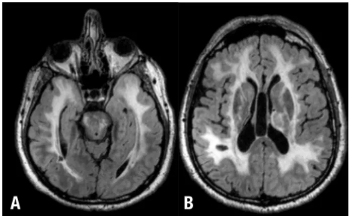
he patient was submitted to MRI which revealed, on FLAIR, extensive areas of hypersignal in subcortical white matter, predominantly frontal, temporal and pa-rietal, in addition to compromised external and internal capsules, brain stem and presenting lacunar infarcts in the temporal and right parietal regions (Figure 1).
Morphometric analysis was also performed using segmentation by the signal intensity technique, evi-dencing the percentage of frontal lobe lesions (41.8%) (Table 2).
Genetic analysis was carried out (Laboratoire Géné-tique Moléculaire de l’ Hôpital Lariboisière – Paris, Prof. Elisabeth Tournier-Lasserve) based on the direct DNA sequencing of exons 3 and 4 of the Notch 3 gene
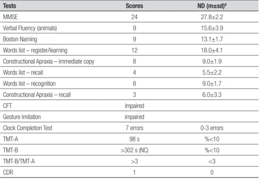
(chro-Table 1. Results of CERAD neuropsychological assessment and supplementary tests.
Tests Scores ND (m±sd)9
MMSE 24 27.8±2.2
Verbal Fluency (animals) 9 15.6±3.9
Boston Naming 9 13.1±1.7
Words list – register/learning 12 18.0±4.1
Constructional Apraxia – immediate copy 8 9.0±1.9
Words list – recall 4 5.5±2.2
Words list – recognition 8 9.0±1.7
Constructional Apraxia – recall 3 6.0±3.3
CFT impaired
Gesture Imitation impaired
Clock Completion Test 7 errors 0-3 errors
TMT-A 98 s %<10
TMT-B >302 s (NC) %<10
TMT-B/TMT-A >3 <3
CDR 1 0
■ Dement Neuropsychol 2012 September;6(3):188-191
190 da Silva JCV, et al. CADASIL
mosome 19), which revealed a nucleotide substitution of Arginine (CGC) to Cysteine (TGC) at position 153 in exon 4 (c.535 C >T: R153C), consistent with CADASIL diagnosis, conirming the etiology of the disease. hree of his siblings were later conirmed as carrying the same mutation. Recently, these individuals were included in a study assessing cognitive and neuroimaging proile.15
DISCUSSION
Four large studies encompassing a total of 175 individu-als have investigated the proile of cognitive decline in CADASIL.16,17 Of these studies, two focused on the
rela-tionship of the age efect and disease stage with cogni-tive proile.17,18
In the present case, changes were evident in global performance (MMSE) and in language, memory, aprax-ia and executive function domains.
In the language domain, both semantic verbal luen-cy (animals category) and naming ability were compro-mised. Deicit in verbal luency is frequently observed in studies on CADASIL.16,19 In the study by Bufon et.
al.,18 verbal luency (semantic category) was found to be
reduced.
Memory showed compromised register/learning yet better performance for recognition compared to spon-taneous recall. Memory in patients with CADASIL tends to by relatively preserved, where patients may present compromise in immediate memory and free recall. On the other hand, both recall with cues and recognition are invariably preserved, suggesting that the encoding process is preserved.16,19
Results on the Complex Figure Copying and Gesture Imitation tests revealed the presence of constructional and ideomotor apraxia, respectively. his inding may be of particular importance given that it has been little dis-cussed in the specialized literature. Some studies20 have
reported ideomotor apraxia in 15% of individuals with lesions conined to the thalamic or lenticular region. Ragno et. al.,21 studied 12 individuals from two
fami-lies and found that only one had deicit in ideomotor apraxia. Trojano et. al.,22 suggested that constructional
and ideomotor apraxia can appear in some patients with cortical lesions. Peter et al.,23 in search of evidence,
carried out a meta-analysis of reports published in the literature between 1994 and 1996, which included 82
patients and focused on apraxias associated with le-sions in deep brain structures, such as the basal gan-glia, thalamus and internal capsule. he study revealed that lesions to periventricular deep white matter play a crucial role in the development of apraxias, particularly ideomotor.
Executive function was also impaired, evidenced by reduced verbal luency, planning diiculties and prob-lems in space usage on the Clock Completion Test, slow-ness on the TMT-A (also relecting attention deicit), incomplete TMT-B (also relecting deicit in shifts in attention) and TMT-B/TMT-A >3, suggesting impaired cognitive lexibility. In line with indings of previous studies, executive dysfunction was clearly evident. Buf-fon et al.,18 in a study of 42 individuals with CADASIL,
found executive dysfunction in almost 90% of individu-als under 50 years of age, and suggested this inding may be explained by a decline in attention and memory performance consistent with some degree of frontal subcortical dysfunction.
Despite concerted research eforts, the mechanisms underlying cognitive dysfunction in CADASIL remain unclear. However, evidence suggests these mecha-nisms may be related to disruption of corticosubcorti-cal/or corticocortical connections due to progressive damage to white matter18 and that cognitive decline in
CADASIL is likely related to accumulated lacunar in-farcts and augmented ventricular volume, but not to brain atrophy24,25.
A
B
Figure 1. MRI FLAIR sequence in axial plan at temporal lobe [A] and basal ganglia [B] levels.
Table 2. Calculation of hyperintense brain lesion volume on FLAIR sequence.
Total Volume % Total FL % Total OL % Total PL % Total TL % Total Sum % Dif.
287.0 100.0 119.9 41.8 3.8 1.3 26.7 9.3 57.4 20.0 207.8 27.6
Dement Neuropsychol 2012 September;6(3):188-191 ■
CADASIL da Silva JCV, et al. 191 Conclusion. he CADASIL case reported, in addition to
exhibiting a characteristic neuroimaging pattern, was diagnostically conirmed by Notch-3 gene analysis. he neuropsychological indings were consistent with those reported in the literature, most notably the pres-ence of apraxias, seldom mentioned in the specialized literature.
It is hoped that this individual and the other
mem-bers of this and other families can beneit from the fu-ture development of protocols for pharmacological in-tervention and cognitive rehabilitation.
Acknowledgements. We would like to thank Prof. Elisa-beth Tournier-Lasserve, head of the service of the Labo-ratoire Génétique Moléculaire de l’Hôpital Lariboisière – Paris, for carrying out the genetic analysis.
REFERENCES
1. Joutel A, Corpechot C, Ducros A, et al. Notch3 mutations in CADASIL, a hereditary adult-onset condition causing stroke and dementia. Nature 1996;383(6602):707-710.
2. Chabriat H, Vahedi K, Iba-Zizen MT, et al. Clinical spectrum of CADA-SIL: a study of 7 families. Cerebral autosomal dominant arteriopathy with subcortical infarcts and leukoencephalopathy. Lancet. 1995;346 (8980):934-939.
3. Tournier-Lasserve E, Joutel A, Melki J, et al. Cerebral autosomal domi-nant arteriopathy with subcortical infarcts and leukoencephalopathy maps to chromosome 19q12. Nat Genet 1993;3:256-259.
4. Chabriat H, Levy C, Taillia H et al. Patterns of MRI lesions in CADASIL. Neurology 1998;51:452–457.
5. Markus HS, Martin RJ, Simpson MA, Dong YB, Ali N, Crosby AH, et al. Diagnostic strategies in CADASIL. Neurology 2002;59:1134-1138. 6. Tomimoto H, Ohtani R, Wakita H, Lin JX, Miki Y, Mizuno T. [Distribution
of ischemic leukoaraiosis in MRI: a difference from white matter lesions in CADASIL]. No To Shinkei. 2005;57:125-130.
7. Schultz A, Santoianni R, Hewan-Lowe K. Vasculopathic changes of CA-DASIL can be focal in skin biopsies. Ultrastruct Pathol 1999;23:241-247. 8. Morris JC, Heyman A, Mohs RC, et al. The Consortium to Establish a Reg-istry for Alzheimer’s Disease (CERAD). Part I. Clinical and neuropsycholog-ical assessment of Alzheimer’s disease. Neurology 1989;39: 1159-1165. 9. Bertolucci PHF, Okamoto, IH, Toniolo Neto J, Ramos LR, Brucki SMD.
Desempenho da população brasileira na bateria neuropsicológica do Consortion to Establish a Registry for Alzheimer´s Disease (CERAD). Rev Psiq Clin 1998;25:80-83.
10. Reitan RM, Wolfson D. The Halstead-Reitan neuropsychological test battery: theory and clinical interpretation. 2. ed. Tucson, AZ: Neuropsy-chology Press; 1993:912.
11. Watson YI, Arfken CL, Birge SJ. Clock completion: an objective screen-ing test for dementia. J Am Geriatr Soc 1993;41:1235-1240. 12. Barbize J, Duizabo P. Manual de Neuropsicologia. São Paulo: Ed.
Mas-son do Brasil; 1985:158.
13. Morris JC. The Clinical Dementia Rating (CDR): current version and scoring rules. Neurology 1993;43:2412-2414.
14. Montano MB, Ramos LR. [Validity of the Portuguese version of Clinical Dementia Rating]. Rev Saude Publica 2005;39:912-917.
15. Silva, JCV, Emerson L. Gasparetto, André C. Cognitive and neuroimag-ing profile of a Brazilian family with CADASIL. Arq Neuropsiquiatr 2011; 69:436-440.
16. Charlton RA, Morris RG, Nitkunan A, Markus HS. The cognitive profiles of CADASIL and sporadic small vessel disease. Neurology 2006;66: 1523-1526.
17. Amberla K, Waljas M, Tuominen S, Almkvist O, Poyhonen M, Tuisku S, Kalimo H, Viitanen M. Insidious cognitive decline in CADASIL. Stroke 2004;35:1598-1602.
18. Buffon F, Porcher R, Hernandez K, Kurtz A, Pointeau S, Vahedi K, et al. Cognitive profile in CADASIL. J Neurol Neurosurg Psychiatry 2006;77: 175-180.
19. Peters N, Opherk C, Danek A, Ballard C, Herzog J, Dichgans M. The pattern of cognitive performance in CADASIL - a monogenic condition leading to subcortical ischemic vascular dementia. Am J Psych 2005; 162:2078-2085.
20. Basso A, Faglioni P, Luzzatti C. Methods in neuroanatomical research and experimental study of limb apraxia. In: Roy EA (editor). Neuropsy-chological studies of apraxia and related disorders. Amsterdam: North-Holland;1985:179-202.
21. Ragno M, Fabrizi GM, Cacchiò G, et al. Two novel Italian CADASIL families from Central Italy with mutation CGC-TGC at codon 1006 in the exon 19 Notch3 gene. Neurol Sci 2006;27:252-256.
22. Trojano L, Ragno M, Manca A, Caruso G. A kindred affected by cerebral autosomal dominant arteriopathy with subcortical infarcts and leuko-encephalopathy (CADASIL). A 2-year neuropsychological follow-up. J Neurol 1998;245:217-222.
23. Peter P. Pramstaller and C. David Marsden. The basal ganglia and apraxia. Brain 1996;119:319-340.
24. Yousry TA, Seelos K, Mayer M, et al. Characteristic MR lesion pattern and correlation of T1 and T2 lesion volume with neurologic and neuro-psychological findings in cerebral autosomal dominant arteriopathy with subcortical infarcts and leukoencephalopathy (CADASIL). AJNR Am J Neuroradiol 1999;20:91-100.