Six-month bracket failure rate with a flowable
composite: A split-mouth randomized controlled trial
Sindhuja Krishnan1, Saravana Pandian2, R. Rajagopal3
Introduction: The use of flowable composites as an orthodontic bonding adhesive merits great attention because of their adequate bond strength, ease of clinical handling and reduced number of steps in bonding. Objective: The aim of this Randomized Controlled
Trial was to comparatively evaluate over a 6-month period the bond failure rate of a flowable composite (Heliosit Orthodontic, Ivoclar Vivadent AG, Schaan) and a conventional orthodontic bonding adhesive (Transbond XT, 3M Unitek). Methods: 53 consecutive patients (23 males and 30 females) who fulfilled the inclusion and exclusion criteria were included in the study. A total of 891 brackets were analyzed, where 444 brackets were bonded using Heliosit Orthodontic and 447 brackets were bonded using Transbond XT. The survival rates of brackets were estimated with the Kaplan-Meier analysis. Bracket survival distributions for bonding adhesives, tooth location and dental arch were compared with the log-rank test. Results: The failure rates of the Transbond XT and the Heliosit Orthodontic groups were 8.1% and 6% respectively. No significant differences in the survival rates were observed between them (p = 0.242). There was no statistically significant difference in the bond failure rates when the clinical performance of the maxillary versus the mandibular arches and the anterior versus the posterior segments were compared. Conclusions: Both systems had clinically ac-ceptable bond failure rates and are adequate for orthodontic bonding needs.
Keywords: Bond failure. Flowable composite. Orthodontic bonding. Bracket survival. Adhesive. Debonding rate.
1 Postgraduation program, Department of Orthodontics and Dentofacial
Orthopaedics, Saveetha Dental College, Saveetha University, Chennai, Tamil Nadu, India.
2 Senior Lecturer, Department of Orthodontics and Dentofacial Orthopaedics,
Saveetha Dental College, Saveetha University, Chennai, Tamil Nadu, India.
3 Professor, Department of Orthodontics and Dentofacial Orthopaedics, Saveetha
Dental College, Saveetha University, Chennai, Tamil Nadu, India.
» The authors report no commercial, proprietary or financial interest in the products or companies described in this article.
DOI: http://dx.doi.org/10.1590/2177-6709.22.2.069-076.oar
How to cite this article: Krishnan S, Pandian S, Rajagopal R. Six-month bracket failure rate with a flowable composite: A split-mouth randomized con-trolled trial. Dental Press J Orthod. 2017 Mar-Apr;22(2):69-76.
DOI: http://dx.doi.org/10.1590/2177-6709.22.2.069-072.oar
Submitted: July 19, 2016 - Revised and accepted: September 27, 2016
Contact address: Sindhuja Krishnan
Department of Orthodontics and Dentofacial Orthopaedics, Madha Dental College, MGR University, Chennai, Tamilnadu, India
E-mail: [email protected]
Introdução: o uso de resinas compostas fluidas como agentes de cimentação em Ortodontia tem merecido grande atenção, em função de sua adequada capacidade adesiva, facilidade de uso clínico e número reduzido de etapas de colagem. Objetivo: o objetivo deste estudo randomizado controlado foi avaliar o índice de falhas nos 6 meses após a colagem com uma resina composta fluida (Heliosit Orthodontic, Ivoclar Vivadent AG, Schaan), em comparação com um adesivo ortodôntico convencional (Transbond XT, 3M Unitek).
Métodos: 53 pacientes consecutivos (23 homens e 30 mulheres) que se enquadravam nos critérios de inclusão adotados foram incluídos no presente estudo. No total, 891 braquetes foram analisados, sendo 444 colados com o Heliosit Orthodontic e 447 colados com o Transbond XT. As taxas de sobrevivência dos braquetes foram estimadas por meio da análise de Kaplan-Meier. As distribuições das taxas de sobrevi-vência dos braquetes em função do adesivo usado, do dente e da arcada dentária em questão foram comparadas por meio do teste de log-rank.
Resultados: os índices de falhas para os grupos Transbond XT e Heliosit Orthodontic foram, respectivamente, de 8,1% e 6%. Não foram observadas diferenças significativas entre os grupos quanto às taxas de sobrevivência dos braquetes (p = 0,242). Também não foram observadas diferenças estatisticamente significativas quanto aos índices de falhas quando se comparou a performance clínica nas arcadas dentárias superior e inferior, e nos segmentos anterior e posterior da boca. Conclusões: ambos os sistemas apresentaram índices de falhas clinicamente aceitáveis, podendo ser considerados adequados para a colagem ortodôntica.
INTRODUCTION
In orthodontics, it’s a standard clinical practice to bond brackets to etched teeth using chemical or light-curing adhesive systems. The high initial bond strength, the minimal extent of oxygen inhibition and the extended working time for optimal brack-et placement have contributed to the popularity of light-curing adhesives.1,2,3
The majority of adhesives currently used for orth-odontic bonding are complex materials composed of synthetic polymers such as bisphenol-A glycol di-methacrylate (Bis-GMA) with either ethylene glycol dimethacrylate (EGDMA) or triethylene glycol di-methacrylate (TEGDMA) as a diluent to reduce the viscosity of Bis-GMA. In addition to the above, mol-ecules that promote or modify the polymerization re-action are incorporated.4
Filler particles are incorporated into a resin matrix to improve its mechanical properties.
The primary purpose of the iller particles is to in-crease the strength of the composite and reduce the amount of matrix material. The illers provide rein-forcement of matrix material, reduction in polymeriza-tion shrinkage and reducpolymeriza-tion in thermal expansion and contraction (dimensional changes). It results in reduced microleakage, improved workability with increased vis-cosity, reduction in water sorption and sotening.5,6,7
Among the composite resins that could be used as orthodontic adhesives, lowable composites merit great attention due to their clinical handling characteristics.8
Flowable composites exhibit two desirable clinical han-dling characteristics that have not existed for composites until very recently:9
1) no stickiness; 2) luid injectability.
The other desirable characteristics include adequate bond strength, suicient working time, shorter curing time, and improved ease of use.
Flowable composites retain the same small particle sizes of traditional hybrid composites, but have less iller content, thus, reducing the viscosity of the mixture. Heliosit Orthodontic (Ivoclar Vivadent AG, Schaan) is a light-curing, highly translucent single-component bonding material for brackets and is supplied in con-venient syringes. The monomer matrix consists of urethane dimethacrylate, Bis-GMA and decandiol di-methacrylate (85 wt %). The iller consists of a highly
dispersed silicon dioxide (14 wt %). The additional contents are catalysts and stabilizers (1 wt %).10 Heliosit
Orthodontic was developed to ease the bonding pro-cedure of orthodontic attachments by eliminating the need for primer application both on the bracket base and on etched tooth surface.
The purpose of this randomized clinical trial was to compare the bond failure rates of brackets bonded with a lowable composite (Heliosit Orthodontic, Vivadent Ivoclar, Schaan) and a conventional multi-step system (Transbond XT, 3M Unitek, Ca, USA) over a 6-month period. The secondary aim was to investigate factors contributing to bracket failure, as tooth location and dental arch. The null hypothesis is that there is no dif-ference in the failure rates of brackets bonded with He-liosit Orthodontic or Transbond XT during the irst 6 months of preadjusted edgewise appliance therapy.
MATERIAL AND METHODS
Estimation of power and sample size
The sample size of the study was estimated by the number of brackets bonded either with Trans-bond XT or Heliosit Orthodontic. In this study, each bonded bracket was the unit of measurement. To obtain an adequate power of 80%, the sample size was determined to be 813 brackets. For 813 brack-ets, approximately 53 patients were required. A buffer of 20% was included in order to compensate for any loss of patients during follow-up. Institutional review board clearance and approval from human ethical committee were obtained from the Saveetha Univer-sity for this single-centered study and patients gave their written consent for participation.
Inclusion criteria
» Age group: 13 to 30 years. » Complete permanent dentition.
» Patient requiring ixed appliance therapy in both arches.
» Both extraction and non-extraction cases. » Absence of labial or buccal restoration.
Exclusion criteria
» Patient with congenital enamel defects, illings or hypoplasia.
Table 1 - Quadrant-wise distribution of adhesive.
Table 2 - Sample characteristics. » Surgically exposed teeth.
» Dentition with occlusal interferences.
Thus, 53 consecutive patients (23 males and 30 fe-males) who fulilled the inclusion criteria were selected for this study. Patients were selected from those seeking orthodontic treatment at the Orthodontics Department of Saveetha University.
Study design
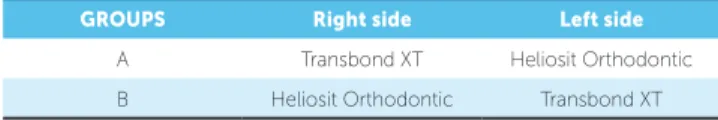
The study design was a single blinded, split-mouth, cross-arch Randomized Controlled Trial. Trans-bond XT was used as the control material and Heliosit Orthodontic was used as the experimental material.
The patients were unaware of the material and the side of the mouth chosen for bonding of the brack-ets. The decision as to the choice of material for each side was allocated by using a Random Number Table. The adhesives were separated based on group category. In Group A, the brackets were bonded with Trans-bond XT on the right side and Heliosit Orthodontic on the let side of maxillary and mandibular arches, and vice versa in Group B (Table 1). This was done so that both materials would be equally distributed on maxil-lary and mandibular right and let quadrants.
The total number of patients involved in the study was 53 (23 males and 30 females). For 53 patients, the total number of brackets bonded was 901. All the pa-tients were bonded with 0.022-in slot MBT prescrip-tion (Gemini stainless steel brackets, 3M Unitek). Two patients were excluded from the study as they did not come regularly for the monthly follow-up. Another patient discontinued treatment ater the third month of bonding. By the end of the 6-month trial, the to-tal number of patients analyzed was 50 and the number of brackets analyzed was 891. The characteristics of the study sample are described in Table 2.
Bonding procedure
It was not possible to blind the operators to the bonding system being used because the two systems had different forms of application. The teeth were cleaned using a rubber cup with pumice and water slurry, rinsed, isolated with cheek retractors and a low-volume suction evacuator, and dried with oil-free air. For the teeth to be bonded using Transbond XT, 37% phosphoric acid was applied to the enam-el surface for 15 seconds before rinsing with water
and drying until the enamel became frosty white. Transbond XT primer was then applied to the etched enamel according to the manufacturer’s instructions and given a gentle burst of air. Transbond XT adhe-sive paste was placed on the back of the brackets.
For the teeth to be bonded using Heliosit Orth-odontic, 37% phosphoric acid was applied to the enamel surface for 15 seconds before rinsing with water and drying until the enamel was frosty white. As per the manufacturer’s recommendation no inter-mediate primer was applied on the surface to be bond-ed. Then Heliosit Orthodontic was placed on the back of the brackets and the brackets were placed onto the etched enamel surface.
In both groups the brackets were positioned along the long axis of the teeth with the help of a bracket positioning gauge, according to the MBT bracket positioning chart. Suicient pressure was applied to squeeze out excess adhe-sive, which was removed from the margins of the bracket
GROUPS Right side Left side
A Transbond XT Heliosit Orthodontic
B Heliosit Orthodontic Transbond XT
n %
Number of patients 53
-Number of brackets 891
-Distribution of brackets by bonding material
Transbond XT 447 50.1%
Heliosit Orthodontic 444 49.9%
Distribution of brackets bydental arches
Maxillary 445 49.9%
Mandibular 446 50.1%
Distribution of brackets by tooth type
Anterior 601 67.45%
Posterior 290 32.55%
Distribution of brackets by side of the arches
Right side 438 49.15%
base with an explorer before polymerization. When satis-ied with the bracket positioning, the adhesive was cured using a Halogen light curing unit (QHL75, Dentsply). The adhesive was cured from the occlusal, gingival, mesial and distal aspects for 10 seconds each.
Standardization was achieved by bonding all the brackets in one appointment by the same operator. Aligning archwire of choice was either a 0.014-in NiTi wire or a 0.016-in NiTi wire, depending on the initial degree of alignment and crowding. These wires were tied approximately 10 minutes ater bonding, using elastomeric modules. No bite planes were used during treatment. The study was concluded before the addition of edgewise archwires. Verbal and written instructions about diet and care were given immediately to the pa-tient ater itting the appliances.
Observation and follow-up
The bonding, follow-up and assessment of bond failure rate of the brackets was done by a single opera-tor (investigaopera-tor). Patients were followed for a period of 6 months. If a bond failed, the following informa-tion was recorded: (1) site of bond failure, (2) number of failed brackets (3) date of bond failure, and (4) pos-sible reason for bond failure. The patients were treat-ed at 3-4 week intervals but were requesttreat-ed to come as soon as possible in case of a bond failure. When the patient was unaware of a bracket failure, the date of the appointment was recorded as the date of failure. Based on the tooth location, they were divided into anterior and posterior segments. The first and second premolars brackets were evaluated in the posterior segment as the molars were banded. Failed brackets were rebonded with the conventional adhesive, but not included in the further study.
Statistical analysis
Statistical data analysis was carried out by us-ing the software SPSS v. 4.0 (SPSS Inc., Chicago, USA), at the 5% level of significance. The survival rates of the brackets were estimated by using the Ka-plan-Meier test. KaKa-plan-Meier estimates of bracket survival curves were plotted. Bracket survival distri-butions with respect to bonding procedure, dental arch (maxillary and mandibular) and tooth location (anterior and posterior) were compared using the log-rank test (p < 0.05).
RESULTS
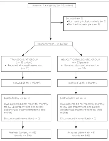
The low chart of the trial is given in Figure 1. During the 6-month observation period , 63 (7.1%) brackets failed: 27 (6%) in the Heliosit Orthodontic group and 36 (8.1%) in the Transbond XT group (Table 3). The corresponding bracket survival curves were plotted by using the Kaplan-Meier product-limit estimate (Fig 2). There was no signif-icant diference in terms of bracket failure risk over the 6 months between groups (p = 0.242, hazard ratio = 0.69; 95%
conidence interval 0.35-1.40; log rank test P = 0.251).
The maxillary arches had a 3.3% failure rate, and the mandibular arches a 3.8% failure rate; these were not statistically signiicant according to the log-rank test (P = 0.518; hazard ratio = 0.71; 95% conidence
level = 0.36-1.43). The inluence of the dental arches on bracket survival rate is shown in Fig 3.
Posterior brackets (premolars) showed lesser (2.6%) failure rates than anterior brackets (4.5%). Figure 4 shows the inluence of tooth location on bracket sur-vival rate. The log-rank test showed no signiicant dif-ferences between anterior and posterior brackets in terms of survival rate (P = 0.0492; hazard ratio = 0.42;
95% conidence level = 0.20-0.83).
Assessed for eligibility (n= 53 patient)
Randomized (n= 53 patient) Excluded (n= 0)
• Not meeting inclusion criteria (n= 0) • Declined to participate (n= 0)
HELIOSIT ORTHODONTIC GROUP (n= 53 patient) • Received allocated intervention
(n= 53)
Analyzes (patient, n= 48) (bonds, n= 891) TRANSBOND XT GROUP
(n= 53 patient) • Received allocated intervention
(n= 53)
Lost to follow-up (n= 3)
(Two patients did not report for monthly follow-ups properly and one patient discontinued treatment from the third month)
Discontinued intervention (n= 0) Lost to follow-up (n= 3)
(Two patients did not report for monthly follow-ups properly and one patient discontinued treatment from the third month)
Discontinued intervention (n= 0)
Followed up for 6 months Followed up for 6 months
Analyzes (patient, n= 48) (bonds, n= 891)
DISCUSSION
There was no statistically significant difference in bond failure rates between the Transbond XT and the Heliosit Orthodontic groups at p < 0.05. The overall bond failure in this study was 63 brackets with 7.1% failure rate. The bond failure of Transbond XT and Heliosit Orthodontic was 36 (8.1%) and 27 (6.0%), respectively (Table 3). Studies by O’Brien et al,11
Adolfsson et al12 and Cal-Neto et al13 reported failure
rates of 4.7-6.6%, 7.2% and 5.48%, respectively, for various adhesive-bracket combinations.
Bond failure rates below 10% are generally con-sidered clinically acceptable.14 It is difficult to make
a direct comparison between studies due to the vari-ety of techniques, materials, research designs and trial durations.15
In any time scale, the overall failure rates for a clinical sample can be calculated. This could provide a straight-forward statement of the overall percentage of failures in a sample over a certain time, or it can be used to compare variables in a sample. Failure rates are a widely accepted means of representing the performance of brackets.16
In in vivo studies, socioeconomic and dental status of patients, malocclusion classiication and resultant mechanotherapy may afect the outcomes. Further-more, masticatory forces varying with facial type, cul-turally inluenced dietary habits, and sex diferences may also inluence the results.17
Heliosit Orthodontic was developed to facilitate orthodontic bonding by eliminating the need for primer application both on the bracket base and the etched tooth surface. It is a Bis-GMA-based light-curing orthodontic adhesive designed for bonding ceramic and metal orthodontic brackets. Its mono-mer matrix consists of urethane dimethacrylate, Bis-GMA and decandiol dimethacrylate (85 wt%). The filler consists of highly dispersed silicon dioxide (14 wt%). Additional contents are catalysts and stabi-lizers (1 wt%). Although Heliosit Orthodontic was initially developed for bonding of brackets, its applica-tion has been as an adhesive for bonding lingual retain-ers,28,29 and even as a luting cement for prosthesis.18
Heliosit Orthodontic has not been widely studied to clinically assess its bonding eicacy. Reynolds19 stated
that bond strengths of 5.9 to 7.8 MPa were clinically acceptable and Heliosit Orthodontic adhesive had shear bond strength within the already-mentioned clinically
Figure 2 - Overall Kaplan-Meier survival plot comparing bond failure between Transbond XT (3M Unitek) and Heliosit Orthodontic (Ivoclar Vivadent).
Figure 3 - Overall Kaplan-Meier survival plot comparing bond failure in maxil-lary and mandibular arches.
Figure 4 - Overall Kaplan-Meier survival plot comparing bond failure in ante-rior and posteante-rior segments.
Survival Functions
Days
Cumula
tive Survival
Group Transbond XT Heliosit Orthodontic Transbond XT censored Heliosit Orthodontic censored
1.0
0.8
0.6
0.4
0.2
0.0
0 50 100 150 200 250
Days
Cumula
tive Survival
Arch
1.0
0.8
0.6
0.4
0.2
0.0
0 50 100 150 200 250
Maxillary Mandibular Maxillary-censored Mandibular-censored
Survival Functions
Days
Cumula
tive Survival
Segment
1.0
0.8
0.6
0.4
0.2
0.0
0 50 100 150 200 250
Anterior Posterior Anterior-censored Posterior-censored
acceptable range. Few in vitro studies have been
car-ried out to evaluate its bond strength. While compar-ing Heliosit Orthodontic with Transbond XT, studies concluded that Transbond XT exhibited higher bond strengths in all the studies. However , the bond strengths of Heliosit Orthodontic were clinically acceptable.20,21,22
Manufacturers claim that its lexural strength is 80 MPa and that the shear bond strength of brackets on etched enamel is 10 MPa, for ceramic brackets and 12 MPa for metal brackets.
Unlike orthodontic bonding systems, such as Trans-bond XT, Heliosit Orthodontic can be applied to acid-etched enamel without the use of intermediate bonding resin due to its low iller loading and improved lowabil-ity. By reducing the number of steps during bonding, cli-nicians can save time and reduce potential errors related to contamination during the bonding procedure. It also allows easier and more even application to the mesh base of the brackets. Another added advantage of using this lowable composite is that it proves to be more cost ef-icient as it does not require an intermediate primer. Thus, as Heliosit Orthodontic guarantees clinically ac-ceptable survival rate, it is undoubtedly advantageous for orthodontic bracket bonding.
Since not all brackets failed by the end of the study period, a survival analysis was done. This analysis dif-fers from other types of statistics because it can use partial or censored information. This analysis was used here because non-failed attachments were all censored at the conclusion of the treatment, and it is impossible
to follow all the brackets to failure. The Kaplan-Meier survival analysis showed that the mean survival time for brackets bonded with Transbond XT (178.83 days) was similar to Heliosit Orthodontic (179.03 days) (Fig 2).
Another factor observed in relation to survival time was that, in the present study, the maximum number of bond failures occurred during the initial 3 months of treatment. The most common reasons cited by the patients for the bond failures were hard brushing and biting on a hard food substances.
O’Brien et al11 presented three possible reasons for
the increased failure rate during the irst 6 months of treatment:
1) They suggested that any deiciencies in the bond strength of any individual bracket/adhesive combina-tion would become evident within this initial period of treatment.
2) The initial period of treatment is also a time of acclimatization and experimentation for patients con-cerning the type of food that can be tolerated by ixed orthodontic appliances.
3) The initial phase of treatment may involve a pe-riod of overbite correction and, therefore, heavy oc-clusal forces may be applied to many of the bonded attachments.
In the present study, failure rates demonstrated no sig-niicant diferences between maxillary and mandibular brackets, with mandibular bonds failing more frequent-ly (maxillary = 3.3% bond failure; mandibular = 3.8% bond failure) at p = 0.304 (Table 3). Similar results have
Variable Number Bracket failures Failure rate (%) p value log-rank
Material used for bonding procedure
Transbond XT 444 36 8.1%
0.242 .251
Heliosit Orthodontic 447 27 6%
Dental arch
Maxillary arch 445 29 3.3%
0.601 .518
Mandibular arch 446 34 3.8%
Bracket location
Anterior region 601 40 4.5%
0.488 .492
Posterior region 290 23 2.6%
been obtained in other studies which found that the fail-ure rate of maxillary brackets was less than the failfail-ure rate of mandibular brackets.23-26
Potential reasons for this could include increased masticatory load on mandibular brackets. In patients with a normal transverse arch relationship, brackets bonded to mandibular teeth have potential antagonists in centric relation, whereas maxillary brackets do not.
It is evident that posterior teeth failed less frequently than anterior teeth. This was true for the overall sample (posterior = 2.6% bond failure and mean survival time of 178.33 days; anterior = 4.5% bond failure and mean survival time of 179.22 days). However, the indings were not statistically signiicant at p < 0.05 (Table 3).
Many studies report that posterior teeth sufer more bracket failures than do brackets on anterior teeth. The higher occlusal forces on posterior teeth, the diiculty of access and moisture control and the more aprismatic enamel on premolars could be pos-sible reasons for this scenario.11,27
The literature on bonding has shown that the pat-tern of orthodontic bond failure in vivo is not uniform
for all the teeth in either dental arch and also between the arches. This occurs even though all the teeth are bonded by the same operator using the same adhesive and a standard protocol, thus emphasizing that only cer-tain sites in the mouth have a greater predilection for failures than others. This can be due to some factors within the oral cavity like tooth morphology, mastica-tory forces and chewing pattern, which predispose cer-tain sites to a greater rate of bond failure.
Present results indicate that both Transbond XT and Heliosit Orthodontic can be eiciently used for bond-ing orthodontic appliances. From a clinical standpoint, the use of Heliosit Orthodontic can be more advanta-geous because it reduces the number of clinical steps re-quired to bond brackets and, thus, saves chair time. It is also cost efective. Undoubtedly, it improves the quality of bonding and eiciency of the operator by reducing the risk of salivary contamination during the bonding procedure. A disadvantage to this technique of using lowable composites as orthodontic bonding adhesive is that it denies application of a illed sealant that protects the enamel from white spot lesions.
However, the choice of a particular orthodontic bonding adhesive will depend on the clinical preference of the operator.
CONCLUSIONS
1) In this randomized controlled trial, the conven-tional adhesive (Transbond XT) and the lowable com-posite (Heliosit Orthodontic) had similar and clinically acceptable bond failure rates.
2) Heliosit Orthodontic is a more desirable compos-ite because it reduces the number of clinical steps. It re-duces chair time, is cost efective and rere-duces the risk of salivary contamination.
Formatting of funding sources
1. Buyukyilmaz T, Usumez S, Karaman AI. Efect of self-etching primers on bond strength—are they reliable? Angle Orthod. 2003 Feb;73(1):64-70.
2. Eliades T, Eliades G, Brantley WA, Johnston WM. Polymerization
eiciency of chemically cured and visible light-cured orthodontic adhesives: degree of cure. Am J Orthod Dentofacial Orthop. 1995 Sept;108(3):294-301.
3. Uysal T, Basciftci FA, Uşümez S, Sari Z, Buyukerkmen A. Can previously
bleached teeth be bonded safely? Am J Orthod Dentofacial Orthop. 2003 June;123(6):628-32.
4. Faltermeier A, Rosentritt M, Faltermeier R, Reicheneder C, Müssig D.
Inluence of iller level on the bond strength of orthodontic adhesives. Angle Orthod. 2007 May;77(3):494-8.
5. Brantley WA, Eliades T. Orthodontic materials. Scientiic and clinical
aspects. New York: Thieme; 2001.
6. Bishara SE, Aljouni R. Evaluation of the orthodontic application of two
new restorative systems. Hellenic Orthod Review. 2004;(7):25-32.
7. Anusavice K, Shen C, Rawls R. Phillips Science of Dental Materials.
12th ed. St. Louis: Saunders; 2012.
8. Elaut J, Asscherickx K, Vande Vannet B, Wehrbein H. Flowable
composites for bonding lingual retainers. J Clin Orthod. 2002 Oct;36(10):597-8.
9. Tecco S, Traini T, Caputi S, Festa F, de Luca V, D’Attilio M. A new one-step
dental lowable composite for orthodontic use: an in vitro bond strength study. Angle Orthod. 2005 July;75(4):672-7.
10. Ivoclar Vivadent. Heliosit Orthodontic product proile. Liechtenstein: Ivoclar Vivadent; 2003.
11. O’Brien KD, Read MJ, Sandison RJ, Roberts CT. A visible light-activated direct-bonding material: an in vivo comparative study. Am J Orthod Dentofacial Orthop. 1989 Apr;95(4):348-51.
12. Adolfsson U, Larsson E, Øgaard B. Bond failure of a no-mix adhesive during orthodontic treatment. Am J Orthod Dentofacial Orthop. 2002 Sept;122(3):277-81.
13. Cal-Neto JP, Miguel JA, Zanella E. Efect of a self-etching primer on shear bond strength of adhesive precoated brackets in vivo. Angle Orthod. 2006 Jan;76(1):127-31.
14. Mavropoulos A, Karamouzos A, Kolokithas G, Athanasiou AE. In vivo evaluation of two new moisture-resistant orthodonticadhesive systems: a comparative clinical trial. J Orthod. 2003 June;30(2):139-47; discussion 127-8.
REFERENCES
15. Littlewood SJ. Investigation of a hydrophilic primer for orthodontic bonding: an in vitro study. J Orthod. 2000 June;27(2):181-6.
16. Millett DT. A comparative clinical trial of a compomer and a resin adhesive for orthodontic bonding. Angle Orthod. 2000 June;70(3):233-40. 17. Pandis N, Theodore Eliades. A comparative in vivo assessment of the
long term failure rate of 2 self etching primers. Am J Orthod Dentofacial Orthop. 2005;128(1):96-8.
18. Breeding LC, Dixon DL, Caughman WF. The curing potential the curing potential of light-activated composite resin luting agents. J Prosthet Dent. 1991 Apr;65(4):512-8.
19. Reynolds IR. A review of direct orthodontic bonding. Br J Orthod. 1975;2(3):171-8.
20. Murray SD, Hobson RS. Comparison of in vivo and in vitro shear bond strength. Am J Orthod Dentofacial Orthop. 2003 Jan;123(1):2-9. 21. Durrani OK. In vitro comparison of shear bond strength of Transbond XT
and Heliosit Orthodontic as Direct bracket bonding adhesives. Pakistan Oral Dent J. 2008:28(2):203-6.
22. Bradburn G, Pender N. An in vitro study of the bond strength of two light-cured composites used in the direct bonding of orthodontic brackets to molars. Am J Orthod Dentofacial Orthop. 1992 Nov;102(5):418-26. 23. Newman GV. Epoxy adhesives for orthodontic attachments: progress
report. Am J Orthod Dentofacial Orthop. 1965;51(12):901-12. 24. Zachrisson BU, Brobakken BO. Clinical comparison of direct versus
indirect bonding with diferent bracket types and adhesives. Am J Orthod. 1978 July;74(1):62-78.
25. Trimpeneers LM, Dermaut LR. A clinical trial comparing the failure rates of two orthodontic bonding systems. Am J Orthod Dentofacial Orthop. 1996;110(5):547-50.
26. Linklater RA. Bond failure patterns in vivo. Am J Orthod Dentofacial Orthop. 2003 May;123(5):534-9.
27. Sunna S, Rock WP. Clinical performance of orthodontic brackets and adhesive systems: a randomized clinical trial. Br J Orthod. 1998 Nov;25(4):283-7.
28. Radlanski RJ, Zain ND. Stability of the bonded lingual wire retainer-a study of the initial bond strength. J Orofac Orthop. 2004;65:321-35. 29. Stormann I, Ehmer U. A prospective randomized study of diferent