Fernanda Angelieri*, Marinês Vieira da Silva Sousa**, Lylian Kazumi Kanashiro*, Danilo Furquim Siqueira*, Liliana Ávila Maltagliati*
Effects of low intensity laser on pain
sensitivity during orthodontic movement
Objective: To evaluate the efficiency of infrared diode laser on pain reduction during
canine initial retraction. Methods: Twelve patients in need of canine retraction were selected. The canines were retracted with closed NiTi coil springs activated to 150 g per side. One canine of each patient was randomly selected for laser irradiation imme-diately after activation and after 3 and 7 days. The contralateral canines were taken as control group and were submitted only to simulation of laser application. Diode laser (ArGaAl) was employed at wavelength of 780 nm, power of 20 mW and energy density in the target tissue of 5 J/cm2, for 10 seconds per point, delivering an energy of 0.2 J
per point and total energy (TE) of 2 J. The analgesic effect was evaluated with aid of a visual analogue scale (VAS), on which the patients indicated 0 to 10 according to the pain experienced at 12, 24, 48 and 72 hours after coil spring activation and laser ap-plication. The procedure was repeated after one month, upon reactivation of canine retraction. Results and Conclusions: There was no statistically significant difference between irradiated side (LG) and control side (CG). Thus, utilization of infrared diode laser (780 nm) according to the present protocol was not statistically effective to reduce pain sensitivity caused by orthodontic movement.
Abstract
Keywords: Diode laser. Low intensity laser. Orthodontic movement. Pain.
INTRODUCTION AND LITERATURE REVIEW
In clinical practice, there is almost a consen-sus that therapy with lowlevel laser (LLL) or la-ser therapy (LT) causes analgesic effect.3,10,13,15,19,24
Thus, laser could become an important aid for orth-odontic treatment6,9,13,14,20 since fear of feeling pain
is one of the main factors which discourages many patients to undergo thiskind of treatment.1,4,25
* PhD in Orthodontics, Bauru School of Dentistry, University of São Paulo. Professor of Dentistry Postgraduation, Orthodontics area, Methodist University of São Paulo. ** MSc, Program of Dentistry, Orthodontics area, Methodist University of São Paulo.
» The authors report no commercial, proprietary, or inancial interest in the
products or companies described in this article.
How to cite this article: Angelieri F, Sousa MVS, Kanashiro LK, Siqueira DF,
Low level laser reduces pain through two different mechanisms: Stimulating the pro-duction of beta-endorfine, a natural media-tor produced by the organism to reduce pain, and inhibiting the release of arachidonic acid, from the damaged cells, which would gener-ate metabolites that interact with the pain re-ceptors. While arachidonic acid produces local effect, beta-endorfine produces analgesic ef-fect throughout the body.17,18 Another type of
laser action on the mechanisms for suppress-ing pain, consists in the repression of the con-duction of nervous impulses, at the peripheral nerve endings, acting upon the mechanism of “sodium-potassium pump”, in order to impair the transmission of local painful impulse.12
Other mechanisms of pain suppression by the employment of laser therapy have been re-ported in the literature, as consequence of the enhancement of endorphin synthesis, demon-strated by Benedicenti in 1982 (apud Geno-vese8), or even the inhibition of prostraglandines
E2 (PG-E2) and interleukine 1-β (IL-1β),23
well-known pain mediators, produced during the in-flammatory process. Ataka2 observed a decrease
of nociceptive transmitters, such as bradykinin and serotonin, after low level laser irradiation. The nociceptors are the receptors present on the nervous terminals, free nerve endings of primary afferent neuron axon (thin mielinic A-delta fibers and amielinic C-fibers), capable to detect painful stimuli when the nociceptive transmitters are present.16
Some clinical research studies have also demonstrated the efficiency of laser to sup-press pain.5,11,13,22,24 Lim et al13 in 1995,
in-serted elastic separators to induce pain in the interproximal contacts of premolars of 39 subjects. The diode laser (ALGaAs), 830 nm, 30 mW, 59.7 mw/cm², was applied on the middle third of the dental root and the sub-jects were divided into four groups: Three of them in accordance to the different periods of
laser application (15, 30 and 60 seconds) and one group with placebo laser therapy for 30 seconds. The procedure was performed during five days and a scale was employed to quantify the pain experimented in each quadrant (Vi-sual Analogue Scale –VAS). The authors con-cluded that the group treated with laser, dem-onstrated lower levels of pain, when compared to the placebo group, suggesting that low level laser reduces the intensity of pain caused by orthodontic movement.
Turhani et al23 evaluated the effect of diode
laser (670 nm; 75 mW; 30 seconds per tooth) in 38 patients undergoing orthodontic treatment, with fixed appliances. The laser was immedi-ately applied over the middle third of the teeth and also after 6, 30, and 54 hours after inser-tion of the orthodontic arches. A control group received placebo laser therapy only. The results showed lower levels of pain for the group that received laser irradiation until 30 hours after the orthodontic activation. The authors concluded that laser therapy may have positive effects sup-pressing pain during orthodontic treatment.
Recently, Fujiyama et al7 showed that the
use of CO2 laser seems to be efficient to con-trol inhibitory effects on analgesia during the four first days, after force application, with-out interference on the amount of orthodontic movement, when 20 pulses of 2 W, 5 pulses per 1000 seconds, were applied at 2 mm focus.
It appears that there are still few studies which investigated the effects of low level la-ser for suppressing pain in orthodontics, and the protocols for laser application are still very variable. Thus, the purpose of this study was to evaluate the efficiency of diode laser, which presented wavelength at the infra-red spectrum (λ=780 nm, power output of 20 mW, with en-ergy density 5 J/cm2, 0,2 J of energy per point,
1
2
3
4
5 MATERIAL AND METHODS
Material
A total of 12 patients were selected, from the Postgraduation in Orthodontics of the Brazil-ian Association of Dental Surgeons (ABCD, São Paulo, Brazil), with mean age of 12.66 years. The inclusion criteria were:
» need of extraction of first premolars, due to double protrusion or dental crowding; » presence of permanent dentition; » absence of systemic diseases;
» no use of any medication and no previous orthodontic treatment.
The diode laser (Twin Laser, MMOptics, São Paulo, Brazil) was employed to irradiate maxillary and mandibular canines, submitted to orthodontic retraction, and the painful sen-sitivity decurrent from this orthodontic move-ment was evaluated by a visual scale (Visual Analogue Scale – VAS).13
This protocol was approved by the Research Ethics Committee at the São Paulo Methodist University, protocol nº 105303/06.
Methods
Orthodontic treatment
Metallic brackets, Andrews prescription, 0.022 x 0.028-in slot (Ormco Corp. Orange, CA, USA), were installed on canines and second premolars, while the first molars were properly banded. Bi-laterally, segmented stainless steel 0.016-in arch-wires were adapted to each side of the dental arch, in conjunction with a closed nickel-titani-um spring (12 mm in length, Morelli Ltda, Brazil) for the initial retraction of the canines (right and left). The intensity of the force released by the spring was measured by a dynamometer (Morelli Ltda), delivering force of 150 g.
Laser irradiation and evaluation of pain sensitivity
After the activation of the spring, only one of the canines (left or right) of each patient was
selected at random for application of diode laser Aluminum-Gallium-Arsenide (ArGaAl), low in-tensity, wavelength in the infra-red spectrum. For the canines on the opposite side, only one simula-tion was performed for laser applicasimula-tion. The ir-radiated side was considered as laser group (LG) and the non irradiated side, as control group (CG).
The diode laser (Twin Laser, MMOptics, São Carlos, Brazil), with light emission at 780 nm wavelength, power output 20 mW, energy den-sity on the surface tissue at 5 J/cm2, was
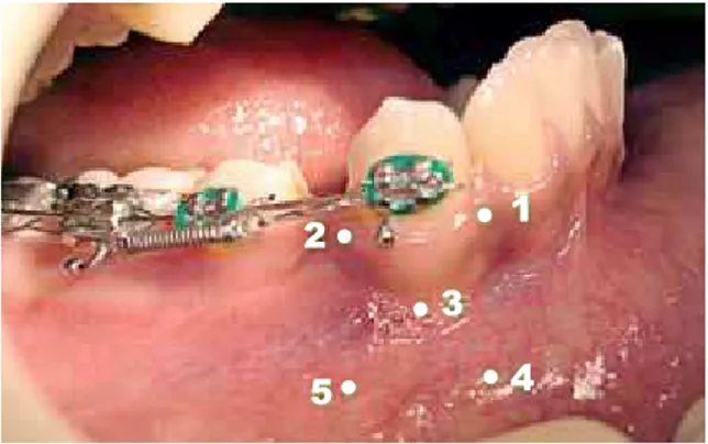
em-ployed for 10 seconds per point, resulting in 0,2 J of energy per point. As 10 points per tooth (5 bucally and 5 lingually) received irradiation (Fig 1), the total energy (TE) surrounding ca-nine roots was 2 J. The irradiations were per-formed by one operator, per points, employing light beam focused perpendicularly and in con-tact with the mucosa, which was kept clean and dry, through relative isolation.
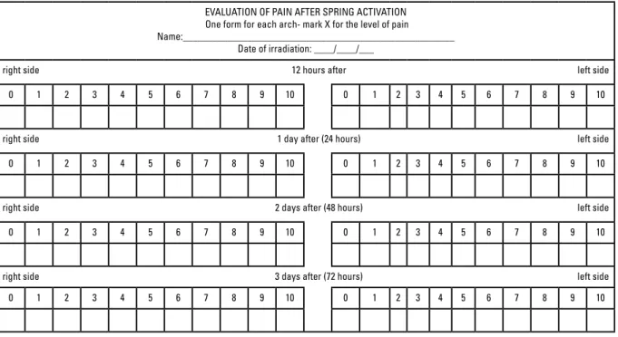
Laser irradiation was performed immediate-ly after the closed NiTi coil spring was activated (day 0), 3 and 7 days after the first application, resulting in 6 J/month of energy. The evalua-tion of the analgesic effect of laser irradiaevalua-tion was performed by the employment of a visual scale (Visual Analogue Scale – VAS)13 provided
to each patient (Fig 2). According to this scale, the patient was oriented to mark from 0 to 10, according to the intensity of pain experimented, differentiating the left and right sides, after 12 (T1), 24 (T2), 48 (T3) and 72 (T4) hours of laser irradiation.
After 30 days, the patients were submitted to a new activation of the closed springs, in order to keep the force exertion at 150 g/side, previously established. A new laser irradiation, following the same protocol, was performed on the same tooth which had been already irradiated, and one more time, the sensitivity was evaluated on the four periods described, employing the same scale. The outcomes were computed into an Excel table, for statistical analysis.
Statistical analysis
For comparison between the “irradiated” and “non-irradiated” sides, over several periods of evaluation, the parametric Wilcoxon test was em-ployed (p<0.05).
RESULTS
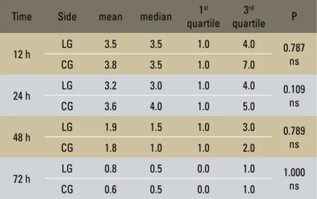
Tables 1 and 2 show the outcomes obtained on the first and second months, respectively, in the four evaluated moments (T1=12 h, T2=24 h, T3=48 h and T4=72 h) after laser application on the irradiated and non-irradiated sides.
The results presented on the first and on the sec-ond months, demonstrated that there is no statisti-cally significant difference between the irradiated and control sides, that is, the irradiation employing infra-red diode laser (780 nm) with the application protocol of 20 mW-10 sec-5J/cm2, 0,2 J per point,
Et=2 J, was not statistically significant for the reduc-tion of sensitivity to pain, caused by orthodontic movement during the initial retraction of canines.
DISCUSSION
In the literature, it is clear that almost all pa-tients submitted to fixed orthodontic treatment suffer some type of discomfort, considering the separation of teeth for posterior orthodontic banding or during the arch insertion, reaching
FIGURE 2 - Visual Analogue Scale (VAS).
EVALUATION OF PAIN AFTER SPRING ACTIVATION One form for each arch- mark X for the level of pain Name:_______________________________________________________
Date of irradiation: ____/____/___
right side 12 hours after left side
0 1 2 3 4 5 6 7 8 9 10 0 1 2 3 4 5 6 7 8 9 10
right side 1 day after (24 hours) left side
0 1 2 3 4 5 6 7 8 9 10 0 1 2 3 4 5 6 7 8 9 10
right side 2 days after (48 hours) left side
0 1 2 3 4 5 6 7 8 9 10 0 1 2 3 4 5 6 7 8 9 10
right side 3 days after (72 hours) left side
levels of pain which may also discourage them to continue the treatment, or even giving up at the beginning of the process.13,24,25
The perception of pain varies considerably from patient to patient.20 Thus, painis a highly
subjective sensation and due to this fact, it be-comes very difficult to quantify it in scientific re-searches.13,20,24
The Visual Analogue Scale (VAS) was pro-posed by Huskisson in 1974 in order to quantify pain.20 It is a 10 cm line which comprehends a
scale from 0 to 10 representing the thresholds of pain experimented, so that 0 represents absence of pain and 10 intense pain. For employing it, the patients are oriented to mark on the line the site correspondent to the intensity of pain experi-mented by them.20
The VAS has been employed by several au-thors13,20 who affirm that this method is
reli-able, safe and easy to comprehend. Although being a subjective method, it is also employed with five-year-old children, who get used to it very easily.20 Due to this fact, the VAS was
ad-opted to quantify pain in the present study. In order to facilitate the measurement of pain sen-sitivity, the line was substituted by a scale13,20
where the patients should mark an “X” on the score from 0 to 10 according to Figure 2.
The patients were oriented to mark the pain at 12, 24, 48 and 72 hours after the orthodon-tic activation, either on the right and left sides, and to prevent bias on the outcomes, one laser irradiation was simulated on the contra-lateral side (placebo).
Normally, pain during orthodontic treatment is noticeable, mainly on the first three days, reach-ing its maximum level in 24 hours, and decreasreach-ing after the third day of activation.20,22,24 These data
are according with the present study, and a higher average of pain can be observed on the periods of 12 and 24 hours after orthodontic activation, decreasing considerably after 48 and 72 hours of orthodontic activation.
Due to discomfort caused by pain during orthodontic treatment, several ways of mini-mizing it were proposed in the literature. The main way consists on the employment of anal-gesic and anti-inflamatory medication. However, studies have shown, that, besides the side effects inherent to medicine, dental movement may be inhibited by the administration of non-steroidal anti-inflammatory drugs.1,4,13 Thus, laser therapy
has been subject of many speculations concern-ing pain inhibition, because there are few contra indications and no side effects.3,10,19
Researchers and clinicians who employ this
TABLE 1 - Outcomes obtained on the first month, during the four mo-ments (T1=12 h, T2=24 h, T3=48 h and T4=72 h) after laser application on the irradiated and non-irradiated sides.
Time Side mean median 1
st
quartile 3rd
quartile P
12 h LG 3.8 3.5 2.0 4.5 0.173
ns
CG 4.2 4.0 2.5 5.0
24 h LG 4.0 4.0 1.0 7.0 0.624
ns
CG 3.9 3.5 1.5 5.5
48 h LG 1.9 2.0 0.5 3.0 1.000
ns
CG 1.8 1.0 0.5 3.0
72 h LG 0.3 0.0 0.0 0.0 1.000
ns
CG 0.3 0.0 0.0 0.0
ns – not statistically significant.
TABLE 2 - Comparison of the values obtained, referring to pain intensity, between the sides irradiated and control, on the second month of treat-ment (Wilcoxon test).
ns – not statistically significant.
Time Side mean median 1
st
quartile 3rd
quartile P
12 h LG 3.5 3.5 1.0 4.0 0.787
ns
CG 3.8 3.5 1.0 7.0
24 h LG 3.2 3.0 1.0 4.0 0.109
ns
CG 3.6 4.0 1.0 5.0
48 h LG 1.9 1.5 1.0 3.0 0.789
ns
CG 1.8 1.0 1.0 2.0
72 h LG 0.8 0.5 0.0 1.0 1.000
ns
resource during daily practice, confirm that laser therapy inhibits pain, totally or partial-ly.5,8,10,13,17,18,20 Nevertheless in the literature the
application protocols still generate some contro-versy, because different dosimetry can be found for the same procedure. In orthodontics, for pain treatment, employing laser Aluminum-Gallium-Arsenide diode laser, Neves et al19 suggested
en-ergy density of 2 J/cm2 on the root tip and three
points along the axis of the root with energy den-sity of 1 J/cm2, applying frequency of application
once or twice a week. Genovese8 indicated the
application of 4 J/cm2 on the upper teeth and
5 J/cm2 for the lower teeth, in two or three points,
following the long axis of the roots.
In order to compare laser application protocols, energy density is not enough, it would be necessary that authors provided other data, such as the ap-plication time, power output and point size of the laser equipment employed (in case of application by points and by contact) and the number of irradi-ated points, so that the energy per point of applica-tion may be calculated and outcomes compared.
In the orthodontic literature the recommenda-tion of high doses of laser for pain treatment can be observed after the activation of orthodontic/ orthopedic appliances and the infra-red is the most indicated wavelength. For example, Liz-zareli15 in 2007, suggested a dose of 35 J/cm2 or
1,4 J per point (79 mW and 20 seconds) in three points along the axis of the tooth buccally: a cer-vical point, a point in the center of the root and another at the apex of the root. With this proto-col, there would be a total dose of energy of 4,2 J per tooth.15
However, Lim et al13 found effective
out-comes, employing infra-red wavelength, power
output 30 mW, power density 59,7 mW/cm2,
in three distinct periods: 15, 30 and 60 seconds, providing energy per point corresponding to 0.45 J, 0.9 J and 1.8 J, respectively. As only one point was irradiated, the total energy per tooth would be equal to the energy per point, that is,
much inferior to the protocol applied by Liz-zarelli,15 4.2 J per tooth.
Turhani et al23 observed satisfactory
out-comes for analgesy during orthodontic treat-ment, with higher dosimetry. During the study, they employed 670 nm wavelength, power out-put 75 mW, power density 14 mW/cm2 for 30
seconds. Thus, when energy was calculated per point, value 2.25 J was obtained for each tooth. Since one point was irradiated in the middle third of the root of all the teeth involved in the orth-odontic mechanism, there would be a total of en-ergy per dental arch of 27 J. Probably the positive effects towards analgesy obtained from this study were due to the sommatization of the irradiation effects throughout the dental arch innervations.
The present study employed infra-red wave-length, power output 20 mW, energy density 5 J/cm2, 0.2 J per point, which are very similar
values to the successfully employed by Lim et al13 and by Turhani et al.23 However, the
out-comes obtained from the present study, dem-onstrated that there is no statistically significant difference between the data for irradiated and non-irradiated (placebo) teeth. This is probably due to the differences concerning the amount of applications. In the study of Turhani et al23
the application was done throughout the den-tal arch, thus, the toden-tal energy accumulated was much higher than the present study. Besides that, in the study of Lim et al13 although the
energy per point is inferior than that applied in this study, the irradiation was done right after the insertion of the elastic separators, 1 day af-ter and sequentially for three more days, that is, a total of five applications, so the total accu-mulated energy was higher than in the present study. Probably, these factors have influenced directly on the positive outcomes achieved by these authors, that is, a higher total laser do-simetry. This corroborates with the outcomes of Lizarelli,15 who employed successfully higher
This protocol was selected for the fact ob-served in the literature, that low intensity laser in accordance with this dosimetry (780 nm / 20 mW / 5 J/cm2 / 0.2 J per point / ET=2.0 J)
promotes an enhancement on the dental move-ment speed in patients with fixed orthodon-tic appliance.6,9 Due to this fact, a lower
do-simetry was used to evaluate if a dodo-simetry indicated to get faster orthodontic movement would be able to decrease the pain sensibility. Nonetheless, due to the outcomes obtained, it is possible to suggest that, clinically it will be necessary to choose between a faster treatment applying lower dosimetry, or a treatment not so painful applying laser in higher dosimetry.
It is true that the ideal solution would be the em-ployment of a dose capable of enhancing the speed of dental movement and to reduce pain sensitivity. Additional studies are necessary to achieve the ideal dosimetry for laser application, in order to establish a faster and less painful orthodontic treatment.
CONCLUSION
Based on the outcomes, it was concluded that laser diode irradiation (ArGaAl) 780 nm wave-length, power output 20 mW, energy density 5 J/ cm2, 0.2 J per point and total energy 2 J per tooth,
Contact address
Marinês Vieira da Silva Sousa Av. Portugal, 237 Jd. Pilar CEP: 09.370-000 – Mauá/SP, Brazil E-mail: [email protected] REfERENCES
Submitted: November 12, 2007 Revised and accepted: April 30, 2009
1. Arias OR, Marquez-Orozco MC. Aspirin, acetaminophen, and ibuprofen: their effects on orthodontic tooth movement. Am J Orthod Dentofacial Orthop. 2006;130(3):364-70.
2. Ataka I. Studies of Nd:YAG low power laser irradiation
on stellate ganglion. In: Ataka I. Laser in dentistry. 1st ed.
Amsterdam: Elsevier; 1989. p. 271-6.
3. Brugnera AJ, Genovese WJ, Villa R. Laser em Odontologia. 1ª ed. São Paulo: Pancast; 1991.
4. Chumbley AB, Tuncay OC. The effect of indomethacin (an aspirin-like drug) on the rate of orthodontic tooth movement. Am J Orthod Dentofacial Orthop. 1986;89(4):312-4.
5. Ciconelli KPC. Bioestimulação óssea utilizando laser de baixa densidade de potência diodo semicondutor 830nm em caso de mini-implante. JBC: J Bras Odontol Clin. 2000;2(11):40-2.
6. Cruz DR, Kohara EK, Ribeiro MS, Wetter NU. Effects of low-intensity laser therapy on the orthodontic movement velocity oh human teeth: a preliminary study. Lasers Surg Med. 2004;35(2):117-20.
7. Fujiyama KT, Deguchi T, Murakami T, Fujii A, Kushima K,
Takano-Yamamoto T. Clinical effect of CO2 laser in reducing
pain in orthodontics. Angle Orthod. 2008;78(2):299-303. 8. Genovese WJ. Laser de baixa intensidade: aplicações
terapêuticas em Odontologia. 1ª ed. São Paulo: Ed. Santos; 2007.
9. Goulart CS, Nouer PR, Mouramartins L, Garbin IU, Lizarelli RFZ. Photoradiation and orthodontic movement: experimental study with canines. Photomed Laser Surg. 2006;24(2):192-6.
10. Gutknecht N, Eduardo CP. A Odontologia e o laser. 1ª ed. Berlin: Quintessence; 2004.
11. Harazaki M, Takahashi H, Ito A, Isshiki Y. Soft laser irradiation induced pain reduction in orthodontic treatment. Bull Tokyo Dent Coll. 1998;39(2):95-101. 12. Kasai S, Kono T, Yamamoto Y, Kotani H, Sakamoto T,
Mito M. Effect of low-power laser irradiation on impulse conduction in anesthetized rabbits. J Clin Laser Med Surg. 1996;14(3):107-13.
13. Lim HM, Lew KKK, Tay DKL. A clinical investigation of the
eficacy of low level laser therapy in reducing orthodontic
postadjustment pain. Am J Orthod Dentofacial Orthop. 1995;108(6):614-22.
14. Limpanichkul W, Godfrey K, Srisuk N, Rattanayatikul C. Effects of low-level laser therapy on the rate of orthodontic tooth movement. Orthod Craniofac Res. 2006;9(1):38-43. 15. Lizarelli RFZ. Protocolos clínicos odontológicos: uso do laser de baixa intensidade. 3ª ed. São Carlos: Gorham Design; 2007.
16. Maciel RN. Fisiopatologia da dor. In: Maciel RN. ATM e
dores craniofaciais: isiopatologia básica. 1ª ed. São Paulo:
Ed. Santos; 2005. p. 215-35.
17. Mateos SB. Uma luz poderosa. Rev Assoc Paul Cir Dent. 2005;59(6):407-14.
18. Mello JB, Mello GPS. Tipos de lasers e indicações. In: Mello JB, Mello GPS. Laser em Odontologia. 1ª ed. São Paulo: Ed. Santos; 2001. p. 41-51.
19. Neves LS, Silva CMS, Henriques JFC, Cançado RH, Henriques RP, Janson G. A utilização do laser em Ortodontia. Rev Dental Press Ortod Ortop Facial. 2005;10(5):149-56.
20. Ngan P, Kess B, Wilson S. Perception of discomfort by patients undergoing orthodontic treatment. Am J Orthod Dentofacial Orthop. 1989;96(1):47-53.
21. Sergl HG, Klages U, Zentner A. Pain and discomfort during orthodontic treatment: causative factors an effects on compliance. Am J Orthod Dentofacial Orthop. 1998;114(6):684-91.
22. Shimizu N, Yamaguchi M, Goseki T, Shibata Y, Takiguchi H, Iwasawa T, et al. Inhibition of prostaglandin E2 e
interleukin 1β- production by low-power laser irradiation
in stretched human periodontal ligament cells. J Dent Res. 1995;74(7):1382-8.
23. Turhani D, Scheriau M, Kapral D, Benesch T, Jonke E, Bantleon HP. Pain relief by single low-level laser irradiation
in orthodontic patients undergoing ixed appliance therapy.
Am J Orthod Dentofacial Orthop. 2006;130(3):371-7. 24. White LW. Pain and cooperation in orthodontic treatment. J Clin