online | memorias.ioc.fiocruz.br
Leptospirosis is a widespread zoonosis in which the most important and life-threatening complications are acute renal failure and pulmonary haemorrhage (Cer-queira et al. 2008, Medeiros et al. 2010). The beneficial effect of antibiotics against severe leptospirosis when treatment is initiated within four days after its clinical onset is undisputed. Although antimicrobial treatment is recommended even if it is delayed, the clinical ben-efits in this case are controversial (WHO 2003). When severe manifestations develop, the most important is-sue in clinical management is supportive therapy, in-cluding dialysis and mechanical ventilation (McBride et al. 2005). In clinical practice, leptospirosis is usually not initially suspected or is diagnosed late because its features overlap with other diseases (e.g., hepatitis and dengue) and because of the limited performance of confirmatory serological tests. Thus, despite aggres-sive supportive treatment, fatalities from severe forms of leptospirosis remain high and adjuvant therapies that, in association with antibiotics, could benefit patient out-comes are urgently needed. We previously developed a model of antibiotic therapy initiated late after the
infec-tion of hamsters. In the first evaluated adjuvant therapy, the antioxidant effects of N-acetylcysteine did not yield any additional benefit compared with ampicillin treat-ment alone (Spichler et al. 2007).
Leptospirosis causes a peculiar form of non-oliguric renal failure [characterised by potassium (K) depletion] that rapidly evolves to an oliguric hyperkalaemic form, indicative of a poor outcome (Cerqueira et al. 2008). Several reports have described patients with severe lep-tospirosis who developed hypomagnesaemia during the acute phase of the disease (Khositseth et al. 2008, Spichler et al. 2008, Craig et al. 2009). Within the kid-neys, the major site of magnesium (Mg) transport is the thick ascending limb (TAL), where 65% of filtered Mg is reabsorbed, while 20-25% returns to the blood through the proximal tubule and 5-10% returns to the distal tu-bule (Berkelhammer & Bear 1985). Regardless of which major molecular targets of leptospirosis lead to tubular dysfunction, impaired ion transport results in sodium (Na) and K wasting (Covic et al. 2003, Cerqueira et al. 2008). The gradient of K and Na is the major driving force for the paracellular reabsorption of Mg in the TAL. Thus, some degree of Mg loss may be expected in K/ Na-wasting states. Because hypomagnesaemia is a com-mon feature of critically ill patients, is correlated with a poor prognosis and has been increasingly recognised in association with severe leptospirosis, we inferred that this ionic imbalance might represent a promising target for adjuvant therapy to treat leptospirosis. Therefore, we were interested in determining whether our hamster model of leptospirosis reproduces ionic changes, such as hypomagnesaemia or hypokalaemia and how these
doi: 10.1590/0074-0276108042013007
Financial support: FAPESB (APP0057/2009), FIOCRUZ-BA + Corresponding author: daa@uf ba.br
Received 26 October 2012 Accepted 26 March 2013
Ionic imbalance and lack of effect of adjuvant treatment
with methylene blue in the hamster model of leptospirosis
Cleiton Silva Santos1,2, Everton Cruz de Azevedo2, Luciane Marieta Soares2,
Magda Oliveira Seixas Carvalho1,2, Andréia Carvalho dos Santos1,2,
Adenizar Delgado das Chagas Júnior1, Caroline Luane Rabelo da Silva1,
Ursula Maira Russo Chagas1, Mitermayer Galvão dos Reis1,2, Daniel Abensur Athanazio1,2/+
1Centro de Pesquisa Gonçalo Moniz-Fiocruz, Salvador, BA, Brasil 2Universidade Federal da Bahia, Salvador, BA, Brasil
Leptospirosis in humans usually involves hypokalaemia and hypomagnesaemia and the putative mechanism underlying such ionic imbalances may be related to nitric oxide (NO) production. We previously demonstrated the correlation between serum levels of NO and the severity of renal disease in patients with severe leptospirosis. Methylene blue inhibits soluble guanylyl cyclase (downstream of the action of any NO synthase isoforms) and was recently reported to have beneficial effects on clinical and experimental sepsis. We investigated the occurrence of serum ionic changes in experimental leptospirosis at various time points (4, 8, 16 and 28 days) in a hamster model. We also determined the effect of methylene blue treatment when administered as an adjuvant therapy, combined with late initiation of standard antibiotic (ampicillin) treatment. Hypokalaemia was not reproduced in this model: all of the groups developed increased levels of serum potassium (K). Furthermore, hypermagnesaemia, rather than mag-nesium (Mg) depletion, was observed in this hamster model of acute infection. These findings may be associated with an accelerated progression to acute renal failure. Adjuvant treatment with methylene blue had no effect on survival or serum Mg and K levels during acute-phase leptospirosis in hamsters.
changes correlate with disease outcomes. Studies on ex-perimental leptospirosis focusing on ionic changes are scarce and have been restricted to in vitro models (Wu et al. 2004), microperfusion analyses (Magaldi et al. 1992) and evaluation of tubular transporter expression via im-munohistochemistry (Spichler et al. 2007). To the best of our knowledge, no study has previously evaluated the possible reproduction of serum Mg changes in experi-mental leptospirosis.
Clinical studies based on clearance tests suggest that the main tubular defect involved in leptospirosis is im-paired function of the Na,2Cl,K cotransporter (NKCC2) in the TAL (Lin et al. 1999, Wu et al. 2004). In vitro, the NKCC2 of murine TAL cells can be inhibited using lep-tospiral outer membrane extracts (Wu et al. 2004). In our hamster model, we demonstrated that NKCC2 expres-sion is reduced in TAL cells during acute infection and the downregulation of NKCC2 can be reversed by anti-microbial therapy (Spichler et al. 2007). Taken together, these data suggest a potential direct toxic effect of lep-tospires on tubular transporters. Furthermore, the renal loss of Mg, Na and K may be related to the production of nitric oxide (NO), which is a known inhibitor of NKCC2 (Ortiz & Garvin 2002, Beltowski et al. 2003). Inducible NO synthase (iNOS) is stimulated in vitro when tubular cells are exposed to leptospirally derived products (Yang et al. 2000, 2002, 2006) and we demonstrated that serum levels of NO correlate with a laboratory marker of renal dysfunction (serum creatinine) in patients (Maciel et al. 2006). Recently, Prêtre et al. (2011) reported increased ex-pression of iNOS in vivo in the kidneys of hamsters and C3H/HeJ mice during acute infection (as determined via immunoblot and immunohistochemistry analyses) as well as elevated nitrite/nitrate concentrations in serum samples from these animals. Thus, inhibition of NO production represents a potential therapeutic target for adjuvant ther-apy in severe leptospirosis. Methylene blue is a known in-hibitor of soluble guanylyl cyclase (which is downstream
of the action of any NOS isoform) that shows encouraging results in patients with sepsis (Kirov et al. 2001, Kwok & Howes 2006, Heemskerk et al. 2008, Paciullo et al. 2010).
The aims of this study were to test the following parameters in our hamster model of leptospirosis (i) whether the acute form of the disease is associated with hypokalaemia and hypomagnesaemia and (ii) whether adjuvant therapy using methylene blue has beneficial ef-fects on survival or ionic imbalance during acute experi-mental leptospirosis in hamsters.
MATERIALS AND METHODS
Bacteria - Leptospires were cultured in liquid El-linghausen-McCullough-Johnson-Harris (EMJH) medi-um (Difco Laboratories, Detroit, MI) at 29ºC and were counted in a Petroff-Hausser counting chamber (Fisher Scientific, Pittsburgh, PA). An isolate from Brazil, Lep-tospira interrogans serovar Copenhageni strain L1-130, was used in all of the assays (Nascimento et al. 2004). This strain was passaged and re-isolated four times from the hamsters and was stored at -70ºC. Frozen aliquots were thawed and passaged in liquid medium eight times prior to use as a low-passage-number isolate in the infec-tion experiments.
Study design - Nine-week-old female Golden Syrian hamsters [Oswaldo Cruz Foundation (Fiocruz), state of Bahia] were used in all of the experiments. The Ethical Committee of the Fiocruz approved the animal proto-cols employed in this study. Based on previous experi-ments in which an inoculum of 103 leptospires was found
to cause 100% lethality, the interval between infection and death was estimated to be 10-14 days. In three pre-liminary experiments, late ampicillin treatment (100 mg/kg/bid) was tested to determine the day on which the initiation of antimicrobial therapy would yield an ap-proximately 50% survival rate (Table I) and that day was chosen to test the adjuvant methylene blue therapy.
TABLE I
Effect of the interval between infection (Leptostpira interrogans strain L1130)
and the initiation of ampicillin (100 mg/Kg/bid) treatment on the survival of nine-week-old hamsters
Experiment 1a 2b 3b
Starting day
Deaths n/N (%)
Days to death
Deaths n/N (%)
Days to death
Deaths n/N (%)
Days to death
Untreated ND - 7/11 (64) 10, 10, 10, 10, 11, 11, 13 7/7 (100) 9, 9, 10, 13, 13, 13, 13
6 0/6 - ND - 0/7
-7 0/6 - ND - ND
-8 0/6 - 0/7 - 0/7
-9 2/6 (33) 10, 10 ND - ND
-10 ND - 2/6 (33) 12, 13 2/5 (40) 10, 15
11 ND - ND - ND
-12 ND - 3/4 (75) 12, 12, 12 ND
The hamsters were inoculated intraperitoneally with 103 bacteria from virulent strain L1130. The experiment
began with 80 infected animals that were observed and euthanised in groups of 20 on days 4, 8, 16 and 28. After treatment was initiated, the 20 animals from each time point were further divided into four groups according to the type of treatment initiated on the 10th day, which was ampicillin alone, methylene blue alone, ampicillin and methylene blue together or no treatment.
In a second experiment, hamsters were infected with a high inoculum dose of 106 leptospires and assigned to
groups (of 9-11 animals) that were treated with ampicil-lin alone, methylene blue alone, ampicilampicil-lin and methyl-ene blue together or received no treatment. The treat-ments were planned to begin when the first death was observed. This study design was used as an alternative strategy to reproduce the late initiation of therapy during acute lethal leptospirosis.
Blood tests - Serum Na, K and Mg levels were mea-sured using a Labmax 240 device (Labtest Diagnostica SA, Minas Gerais, Brazil). Na and K were quantified using ion-selective electrodes, while Mg was measured using a colorimetric method. Creatinine analyses were performed in serum with an immunochemistry assay (A25 system, Biosystems SA, Barcelona, Spain). The reference values presented in Figs 1-4 were obtained from previous clinical chemistry reports on laboratory hamsters (Tomson & Wardrop 1987). Four or five unin-fected animals were also tested to serve as controls.
Histopathological analysis - Necropsies were per-formed immediately after euthanasia. Kidneys were fixed in 4% formalin and embedded in paraffin and 4-5
μm-thick sections were subjected to conventional histo -logical analyses. Semi-quantitative estimation of inter-stitial nephritis was performed as previously described (Bandeira et al. 2011). Briefly, grade + nephritis was de-fined as an infiltrate that was rich in macrophages and lymphocytes restricted to periarterial areas, grade ++
Fig. 1: serum levels of potassium (K) in the hamsters that were treated with ampicillin, methylene blue, both or no treatment. The vertical dashed line indicates the initiation of treatment. Values are expressed as mEq/L. The horizontal dashed lines indicate the previously reported reference values for laboratory hamsters (Tomson & Wardrop 1987).
Fig. 2: serum levels of magnesium (Mg) in the hamsters that were treat-ed with ampicillin, methylene blue, both or no treatment. The vertical dashed line indicates the initiation of treatment. Values are expressed as mg/dL. The horizontal dashed lines indicate the previously reported reference values for laboratory hamsters (Tomson & Wardrop 1987).
Fig. 3: serum levels of sodium (Na) in the hamsters that were treated with ampicillin, methylene blue, both or no treatment. The vertical dashed line indicates the initiation of treatment. Values are expressed as mEq/L. The horizontal dashed lines indicate the previously reported reference values for laboratory hamsters (Tomson & Wardrop 1987).
nephritis was characterised as an infiltrate that extended to other renal parenchymal zones with one-two lesions per field of view at 100X magnification and grade +++ nephritis was characterised by lesions detected in more than two areas per field of view at 100X magnification. In the present study, the regeneration of the tubular epi-thelium was quantified using the criteria for foci of in-terstitial nephritis. Acute tubular damage (tubular cell swelling) was also estimated semi-quantitatively, as mild, moderate or severe.
Statistical analyses - Statistical analyses and graphical presentation of the data were performed using the Prism v4.03 software package (GraphPad Software Inc, La Jol-la, CA USA). Numerical data were compared using the non-parametric Mann-Whitney U test when comparing two groups and using the non-parametric Kruskal-Wallis U test when comparing more than two groups. Survival curves were compared using the log-rank Mantel-Cox test. A p value < 0.05 was considered significant.
RESULTS
We selected the 10th day for the initiation of treatment, as this resulted in the survival rate closest to 50% (Table I). During the second and third preliminary experiments, in which the selected inoculum of 1,000 leptospires was used, ampicillin treatment initiated on day 10 resulted in survival rates of 33% and 40%, respectively.
The first experiment involved 80 hamsters. At both the day 4 and day 8 scheduled time points, 20 animals were euthanised and necropsied. Treatments were initiated on day 10. Another 20 hamsters were evaluated at the day 16 and day 28 time points, now assigned to four different treatment groups. Prior to day 16, deaths were observed in two untreated hamsters (both on day 11), four ampicillin-treated hamsters (on days 10, 10, 10 and 11) and five am-picillin + methylene blue-treated hamsters (on days 10, 10, 10, 11 and 13). At day 16, five animals were evaluated in each group. Then, prior to day 28, two additional animals from the methylene blue-treated group died (both on day 27). Thus, on day 28, all of the survivors were euthanised, which included three in the untreated group, three in the methylene blue only group and one in the ampicillin only group. No hamsters treated with both ampicillin and methylene blue survived until day 28. Blood and tissue samples were only collected from animals that survived until the scheduled time points.
At days 4 and 8, serum K levels were found to be sig-nificantly higher (Mann-Whitney U, p < 0.05) among the infected hamsters compared with uninfected hamsters. At day 16, all of the infected groups displayed higher serum K levels compared with the uninfected animals, regardless of the treatment received. At day 28, the three remaining ani-mals in the methylene blue and no-treatment groups still exhibited significantly higher levels of serum K compared with the controls. The dynamics of the serum K levels in the different treatment groups are shown in Fig. 1.
Both the uninfected and infected animals (regardless of the treatment group) showed higher serum Mg levels compared to historical (literature-based) reference values. For this reason, serum Mg measurements were repeated
twice for each sample and samples from other uninfect-ed hamsters collectuninfect-ed after the end of experiment were found to show consistent serum Mg levels of 4.0-4+5 mg/ dl. In all cases, these measurements confirmed similar results. Infected animals showed higher Mg levels com-pared to uninfected controls on day 4 and there was a trend toward decreasing levels of Mg detected on days 8, 16 and 28 among the infected hamsters. Serum Mg levels were significantly higher among the infected hamsters compared with the uninfected hamsters on day 4 (Mann-Whitney U, p < 0.05). None of the other differences were significant. The dynamics of the serum Mg levels in the different treatment groups are shown in Fig. 2.
Serum Na levels were within the normal range in all hamsters, though there was a trend toward lower Na lev-els among the infected animals compared with the unin-fected controls. The serum Na levels were significantly lower among the infected hamsters compared with the uninfected hamsters on days 4 and 8. At day 16, all of the infected groups exhibited lower serum Na levels compared with the uninfected animals, regardless of the treatment received. At day 28, the three remaining animals in the no-treatment group still presented signifi-cantly lower Na levels compared with the controls and the group receiving methylene blue exhibited a trend to-ward lower Na levels (Mann-Whitney U, p = 0.06). The dynamics of the serum Na levels in the different treat-ment groups are shown in Fig. 3.
The only difference observed between the groups of infected animals was that the methylene blue treatment group presented lower serum Na levels at day 16 compared with the other infected groups (Kruskal-Wallis, p = 0.04).
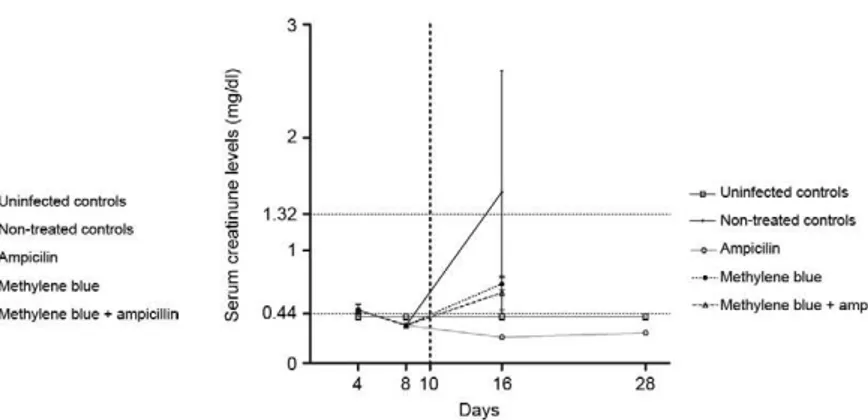
Serum creatinine levels peaked at day 16 and were higher in the untreated group. The serum creatinine levels in the untreated group were higher than normal reference limits and significantly higher than the levels in the hamsters in all of the treatment groups (Kruskal-Wallis, p = 0.02). Ampicillin administration prevented the elevation of serum creatinine at days 16 and 28. The dynamics of the serum creatinine levels in the different treatment groups are shown in Fig. 4.
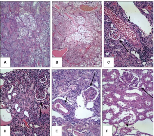
The kidney samples from the hamsters euthanised at day 4 were uniformly normal when examined under light microscopy (data not shown). In contrast, the ham-sters euthanised on day 8 presented the typical features of acute disease, such as diffuse massive tubular cell swelling with mild or no interstitial nephritis. Foci of tubular cell swelling were still detectable at days 16 and 28, but were not as large or as diffusely distributed as on day 8. Regenerative tubules and interstitial inflam-mation were detected to various degrees on days 16 and 28, but not on day 8. The frequency and severity of renal lesions did not differ in the infected hamsters on day 16 and 28. Illustrative images of the renal histopathology of these hamsters are displayed in Fig. 5.
methyl-ene blue-treated and methylmethyl-ene blue + ampicillin-treated groups (Table II). Additional methylene blue administra-tion resulted in no improvement of outcomes, evaluated either in terms of survival or the period between infection and death (Log-rank Mantel-Cox test, p = 0.35).
DISCUSSION
The hamster model employed in this study did not re-produce the hypokalaemia that is commonly observed in human leptospirosis. The rapid and progressive develop-ment of hyperkalaemia most likely reflects experidevelop-mental conditions in which supportive therapy is not feasible, such as venous rehydration. Even the surviving hamsters that were euthanised at day 28 presented elevated levels of serum K (Fig. 1). Antibiotic treatment and/or meth-ylene blue treatment had no effect on serum K levels in the infected hamsters. Serum K levels were significantly higher among the infected hamsters compared with the uninfected hamsters on days 4 and 8. At day 16, all of the infected groups exhibited higher serum K levels than the uninfected animals, regardless of the treatment received. At day 28, the three remaining animals in the methylene blue and no-treatment groups still exhibited significant-ly higher levels of serum K than the controls.
A similar pattern of Mg elevation was observed among the infected hamsters (Fig. 2). Although some Mg depletion might be expected based on clinical studies, rapid progression to severe renal failure could explain the retention of Mg. In contrast to the K dynamics, there was a trend toward decreasing levels of Mg observed on days 8, 16 and 28. The baseline serum Mg levels in the uninfected hamsters were the only measurements that were considerably different (higher) compared with the reference values for laboratory hamsters. The serum Mg levels were significantly higher among the infected hamsters compared with the uninfected hamsters at day 4. None of the other differences were significant.
High baseline serum Mg levels were not expected. Reference values for serum Mg (Fig. 2) in hamsters were obtained from a textbook (Tomson & Wardrop 1987), which were based in two previous studies using 164 and 19 hamsters (mean ± standard deviation of up to 2.5 ± 0.2 mg/dl in males from one study and 1.6 ± 0.4 mg/dl in females from the other). The range suggested from these data is indeed close to the reference serum Mg levels in humans. Another reference textbook indicated values with a considerably wider range, of 1.9-3.5 mg/dl (Gad 2007). However, a more recent textbook apparently
nores these previous data and concludes that reference serum Mg values are not available for hamsters. Strik-ingly, the indicated reference values for other laboratory animals are as high as 2.0-5.4 mg/dl in rabbits, 3.5-4.1 mg/dl in guinea pigs and 3.6-4.0 mg/dl in chinchillas (Washington & van Hoosier 2012). In comparison to the normal range for guinea pigs, the baseline serum Mg lev-els found in the uninfected controls in the present study would considered be normal, while in comparison to the normal range in rabbits, the serum Mg levels found in all of our experimental groups would be within normal limits. For the purpose of the present study, the analysis was based on the comparison between all groups with the serum Mg levels detected in our uninfected controls. These values consistently ranged from 4.0-4.5 mg/dl in samples from the uninfected animals used in this study and from other uninfected hamsters in our laboratory. Although this is beyond the scope of our study, we spec-ulate that reference values for serum Mg in hamsters should be reviewed or further evaluated based on the following observations: (i) the available data in the lit-erature are scarce and show a wide range, (ii) the present results, both from animals evaluated during the study period and in different independent experiments after-wards, indicate considerably higher levels than have been previously reported and (iii) some related labora-tory rodents are known to display considerably higher serum Mg levels.
There was a trend toward lower Na levels among the infected animals compared with the uninfected controls. However, all of the measurements were within the nor-mal range of serum Na levels for hamsters (Fig. 3). The serum Na levels were significantly lower among the in-fected hamsters compared with the uninin-fected hamsters at days 4 and 8. On day 16, all of the infected groups ex-hibited lower serum Na levels compared with the unin-fected animals, regardless of the treatment received. At day 28, the three remaining animals in the no-treatment group still presented significantly lower Na levels com-pared with the controls and the group receiving methyl-ene blue exhibited a trend toward lower Na levels.
The methylene blue treatment group presented low-er slow-erum Na levels on day 16 compared with the othlow-er infected groups (Fig. 3). Any discussion regarding this finding is merely speculative, but it might be explained
based on the combination of the following factors: (i) a syndrome of inappropriate antidiuretic hormone hyper-secretion causes hyponatremia and is known to occur in some severe infectious diseases such as pneumonia and meningitis (Mendoza 1976) and (ii) local inhibition of NO production in the kidney interferes with Na reab-sorption and may result in either depletion or retention (Manning & Hu 1994).
In this study, the dynamics of ionic changes were not found to be associated with any peculiar histopathologic features in the kidneys. The kidney samples from the hamsters that were euthanised at day 4 were uniformly normal when examined via microscopy (data not shown), despite the high serum levels of Mg detected in these ani-mals (Fig. 2). The hamsters that were euthanised at day 8 presented the typical features of acute disease, such as diffuse and massive tubular cell swelling with mild or no interstitial nephritis. At this time point, marked hyperka-laemia was observed (Fig. 1); however, it was even higher on day 28, when the tubular changes were resolved and more severe manifestations of interstitial nephritis domi-nated the microscopic observations (Fig. 5E). In the pres-ent study, severe ionic changes persisted after recovery from the acute tubular changes. This last finding con-firmed a previous clinical report, suggesting that tubular defects, such as impaired urinary concentration capacity, may last for months in patients (Daher Ede et al. 2004).
The hamsters that were euthanised on days 16 and 28 exhibited variable degrees of acute tubular changes, regeneration of tubular epithelia and interstitial nephri-tis. In a previous report on experimental leptospirosis in guinea pigs, de Arriaga et al. (1982) did not observe any association between the frequency or severity of inter-stitial nephritis and renal failure. In the same study, re-generative tubular changes were found to be associated with renal dysfunction, which may imply previous tubu-lar necrosis. As shown in Fig. 4, serum creatinine lev-els peaked at day 16, when regenerative changes in the epithelial tubular cells were first detected. The serum creatinine levels in the untreated group were higher than normal reference limits and significantly higher than the levels in the hamsters from all of the treatment groups. In contrast, serum creatinine levels were not significant-ly different from other treatment groups. In this study, the measured serum creatinine levels were likely biased
TABLE II
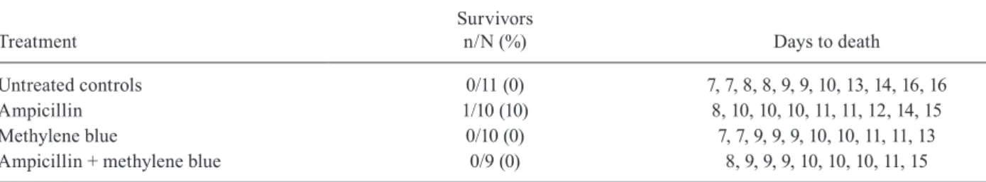
Outcome of hamsters infected by Leptospira interrogans strain L1130 and treated with ampicillin, methylene blue, both or no treatment
Treatment
Survivors
n/N (%) Days to death
Untreated controls 0/11 (0) 7, 7, 8, 8, 9, 9, 10, 13, 14, 16, 16
Ampicillin 1/10 (10) 8, 10, 10, 10, 11, 11, 12, 14, 15
Methylene blue 0/10 (0) 7, 7, 9, 9, 9, 10, 10, 11, 11, 13
Ampicillin + methylene blue 0/9 (0) 8, 9, 9, 9, 10, 10, 10, 11, 15
because serum samples were only collected from the hamsters that were euthanised at scheduled time points. Hamsters that died before the scheduled euthanasia date likely exhibited more severe ionic imbalances and renal dysfunction. Importantly, the histopathology was similar in all groups at day 16, even though the serum creatinine levels were higher in the untreated animals. We have previously demonstrated that late antibiotic treatment in this hamster model can prevent or reverse the loss of Na+/H+ exchanger 3 and NKCC2 expression in renal tu-bular cells (Spichler et al. 2007), which is a finding that may be linked to the effect of antimicrobial treatment on the renal dysfunction markers observed in this study.
As shown in Table I, deaths were not expected to occur in the treatment assay after 15 days of infection. Thus, we defined survivors as those animals that sur-vived to the 16 and 28 day time points. There was no observable benefit of adding methylene blue to standard ampicillin treatment. We previously reported a similar lack of an effect for the antioxidant N-acetylcysteine as an adjuvant therapy (Spichler et al. 2007). However, the strict methodology applied in these studies is the best way to reproduce the clinical conditions related to late treatment of patients. Models of delayed experimental treatment avoid obtaining promising results associated with experimental treatments that may be initiated too early during experimental infection to yield reproduc-ible benefits in clinical practice.
Additionally, we have previously reported that inos -knock-out C57Bl/6 mice display lower rates and severi-ties of interstitial nephritis and that the absence of iNOS is not associated with increased bacterial dissemination in tissues (Bandeira et al. 2011). Prêtre et al. (2011) have reported that treatment with another NOS inhibitor, 4-aminopyridine (0.3 mg/kg daily, starting on the day of infection), without co-administration of antibiotics, results in an accelerated lethal outcome in hamsters and a higher mortality rate in C3H/HeJ mice as well as a higher leptospiral burden in their tissues. The design of this previous study was different from the one present-ed here, as the former did not test NOS inhibition as an adjuvant treatment in the late stage of acute infection. In contrast with the effects observed in other models of sepsis (Kirov et al. 2001, Kwok & Howes 2006, Heem-skerk et al. 2008, Paciullo et al. 2010), methylene blue does not appear to be beneficial in this model of lep-tospirosis. Blocking NO production also had no effect on the ionic imbalance observed during acute leptospirosis in the hamster model.
One limitation of this study is the lack of information on the bacterial burden. While it is important to under-stand the dynamics of infection, the examination of lep-tospiral loads does not directly interfere with analyses of survival, ionic changes and the frequency of renal lesions. We have previously shown that ampicillin treatment is as-sociated with the clearance of leptospiral antigens, which parallels the preservation of renal tubule transporter ex-pression (Spichler et al. 2007). In addition, we have dem-onstrated that genetic deficiency of iNOS is not associated with differences in the leptospiral load in tissues from the C57BL/6 mouse model (Bandeira et al. 2011).
The present study also revealed that the hamster model is not practical for reproducing and therefore studying the common ionic disturbances (such as hy-pokalaemia and hypomagnesaemia) that are observed in patients with severe leptospirosis. These ionic changes may occur during experimental infection, but progress so quickly to typical acute renal failure that they could not be detected at the time points selected in the present study. Patients with leptospirosis usually receive aggres-sive supportive therapy (including fluid expansion) that blocks or retards progression to an oliguric/hyperkalae-mic state. Such supportive therapy was not provided in this study, which may explain the rapid progression to a severe form of renal failure.
In our second experiment, a different strategy was employed to reproduce the late initiation of treatment of experimental leptospirosis. Infected hamsters were fol-lowed to detect clinical signs and treatment was initiated one day after the first death was observed. In this model, even antibiotic therapy had almost no effect on surviv-al (only 1 out of 10 animsurviv-als survived) and the addition of methylene blue to the treatment regime provided no improvement of outcomes (Table II). Notably, although antibiotics are highly recommended, even in late-stage leptospirosis in clinical settings, there is no evidence demonstrating positive effects on survival when anti-biotic treatment is initiated in late-stage leptospirosis. Such studies will likely not be performed because of ethical concerns about denying patients antibiotic treat-ment, even if no clinical benefit has been demonstrated. Hypokalaemia was not reproduced in our hamster model: all of the groups developed increased levels of serum K. Furthermore, in this model of acute infection, hypermagnesaemia (rather than Mg depletion) was ob-served. These findings may be associated with an accel-erated progression to acute renal failure. Furthermore, adjuvant methylene blue treatment had no effect on se-rum Mg and K levels during acute-phase leptospirosis in hamsters. Late antibiotic treatment prevented the acceler-ated elevation of serum creatinine levels in this model.
REFERENCES
Bandeira M, Santos CS, de Azevedo EC, Soares LM, Macedo JO, Marchi S, da Silva CL, Chagas-Junior AD, McBride AJ, McBride FW, Reis MG, Athanazio DA 2011. Attenuated nephritis in induc-ible nitric oxide synthase knockout C57BL/6 mice and pulmonary hemorrhage in CB17 SCID and recombination activating gene 1 knockout C57BL/6 mice infected with Leptospira interrogans.
Infect Immun 79: 2936-2940.
Beltowski J, Marciniak A, Wojcicka G, Gorny D 2003. Nitric oxide decreases renal medullary Na+, K+-ATPase activity through cyclic GMP-protein kinase G dependent mechanism. J Physiol Pharmacol 54: 191-210.
Berkelhammer C, Bear RA 1985. A clinical approach to common electrolyte problems. 4. Hypomagnesemia. Can Med Assoc J 132: 360-368.
Cerqueira TB, Athanazio DA, Spichler AS, Seguro AC 2008. Renal involvement in leptospirosis - new insights into pathophysiology and treatment. Braz J Infect Dis 12: 248-252.
to leptospirosis: 58 cases and a review of the literature. Nephrol Dial Transplant 18: 1128-1134.
Craig SB, Smythe LD, Graham GC, McKay DB 2009. Hypomag-nesemia in acute leptospirosis. Am J Trop Med Hyg 80: 1067.
Daher Ede F, Zanetta DM, Abdulkader RC 2004. Pattern of renal function recovery after leptospirosis acute renal failure. Nephron Clin Pract 98: 8-14.
de Arriaga AJD, Rocha AS, Yasuda PH, de Brito T 1982. Morpho-functional patterns of kidney injury in the experimental lep-tospirosis of the guinea-pig (L. icterohaemorrhagiae). J Pathol 138: 145-161.
Gad SC 2007. The hamsters - Toxicology. In SC Gad (ed.), Animal models in toxicology, 2nd ed., CRC Press, Boca Raton, p. 295.
Heemskerk S, van Haren FM, Foudraine NA, Peters WH, van der Ho-even JG, Russel FG, Masereeuw R, Pickkers P 2008. Short-term beneficial effects of methylene blue on kidney damage in septic shock patients. Intensive Care Med 34: 350-354.
Khositseth S, Sudjaritjan N, Tananchai P, Ong-ajyuth S, Sitprija V, Thongboonkerd V 2008. Renal magnesium wasting and tubular dysfunction in leptospirosis. Nephrol Dial Transplant 23: 952-958.
Kirov MY, Evgenov OV, Evgenov NV, Egorina EM, Sovershaev MA, Sveinbjornsson B, Nedashkovsky EV, Bjertnaes LJ 2001. Infu-sion of methylene blue in human septic shock: a pilot, random-ized, controlled study. Crit Care Med 29: 1860-1867.
Kwok ES, Howes D 2006. Use of methylene blue in sepsis: a system-atic review. J Intensive Care Med 21: 359-363.
Lin CL, Wu MS, Yang CW, Huang CC 1999. Leptospirosis associated with hypokalaemia and thick ascending limb dysfunction. Neph-rol Dial Transplant 14: 193-195.
Maciel EA, Athanazio DA, Reis EA, Cunha FQ, Queiroz A, Almeida D, McBride AJ, Ko AI, Reis MG 2006. High serum nitric oxide levels in patients with severe leptospirosis. Acta Trop 100: 256-260.
Magaldi AJ, Yasuda PN, Kudo LH, Seguro AC, Rocha AS 1992. Renal involvement in leptospirosis: a pathophysiologic study.
Nephron 62: 332-339.
Manning Jr RD, Hu L 1994. Nitric oxide regulates renal hemodynamics and urinary sodium excretion in dogs. Hypertension 23: 619-625.
McBride AJ, Athanazio DA, Reis MG, Ko AI 2005. Leptospirosis.
Curr Opin Infect Dis 18: 376-386.
Medeiros FR, Spichler A, Athanazio DA 2010. Leptospirosis-asso-ciated disturbances of blood vessels, lungs and hemostasis. Acta Trop 115: 155-162.
Mendoza SA 1976. Syndrome of inappropriate antidiuretic hormone secretion (SIADH). Pediatr Clin North Am 23: 681-690.
Nascimento AL, Ko AI, Martins EA, Monteiro-Vitorello CB, Ho PL, Haake DA, Verjovski-Almeida S, Hartskeerl RA, Marques MV, Oli- veira MC, Menck CF, Leite LC, Carrer H, Coutinho LL, Degrave
WM, Dellagostin OA, El-Dorry H, Ferro ES, Ferro MI, Furlan LR, Gamberini M, Giglioti EA, Góes-Neto A, Goldman GH, Goldman MH, Harakava R, Jerônimo SM, Junqueira-de-Azevedo IL, Kimu-ra ET, KuKimu-ramae EE, Lemos EG, Lemos MV, Marino CL, Nunes LR, de Oliveira RC, Pereira GG, Reis MS, Schriefer A, Siqueira WJ, Sommer P, Tsai SM, Simpson AJ, Ferro JA, Camargo LE, Ki-tajima JP, Setubal JC, Van Sluys MA 2004. Comparative genomics of two Leptospira interrogans serovars reveals novel insights into physiology and pathogenesis. J Bacteriol 186: 2164-2172.
Ortiz PA, Garvin JL 2002. Role of nitric oxide in the regulation of nephron transport. Am J Physiol Renal Physiol 282: 777-784.
Paciullo CA, Horner MD, Hatton KW, Flynn JD 2010. Methylene blue for the treatment of septic shock. Pharmacotherapy 30: 702-715.
Prêtre G, Olivera N, Cedola M, Haase S, Alberdi L, Brihuega B, Gomez RM 2011. Role of inducible nitric oxide synthase in the pathogen-esis of experimental leptospirosis. Microb Pathog 21: 303-308.
Spichler A, Athanazio DA, Furtado J, Seguro A, Vinetz JM 2008. Case report: severe, symptomatic hypomagnesemia in acute lep-tospirosis. Am J Trop Med Hyg 79: 915-917.
Spichler A, Ko AI, Silva EF, de Brito T, Silva AM, Athanazio D, Silva C, Seguro A 2007. Reversal of renal tubule transporter down-regulation during severe leptospirosis with antimicrobial therapy.
Am J Trop Med Hyg 77: 1111-1119.
Tomson FN, Wardrop KJ 1987. Chemical chemistry and hematology. In GL Van Hoosier, CW McPherson (eds.), Laboratory hamsters, Academic Press Inc, Orlando, p. 51.
Washington IM, van Hoosier G 2012. Clinical biochemistry and he-matology. In MA Suckow, KA Stevens, RP Wilson (eds.), The laboratory rabbit, guinea pig, hamster and other rodents, Aca-demic Press, San Diego, p. 69.
WHO - World Health Organization 2003. Human leptospirosis: guid-ance for diagnosis, surveillguid-ance and control. Rev Inst Med Trop Sao Paulo 45: 292.
Wu MS, Yang CW, Pan MJ, Chang CT, Chen YC 2004. Reduced re-nal Na+-K+-Cl- co-transporter activity and inhibited NKCC2 mRNA expression by Leptospira shermani: from bed-side to bench. Nephrol Dial Transplant 19: 2472-2479.
Yang CW, Hung CC, Wu MS, Tian YC, Chang CT, Pan MJ, Vande-walle A 2006. Toll-like receptor 2 mediates early inflammation by leptospiral outer membrane proteins in proximal tubule cells.
Kidney Int 69: 815-822.
Yang CW, Wu MS, Pan MJ, Hong JJ, Yu CC, Vandewalle A, Huang CC 2000. Leptospira outer membrane protein activates NF-kap-paB and downstream genes expressed in medullary thick ascend-ing limb cells. J Am Soc Nephrol 11: 2017-2026.