Acta Cir. Bras. 2018;33(9):806-815 DOI: http://dx.doi.org/10.1590/s0102-865020180090000009
Adriano Silva SilveiraI, Ricardo Dutra AydosII, Rondon Tosta RamalhoIII, Iandara Schettert SilvaIV,
Ruy de Araujo CaldasV, Abílio Torres dos Santos NetoVI, Camila Tozaki RodriguesVII
Oxidative stress effects in the uterus, placenta
and fetus of pregnant rats submitted to acute
and chronic stress
1Abstract
Purpose: To evaluate the effects of oxidative stress in pregnant rats submitted to acute and chronic stress, relating to alterations in the uterus, placenta and fetus.
Methods: Twenty-four female Wistar albino (Rattus norvegicus), were divided into four groups, for induction of oxidative stress the animals were submitted to cold and physical immobilization. Plasma fasting glucose and MDA were determined in all groups and the fetuses and placentas were measured.
Results: There were no statistical differences in the levels of malonic dialdehyde (MDA), however the averages of chronic stress group were higher compared to control groups, which could explain the observed adverse effects; there was no correlation between puppies’ size, the weight of the placenta and MDA values.
Conclusions: Chronic stress causes adverse effects, when compared to control groups; chronic stress group had fetuses, placentas and number of puppies, significantly lower compared to other groups. The rats exposed to chronic stress, also presented a higher frequency of fetal resorption.
Key words: Oxidative Stress. Lipid Peroxidation. Rats.
IFellow Master degree, Postgraduate Program in Health and Development in the Midwest Region, Universidade Federal
do Mato Grosso do Sul (UFMS), Campo Grande-MS, Brazil. Conception and design of the study; technical procedures; acquisition, interpretation and analysis of data; manuscript writing.
IIPhD, Associate Professor of Surgery, Postgraduate Program in Health and Development in the Midwest Region, UFMS,
Campo Grande-MS, Brazil. Conception and design of the study, interpretation of data, manuscript writing, critical revision, final approval.
IIIFull Professor, Laboratory of Experimental Carcinogenicity, UFMS, Campo Grande-MS, Brazil. Macroscopic and
histopathologic analysis.
IVFull Professor, Postgraduate Program in Health and Development in the Midwest Region, UFMS, Campo Grande-MS,
Brazil. Intellectual and scientific content of the study.
VSenior Visiting Professor, UFMS, Campo Grande-MS, Brazil. English language, manuscript writing.
VIFellow Master degree, Postgraduate Program in Health and Development in the Midwest Region, UFMS, Campo
Grande-MS, Brazil. Technical procedures.
This imbalance may be caused by a number of
factors like inadequate nutrition and animal’s retention in stressful conditions related to higher ROS production and/or reduced antioxidant activity.
Experimental models should provide data for both biological and behavioral
phenomena to understand the pathological processes that apply to the human being5.
The most important experimental
models described in the literature are those of
induced or spontaneous disease. To understand
pathophysiological mechanisms of diseases,
effects of medications and surgical procedures thus experimental animal models can be used in all fields of biological research, they are very important for medical science. Trials in humans
are limited and a series of procedures are
required for ethical reasons to preserve physical and psych emotional integrity. In addition, when a study of an entire population becomes a limiting factor, experimental animal models can provide data statistically comparable to the whole population based on the concept of sampling, since they represent a portion of it5,6.
Studies of lipid peroxidation in
pregnancy are limited to determine the
etiology and pathophysiology of the factors that trigger oxidative stress, animal models have been proposed for understanding the
physiology and pathology when is impossible
to control variables in humans. Many of these experimental models use the induction of
chronic or acute physical stress to establish
and understand relationships between stressor events. It has been reported that stressors elevate blood pressure and the risk for preeclampsia. The stress is associated with complications during pregnancy, premature birth and intrauterine growth restriction7,8.
Rat’s models are appropriate to understand the mentioned stressors because
of the similarity of the placenta and its
trophoblastic invasion between rats and
humans9.
■
Introduction
Reactive oxygen species (ROS)
generating in oxidative stress promotes
physiological and pathological changes in the
female reproductive tract. ROS are detected in the ovaries, uterine tube and embryos which are involved in several physiological processes, such as oocyte maturation, steroidogenesis
and corpus luteum1.
Concerning infertility, it is known that steroidogenesis is related to antioxidant concentration. High concentrations of estrogen contribute to a greater enzymatic antioxidant activity; furthermore oxidative stress has an impact on the production of steroid hormones
produced by granulosa cells and that the
oocyte within the follicle is naturally exposed to oxidative stress leading to lipid peroxidation and influencing the production glycoproteins
produced by the granulosa cells2.
Oxidative stress causes embryos injury may due to peroxidation of membrane phospholipids and chemical modifications of different types of biomolecules. The
consequences of these damages include
mitochondrial activity, embryonic development and apoptosis. Oocytes and embryos are protected from oxidative stress by the presence of antioxidants from follicular and oviduct fluids3.
One of the theories considered is related
to decreased uteroplacental flow, which leads to poor fetal oxygenation. This effect is caused by the inadequate invasion of the intravascular
trophoblast, which maintains the high resistance of the spiral arteries consequently
endothelial injury, elevation of reactive oxygen species (ROS) and reactive nitrogen species (RNS). Low antioxidant capacity of placental tissue leads the development of oxidative
stress4.
In the placental uterus process, it is assumed that oxidative stress is a causal factor in chronic complications and can be measured based on the level of thiobarbituric acid reactive substances (TBARS) and catalase activity10.
Arguably, the most important event at the onset of normal placentation is the establishment of effective maternal circulation,
a process that is linked to the physiological
conversion of maternal spiral arteries into a flaccid sinoid duct with low resistance that
allows the blood supply necessary to the
development of the placenta11.
In contrast, the fetal origin trophoblastic invasion process contributes to the transformation of the uteroplacental circulation into a high capacity, low resistance system that exhibits relative resistance to vasopressor agents12.
This process occurs in humans in two
stages. Initially, the deciduous segments of the spiral arteries are affected by the first wave of trophoblastic invasion followed by a second wave, placental ischemia. Progresses continuously during pregnancy and the narrowed vessels do not provide the demand for the developing placental fetal unit, a hypoxia insurge, generating oxidative stress and strong inflammatory response of maternal origin. There is increased production of placental antiangiogenic factors, specifically soluble forms of tyrosine kinase 1 (sFLT-1) and
soluble endoglin13.
Other factors such as alterations in the renin-angiotensin-aldosterone axis, poor immune adaptation, excessive loss of trophoblasts fragments and genetic factors
seem to contribute to the pathogenesis of
abnormal placentation12,22.
The oxidative stress is in other words, an imbalance between the pro and antioxidant species, and the antioxidant species are at disadvantage13,14.
Basically, there are two ways to measure oxidative stress, by evaluating the concentrations of both by-products and promoters of the oxidative damage; which provides the dimensions of the damage; and by the antioxidant capacity of the medium in response to oxidative stress which determines the adaptation the organism to the unfavorable environment13,15.
The most commonly used markers for
assessing lipid damage by oxidation are the MDA test (the main metabolite of peroxilipid
breakdown), which uses the TBARS assay13.
This test identifies lipid oxidation products,
bile pigments, amino acids and sugars that
can generate interfering chromogens. In view
of this, it is more appropriate to use the term
thiobarbituric acid reactive substances (TBARS). Moreover, the fact that TBA is calibrated with MDA leads the authors to express their results
with the term “amount of MDA”16.
Gestational diabetes were used induced in rats to study different mechanisms that have been proposed for the increase in the production of free radicals in the hyperglycemic
state, among them glucose metabolism as the main source of ROS17,18 the lipid peroxidation
induced by hyperglycemia19 and its advanced
glycosylation end -products (AGEs), which,
through their cellular receptors, modify and
inactivate enzymes20, stimulate the production
of ROS21, as well the transcriptional activity of
NF-κB, a classically inflammatory pathway23.
Thus, chronic hyperglycemia, through oxidative stress, functionally compromises cellular proteins, membrane phospholipids (formation of malondialdehyde) and nucleic acids, in addition to stimulating the production of inflammatory cytokines such as tumor necrosis factor alpha (TNF-α)8,16, events strongly related
to the development of diabetic comorbidities.
The consequences on the body
during pregnancy but also on the life period
studied. It has been shown in rodents that intrauterine exposure to low doses of STZ
during pregnancy is associated with normal weight or macrosomia25,26. On the other
hand, studies carried out with the offspring of mothers submitted to high doses of STZ
showed that the animals had reduced growth in adulthood27.
Newborns, especially preterm infants, are highly susceptible to tissue and organ damage by free radicals, notably those derived from oxygen, with the superoxide anion being the most important of the reactive oxygen intermediates (ROI) and produced in response to hypoxic stimuli - ischemic or inflammatory. In addition, gestation itself is a physiological condition with increased metabolic demand and increased tissue oxygen requirements and, in the event of abnormalities in pregnancy, oxidative imbalance may occur, and excess
free radicals can promote damage to the fetus,
which already have an antioxidant defense
system, it is important to emphasize that the
main intracellular antioxidant component, glutathione, only shows its maximum production at the end of gestation28-30.
In this work, the effects of oxidative stress (lipid peroxidation) in pregnant rats submitted to acute and chronic stress related
to changes in the uterus, placenta and fetuses
were determined.
■
Methods
The study was approved by the Animals Research Ethics Committee of UFMS (protocol CEUA / UFMS number 647/2014) and was performed in accordance with the International Guiding Principles for Biomedical Research Involving Animals, published by the Council for International Organizations of Medical Sciences (CIOMS), as well as with the Brazilian law on the scientific use of animals (Law 11794/2008).
Adult female Wistar rats 45 days old, weighing 150-200 grams, were obtained from the Central Laboratory for Animals (UFMS), were randomized into four groups: non-pregnant rats without stress (G1, n = 6), control pregnant rats (G2, n = 6), pregnant rats subjected to acute stress (G3, n = 6) and pregnant rats subjected to cronic stress (G4, n = 6).
The animals were housed in a polypropylene cage with containing paddy
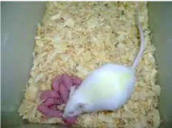
husk as bedding. Each cage of G2, G3 and G4 maintained a ratio of three females to one male, except in G1, which did not contain male in the cage (Figure 1).
Figure 1 - Female with their cubs on the day of
childbirth / euthanasia.
All animals were kept in ventilated rack and maintained at controlled conditions of temperature (23±1°C), humidity .and dark-light cycle (12 hours). The animals had free access to
standard pellet (Nuvilab) and water ad libitum.
The mating was performed and the first
day of pregnancy was considered the third day
of stay of the male in the cage.
carton for 5 minutes on the 7th and 14th day
of pregnancy. The G4 was kept in a separately ventilated shelf to be exposed to cold. Two
liters of liquid nitrogen were used, kept in a
semi-open styrofoam box inside the shelf, from
the 7th day of pregnancy, in order to maintain
the temperature at + 21°C. The polystyrene box with nitrogen was changed every day until the end of the experiment and the temperature of the ventilated shelf was monitored.
The rats were manipulated during the
twenty days of pregnancy/ non-pregnancy and evaluated at 0, 7, 14 and 20 days when euthanasia was performed.
Oxidative stress damage
Malondialdehyde (MDA) levels were measured using thiobarbituric acid reactive substances (TBARS).
Maternal capillary glycemia, fetal weight and size and euthanasia
Fasting blood glucose dosage was
performed on days 0, 7, 14 and 20 of pregnancy
and on non-pregnancies in the equivalent period. Blood collection was performed on the animal’s tail and the glycaemia determined utilizing the Accu-Chek® Active blood glucose
monitoring system.
Fetuses and placentas were weighed on
a QUIMIS® precision scale. The size of newborns
was measured with a ruler graduated in
centimeters (Figure 2).
Figure 2 - Cephalo-anal measurement and fetal
weighing performed in all groups.
All animals were submitted to euthanasia on the 20th day using intraperitoneal injection with a combination of Ketamine (150mg / kg) and Xylazine (75mg/kg). Death was confirmed after cardiac puncture exsanguination.
Statistical analysis
The data were tabulated in Excel version 2013. For the discrimination of the difference between groups, the Mann-Whitney U test was used followed by Student’s t-test. The Pearson correlation test was performed on MDA variables, fetal weight, fetal cephalal-anal size and placental weight. The other results evaluated in this study were described in tables and graphs. The software used was BioEstat 5.0, using p<0.05
■
Results
Table 1 - MDA values, showing oxidative stress, according to groups.
MDA
GNC GPC GPEA GPEC
Table 2 - Average glycemia of females, in mg/dl according to group.
D1 D7 D14 D21
GNC 99.50±14.56 97.50±9.59 80.67±12.69 136.50±7.85
GPC 98.50±8.96 111.83±7.25 90.17±13.01 80.83±21.45
GPEA 91.83±10.83 127±7.77 84.67±3.72 95.00±8.76
GPEC 95.67±4.41 96.50±13.03 85.33±6.12 88.00±8.10
Table 3 - Mean values according to weight, height, placenta weight and number of fetuses/female.
Group newborn newborn Placenta Relationship between
weight stature weight fetus and mother
FPC 3.92±2.48 3.90±1.62 0.50±0.19 12.60±2.07
FPEA 4.30±1.38 4.05±0.87 0.64±0.20 14.17±1.33
FPEC 3.22±0.74 3.21±0.59 0.55±0.24 10.60±2.88
■
Discussion
Oxidative stress and impact of the
stressful stimulus on maternal parameters The average chronic group G4 (629.81ng/ml) was the largest and among the G3 Group (412.37ng/ml) and G4 difference, showing that chronic stress causes adverse effects, when compared to control groups (Table1).
By studying the oxidative stress in
pregnant women with preeclampsia it was
found a reduced antioxidant enzyme activity, however did not identify difference in plasma levels of MDA27. Increase in oxidative status
of pregnancy without complications must be
accompanied by increased ability of pregnant
women to overcome the effects of oxidative
stress without pathological consequences18.
Despite the non-significant difference between acute and chronic stress groups, the average levels of MDA chronic group was higher. Studies that have applied the exercise as a stressor, showed that acute exercise increases oxygen consumption due to mobilization of
energy caused by the acute physical stress,
leading to increased formation of ERO. Second bear and about 2 to 5% of the oxygen used in
the mitochondria for cellular respiration are
converted into free radicals28. While the chronic
exercise reduces basal levels of damage, increases resistance to oxidative stress, since
the body tends to adapt to stressful situations7
Evaluation of maternal glucose levels (mg/dl) of the groups
There was a significant difference
between 7 and 20 days, and in the 7th day the
capillary glycaemia of the acute stress group
(G3) was higher than the non-pregnant (G1) and chronic stress (G4). At the end of the experiment, the glycemia values of G1 and G3
were higher than the control pregnant group
(G2) (Table 2).
In this study there was no increase in MDA, which may explain the non-alteration of glycemia in the studied groups.
Although there was no hyperglycemia at the time studied, there was a significant difference in the capillary glycemia of the acute stress group on control and chronic stress.
The rats in this study showed similar
values of capillary glycaemia, which sought to establish reference values of biochemical,
physiological and morphological parameters
in Wistar rats. The values detected in this
study were considered normal and reported
mellitus28. The reverse process can occur, i.e.
diabetes increases the oxidative stress, reduces the antioxidant system of pregnant rats and can still be related to occurrence of hypertension and abortion13.
In this study there was no increase in MDA levels, which may explain normal glycaemia in all groups. The females in the
chronic stress group had hyper glycaemia
statistically significant differences this can be explained by the fact that stress, fight-or-flight phase (first phase) mobilizes large amount of glucose and oxygen essential to organs. Cortisol liberated into the blood stream inhibits the release of insulin and stimulates processes
such as gluconeogenesis, glycogenolysis and
lipolysis for energy production. When the stressor stimulus becomes chronic, excessive and continuous production of cortisol leads to fat deposit due to the continuous process of catabolism for energy production. During pregnancy glucose is the main energetic source for the fetus; therefore there is an increasing
demand for glucose during pregnancy,
explaining the higher glycemic index in chronic
stress group compared with the control did not pregnant30.
Stress stimulus on fetal-placental parameters
The mean litter size of the rats was
lower in group 2 and the mean of the acute
stress group (G3) was higher. Regarding the number of fetal resorption, the chronic stress group was higher.
Average weight of puppies
The mean litter size of the rats was
lower in group 2 and the mean of the acute
stress group (G3) was higher. Regarding the number of fetal resorption, the chronic stress group was higher.
The weight of the rats subjected to
chronic stress was significantly different when
compared to the offspring of acute stress
group and control. The off springs of acute stress group did not have different weight in the control group. There was no correlation between fetal weight and MDA level.
Evaluation of oxidative stress was carried out in the laboratory of experimental Carcinogenesis after cardiac puncture; blood was collected in pediatric vacuum tube with EDTA and centrifuged. Determination of plasma concentration of malondialdehyde (MDA) was performed through the MDA reaction with thiobarbituric acid (TBAR), forming the complex MDA-TBA in the ratio of 1:2. The tubes were heated in a water bath at 94°C for 1 hour and 15 minutes, after cooling were homogenized with 4 ml of n-butyl alcohol in a Vortex (QL-901) for 30 seconds, following centrifugation at 3,000 rpm
for 20 minutes and the supernatants were read
at 532 nm in a Spectrophotometer(LMR-96, Loccus Company). For the calculation of concentration of MDA, was used an equation obtained by standard curve of absorbance. The concentrations expressed in ng/ml.
Results = Fc x Abs, and Fc = 4406/ABS x MDA
All animals were euthanized on day 20 of pregnancy/ non-pregnant by intraperitoneal Ketamine injection (150 mg/kg) and Xylazine (75 mg/kg) for the removal of the fetus, placenta. The death was confirmed after exsanguination by cardiac puncture, followed by removal of the uterus and in the case of ferns, hypothermia.
The statistical analysis the data were tabulated in Excel version 2013 programme. For the discrimination of the difference between groups was used the Mann-Whitney U test followed by the Student’s t-test. C test was done.
The puppies’ size of chronic stress group was statistically lower than those of other groups. There was no difference between
control. There was no correlation between size of the puppies and MDA of rats.
The placenta weight of the acute
stress group showed a statistically significant difference in comparison to both the control group and the chronic stress group. There was no correlation between the weight of the placenta and levels of MDA. It was possible to observe a correlation between fetus weight and placenta weight. There were identified a minor fetal weight of the group that was exposed to chronic stress, as well as the findings in this survey, where it was observed that chronic
stress group had fetuses, placentas and number
of puppies, significantly lower in relation to
other groups30. Rats exposed to chronic stress,
also presented a higher frequency of fetal
resorption, which was also observed. The inadequate trophoblastic invasion mechanisms
lead to reduced perfusion and placental failure
causing intrauterine growth restriction and if there is severe vasoconstriction leads to fetal
distress and death27 (Table 3).
■
Conclusions
Pregnant rats with regard to malondialdehyde presented the highest levels in the group that suffered chronic stress, as well as blood glucose levels when compared to non-pregnant and acute stress groups.
It was observed that chronic stress
Group had fetuses, placentas and number of
pups, significantly lower compared to other groups. The rats exposed to chronic stress,
also presented a higher frequency of fetal
resorption.
The weight of the puppies subjected to chronic stress was lower when compared to
the acute stress and control group, however
the acute stress group showed no weight
difference in relation to the control group. There was no correlation between the fetus weights with the levels of MDA.
The fetus size, the chronic stress groups
were statistically lower than those of other groups. There was no difference between
the size of the puppies of chronic stress and
control. There was no correlation between size of the puppies and MDA of rats.
■
References
1. Amaral WT, Peraçoli JC. Fatores de risco relacionados à pré-eclâmpsia. Com Ciênc Saúde. 2011;(22 Supl 1):S161-8.
2. Caldas JPS, Vilela MS, Braghini C, Mazzola TN, Marba STM. Uso materno antenatal de corticosteroide e marcadores de estresse oxidativo e de inflamação no sangue de cordão umbilical de recém-nascidos pré-termo de muito baixo peso. J Pediatr. 2012;88(1):61-6. doi: 10.2223/JPED.2158. 3. Barbosa KBF, Costa NMB, Alfenas RG, De
Paula SO, Minim VPR, Bressan J. Estresse oxidativo: conceito, implicações e fatores modulatórios. Rev Nutr. 2010;23(4):629-43. doi: 10.1590/S1415-52732010000400013. 4. Arad I, Bar-Oz B, EZ NA, Barak V. Interleukin-6
and N-terminal pro-brain natriuretic peptide cord blood levels in premature infants: correlations with perinatal variables. Isr Med Assoc J. 2010;12(7):419-23. PMID: 20862823.
5. Fagundes DJ, Taha MO. Modelo animal de doença: critérios de escolha e espécies de animais de uso corrente. Acta Cir Bras. 2004;19(1):59-65. doi: 10.1590/S0102-86502004000100010.
6. Schanaider A, Silva PC. Uso de animais em cirurgia experimental. Uso de animais
em cirurgia experimental. Acta Cir Bras.
2004;19(4):441-7.doi:
10.1590/S0102-86502004000400014.
7. Schneider CD, Oliveira AR. Radicais livres de oxigênio e exercício: mecanismos de formação e adaptação ao treinamento físico. Rev Bras Med Esporte. 2004;10(4):308-13. doi: 10.1590/S1517-86922004000400008. 8. Lykkesleldt J. Malondialdehyde as biomarker
of oxidative damage to lipids caused by smoking. Clin Chim Acta. 2007;380(1-2):50-8. doi: 10.1016/j.cca.2007.01.022007;380(1-2):50-8.
9. Groen B, Uuldriks GA, de Vos P, Visser JT,
invasion and increased numbers of
immune cells at day 18 of pregnancy in the
mesometrial triangle of type 1 diabetic rats. Placenta. 2015;36(2):142-9. doi: 10.1016/j. placenta.2014.12.004.
10. Wietzycoski CR, Marchesini JCD, Al-Themyat S, Meyer FS, Trindade MRM. Improvement in oxidative stress after duodenojejunostomy in an experimental model of type 2 diabetes mellitus. Arq Bras Cir Dig. 2016;29(Supl.1):3-7 doi: 10.1590/0102-62016;29(Supl.1):3-720201600S10002.
11. Halliwell B, Whiteman M. Measuring
reactive species and oxidative damage in vivo and in cell culture: how should you
do it and what do the results mean? Br
J Pharmacol. 2004;142(2):231-55. doi: 10.1038/sj.bjp.0705776.
12. Caldas JPS, Vilela MS, Braghini CA, Mazzola TN, Marba STM. Uso materno antenatal de corticosteroide e marcadores de estresse oxidativo e de inflamação no sangue de cordão umbilical de recém-nascidos pré-termo de muito baixo peso. J Pediatr. 2012;88(1):61-6. doi: 10.2223/JPED.2158.
13. Dotan Y, Lichtenberg D, Pinchuk L.
Lipid peroxidation cannot be used as a universal criterion of oxidative stress. Prog Lipid Res. 2004;43(3):200-27. doi: 10.1016/2003.10.001.
14. Posada D. jModelTest: phylogenetic model averaging. Mol Biol Evol. 2008;25(7):1253-6. doi: 10.1093/molbev/msn083.
15. Nikki C, Tatsuya O, Massimo S, Anne-Marie B, Cameron WB, Dolores H, Eric CH. Perivascular nitric oxide activates notch signaling and promotes stem-like character in PDGF-induced glioma cells. Cell Stem Cell. 2010;6(2):141-52 doi: 10.1016/j. stem.2010.01.001.
16. Souza Jr. TP, Oliveira PR, Pereira B. Exercício físico e estresse oxidativo: efeitos do exercício físico intenso sobre a quimioluminescência urinária e malondialdeído plasmático. Rev Bras Med Esporte. 2005;11(1):91-6. doi: 10.1590/S1517-86922005000100010. 17. Silveira SL. Programação metabólica: estudo
de parâmetros indicadores de resistência à insulina e espécies reativas de oxigênio em ratos [Tese]. Instituto de Ciências Básicas da Saúde, Universidade Federal do Rio Grande do Sul; 2010.
18. Martim AC, Sanders RA, Watkins JB. Diabetes, oxidative stress, and antioxidants: a review.
J Biochem Mol Toxicol. 2003;17(1):24-38. doi: 10.1002/10058.
19. Kawamura M, Heinecke JW, Chait A.
Pathophysiological concentrations of
glucose promote oxidative modification of low density lipoprotein by a superoxide-dependent pathway. J Clin Invest. 1994;94(2):771-8. doi: 10.1172/ JCI117396. 20. Nédic O, Rattan SIS, Grune T, Trougakos IP.
Molecular effects of advanced glycation
end products on cell signalling pathways,
ageing and pathophysiology. Free
Radic Res. 2013;47(Suppl 1):28-38. doi: 10.3109/10715762.2013.806798.
21. Baynes JW, Thorpe SR. Role of oxidative stress in diabetic complications: a new perspective on an old paradigm. Diabetes. 1999;48(1):1-9. doi: 10.2337/diabetes.48.1.1.
22. Mohamed AK, Bierhaus A, Schiekofer S, Tritschler H, Ziegler R, Nawroth PP. The role of oxidative stress and NF-kappaB activation in late diabetic complications. Biofactors. 1999;10(2-3):157-67. PMID: 10609877. 23. Singh R, Kaur N, Kishore L, Kumar GG.
Management of diabetic complications: A chemical constituents based approach. J Ethnopharmacol. 2013;150(1):51-70. doi: 10.1016/j.jep.2013.08.051.
24. Buonocore G, Perrone S, Longini M, Vezzosi P, Marzocchi B, Paffetti P, Bracci R. Oxidative
stress in preterm neonates at birth and
on the seventh day of life. Pediatr Res. 2002;52:46-9. doi: 10.1203/00006450-200207000-00010.
25. Mulay S, Philip A, Solomon S. Influence of maternal diabetes on fetal rat development: Alteration of insulin receptors in fetal liver and lung. J. Endocrinol. 1983;98(3):401-10. doi: 10.1677/joe.0.0980401.
26. Merzouk H, Madani S, Sari DC, Prost J, Bouchenak M, Belleville J. Time course of
changes in serum glucose, insulin, lipids
and tissue lipase activities in macrosomic offspring of rats with streptozotocin-induced diabetes. Clin Sci. 2000;98(1):21-30. doi: 10.1042/cs0980021.
28. Calabrese V, Cornelius C, Dinkova-Kostova AT, Calabrese EJ, Mattson MP. Cellular stress
responses, the hormesis paradigm, and
vitagenes: novel targets for therapeutic intervention in neurodegenerative disorders. Antioxid Redox Signal. 2010;13(11):1763-811.doi: 10.1089/ars.2009.3074.
29. Gracy X. Rosario RA, Toshihiro K, Michael JS. Intrauterine fate of invasive trophoblast cells. Placenta. 2009;30(5):457–63. doi:
10.1016/2009.02.008.
30. Lamb SE, Hansen Z, Lall R, Castelnuovo E, Withers EJ, Nichols V, Potter R, Underwood MR; Back Skills Training Trial investigators. Group cognitive behavioural treatment for low-back pain in primary care: a randomised controlled trial and cost-effectiveness analysis. Lancet. 2010;375 916–23. doi: 10.1016/S0140-6736(09)62164-4.
Correspondence:
Ricardo Dutra Aydos Estrada EW2, 194
79037-802 Campo Grande – MS Brasil Tel.: (55 67)99984-5773
Received: May 06, 2018 Review: July 09, 2018 Accepted: Aug 04, 2018
Conflict of interest: none
Financial sources: FINEP, FAPEC, and FUNDECT
1Research performed at Laboratory of