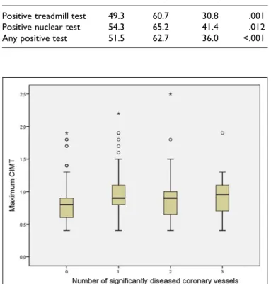
Carotid Intima-Media Thickness and Carotid Plaques Improves Prediction of Obstructive Angiographic Coronary Artery Disease in Women
Texto
Imagem

Documentos relacionados
Porém, não se deve culpabilizar os professores por não ter tal formação, pois, a pesquisa realizada por Rodrigues e Dalla (2007) sobre o Curso PROEJA 4 , que é um curso de
Para a análise da dieta os itens foram agrupados em categorias da seguinte forma: algas unicelulares; algas filamentosas; vegetais superiores (briófita, raiz, caule, folha, flor,
We agree with and reinforce the observation that carotid intima- media thickness (cIMT) measurement is a very well-accepted marker of subclinical atherosclerosis in a variety
Insulin, homeostatic model assessment-insulin resistance (HOMA-IR), FGF-23 levels, CIMT, left ventricular (LV) mass, LV mass index and myocardial performance index (MPI)
Association between carotid intima-media thickness and adiponectin in participants without diabetes or cardiovascular disease of the Brazilian Longitudinal Study of Adult
Objective: To analyze the structural properties (intima-media thickness) and functional properties (distensibility measurement) of the carotid arteries in subjects
detectable indicators of coronary atherosclerosis, or Coronary Artery Disease (i.e., classical risk factors, hs-CRP test results, carotid intima-media thickness,
3.3 Diabetes mellitus tipo 2 nas crianças Sendo os valores de glicemia normais inferiores a 110 mg/dl, os critérios de diagnóstico para a diabetes segundo a Direcção Geral de
