UNIVERSIDADE DE LISBOA
Faculdade de Medicina
The EEG in Acute Ischaemic Cerebrovascular Disease
Carla Cristina Paulo Gabriel Bentes
Orientadores: Prof. Doutor José Manuel Morão Cabral Ferro
Prof.ª Doutora Maria Teresa de Aguiar dos Santos Paiva
Tese especialmente elaborada para obtenção do grau de Doutor em
Medicina, ramo de Neurologia
UNIVERSIDADE DE LISBOA
Faculdade de Medicina
The EEG in Acute Ischaemic Cerebrovascular Disease
Carla Cristina Paulo Gabriel Bentes
Orientadores: Prof. Doutor José Manuel Morão Cabral Ferro
Prof.ª Doutora Maria Teresa de Aguiar dos Santos Paiva
Tese especialmente elaborada para obtenção do grau de Doutor em Medicina, ramo de Neurologia
Júri
Presidente: Doutor José Luís Bliebernicht Ducla Soares, Professor Catedrático em regime de tenure da Faculdade de Medicina da Universidade de Lisboa
Vogais:
- Doutor António Freire Gonçalves, Professor Associado com Agregação da Faculdade de Medicina da Universidade de Coimbra
- Doutor António Martins da Silva, Professor Catedrático Convidado Jubilado do Instituto de Ciências Biomédicas Abel Salazar da Universidade do Porto
- Doutor António José Gonçalves Ferreira, Professor Catedrático da Faculdade de Medicina da Universidade de Lisboa
- Doutor José Manuel Morão Cabral Ferro, Professor Catedrático da Faculdade de Medicina da Universidade de Lisboa
- Doutor Mamede Alves de Carvalho, Professor Catedrático da Faculdade de Medicina da Universidade de Lisboa
Aos meus Pais À Catarina, ao Francisco, ao Afonso e ao Duarte Ao Filipe
I
PREAMBLE
“It is important to learn; but it is much more important to learn how to learn, and to desire to go on learning all through life”
II
O meu gosto pela Neurologia começou em 1988 na Faculdade de Medicina da Universidade de Lisboa, à qual me proponho agora para obtenção do grau de Doutor. O início aconteceu durante a cadeira de Anatomia II com o Professor Doutor António Gonçalves Ferreira, cresceu e amadureceu significativamente na cadeira de Neurologia pela mão do meu assistente, o Dr. Francisco Pinto e fortificou-se no 6.º ano na cadeira de Neurocirurgia por influência marcante do Professor Doutor João Lobo Antunes.
Desde então, não mais parei de me entusiasmar pelas infindáveis potencialidades e mistérios do Sistema Nervoso. Esta paixão alimenta-se dia-a-dia com o apoio de Neurologistas extraordinários com os quais tenho tido o privilégio de percorrer este caminho. Logo nos primeiros anos do internato, no Serviço de Neurologia do Hospital de Santa Maria, a Professora Doutora Maria de Lurdes Salles Luís e o Professor Doutor Mamede de Carvalho deram-me a conhecer a Neurofisiologia e mudaram por completo a minha forma de estar e pensar em Neurologia. O entusiasmo foi tão forte e evidente que a minha diferenciação nesta área foi perfeitamente natural, como se nunca pudesse ter escolhido outro campo de diferenciação na Neurologia. Houve ainda uma influência fundamental do Grupo de Estudos da Epilepsia do Serviço e do Grupo da Cirurgia da Epilepsia, na altura ainda no seu inicio (com o Professor Doutor António Gonçalves Ferreira, Professor Doutor Carlos Garcia, Dr. Francisco Pinto e Professora Doutora Maria Sande Lemos). Alguém muito especial no meu percurso foi, sem dúvida, a Professora Doutora Teresa Paiva, responsável pelo Laboratório de EEG/Sono onde fiz o meu estágio de Neurofisiologia, primeiro obrigatório e depois opcional, e onde continuei a trabalhar desde então. Foi desta forma que durante o internato conheci o mundo aliciante, os factos e os enigmas de duas áreas neurológicas em franco desenvolvimento, a Epilepsia e o Sono, aliados inseparáveis da Neurofisiologia Clinica, e que dei o primeiro passo de uma viagem que me trouxe até aqui. Realço ainda o Professor Doutor Colin Binnie e a Dr. Nandini Mullati com os quais tive oportunidade de trabalhar no King´s College Hospital em Londres, meus mentores no gosto e diferenciação em Neurofisiologia Clínica e estudos neurofisiológicos no doente com epilepsia.
O meu percurso académico esteve lado a lado com a minha formação e vida hospitalar e muitas vezes foi exatamente o mesmo. Durante o curso de Medicina, dei tímidos passos na investigação laboratorial com a Professora Doutora Leonor Parreira e Professora Doutora Carmo Fonseca no Laboratório de Biologia Celular, nessa altura da responsabilidade do Professor Doutor David Ferreira. Posteriormente, tive o privilégio de me formar e depois adquirir um vínculo de trabalho num Serviço onde a investigação, o ensino e clínica viviam
III
e vivem em harmonia, complementando-se numa relação de simbiose autossustentável e gratificante. Foi então essencial, uma vez mais, a influência do Professor Doutor Mamede de Carvalho com quem dei os primeiros passos na comunicação e publicação em Ciência. Com uma mente brilhante e uma motivação inesgotável, o Professor Doutor Mamede de Carvalho será sempre uma referência na minha vida profissional. Agradeço-lhe todos os minutos em que me ensinou Neurofisiologia e Ciência. Sem eles, a minha vida profissional teria seguido, certamente, um rumo diferente. Mais tarde, o Professor Doutor José Pimentel e a Professora Doutora Teresa Paiva incentivaram o meu interesse pela investigação em epilepsia e medicina do sono, respectivamente. Por outro lado, o meu gosto em ensinar (provavelmente genético) e pela formação médica sempre esteve presente. Desde cedo a minha colaboração voluntária nas aulas práticas de neurologia foi um estímulo para o meu próprio crescimento e aprendizagem. Sempre, a Professora Doutora Teresa Paiva e o Professor Doutor José Pimentel incentivaram este meu gosto e proporcionaram-me diferentes experiências curriculares na formação pré e pós-graduada, médica e não médica, dentro e fora da Faculdade de Medicina da Universidade de Lisboa, que enriqueceram a minha própria formação.
Desde o final do internato que tinha a intensão de prosseguir para o doutoramento. Outros projetos e imponderáveis foram protelando a decisão de o iniciar. Achei que precisava de me tornar formalmente neurofisiologista pelo que fiz o Ciclo de Estudos Especiais em Neurofisiologia Clínica, ganhando progressivamente prática na interpretação dos exames neurofisiológicos. Estive 4 anos num hospital distrital, numa “vaga de carenciados”, onde aprendi outra realidade sobre a medicina e em particular sobre a Neurologia em Portugal. No Ribatejo, fiz ainda alguns amigos. Constituí a minha própria família, numerosa por opção. Assumi precocemente a responsabilidade de um Laboratório de EEG/Sono e de uma consulta externa num Departamento de Neurociências de um Hospital Universitário e, por este motivo, decidi fazer uma pós-graduação em liderança e gestão de unidades de saúde. Não foi tempo perdido, foi tempo ganho a adquirir outras capacidades, competências, atitudes e a construir um curriculum, principalmente “escondido”, que muito me tem ajudado a fazer a minha viagem profissional, clinica e académica, e a construir a pessoa que sou hoje.
A decisão de juntar o EEG, a Epilepsia e as Doenças Vasculares Cerebrais numa tese de doutoramento tem naturalmente a influência e a liderança perspicaz do Professor Doutor José Manuel Ferro, Diretor do Departamento de Neurociências onde trabalho. O tema não
IV
era algo totalmente novo no meu percurso clinico e académico, tendo já trabalho publicado desde 2001 não só sobre crises e epilepsia pós-AVC 1,2, sob a orientação do Professor Doutor José Manuel Ferro, mas também sobre recomendações na utilização do EEG em epilepsia 3, com a orientação do Professor Doutor António Martins da Silva. Agora, a vertente neurofisiológica, já mais amadurecida, foi adicionada ao tema. Tive como principal objetivo sair um pouco da minha zona de conforto para conseguir aprender mais sobre investigação clínica e método científico. Queria aprender mais para conseguir um dia ensinar melhor. Começar do início, de uma hipótese, projetar, escrever e desenvolver um projeto original, implementá-lo na prática clínica multi e interdisciplicarmente, utilizar diferentes técnicas e métodos de análise em investigação clinica e neurofisiológica, recolher dados e analisá-los corretamente, escrever, escrever e reescrever, tantas vezes quanto o meu sentido crítico exigisse, aprender a lidar com a revisão por pares ao ponto de se tornar um desafio viciante e gratificante e, no final, conseguir resumir vários anos de trabalho num texto de 1200 palavras ou em 20 minutos de apresentação. Estas foram as minhas prioridades na escolha do tema da tese e do meu orientador. Estava consciente de que para adquirir um novo Saber era preciso “desaprender” primeiro, relativizar algumas coisas que sabia ou pensava que sabia, começar de novo com alguém que me questionasse por tudo, que fosse mais exigente do que eu própria e que me permitisse abandonar alguns hábitos, mas principalmente criar outros. Sendo uma referência Nacional e Internacional, Clínica e Académica, na área da Neurologia e Doenças Vasculares Cerebrais, com um gosto secreto pela Epilepsia (gosto de acreditar que sim...), quem melhor do que o Professor Doutor José Manuel Ferro para me fazer sair da minha zona de conforto? Agradeço-lhe ter-me acolhido nesta área e a confiança que em mim depositou ao aceitar o desafio de ser meu orientador. Peço-lhe desculpa por todos os e-mails ao fim-de-semana e por todos os textos e pedidos de assinaturas para submissão de artigos, que lhe enviei quando estava de férias. A orientação conjunta da Professora Doutora Teresa Paiva surgiu naturalmente. Não teria sentido de outra forma. Muito do que aprendi e continuo a aprender sobre Neurofisiologia, Sono e sobre a Vida em geral devo à Professora Doutora Teresa Paiva. Tenho imenso orgulho em fazer parte da Escola que formou, e continua a formar, e de merecer a sua amizade.
A escolha do tema da tese teve ainda dois outros motivos subjacentes, mutuamente relacionados. Sendo uma tese de doutoramento em Neurologia, fazia-me sentido que englobasse e integrasse diferentes áreas do conhecimento neurológico de uma forma concertada. Por outro lado, tem sido meu objetivo abrir as portas do Laboratório de EEG/Sono onde trabalho a outras áreas da Neurologia, convicta que a Neurofisiologia e a
V
Medicina do Sono são áreas transversais e essenciais às Neurociências (e não ilhas isoladas), e que todas ganham com o contributo multi e interdisciplinar. A colaboração com grupo de Doenças Vasculares Cerebrais do Serviço de Neurologia, ao qual agradeço a oportunidade que me foi dada, foi um exemplo bem sucedido disso mesmo. Este projeto de doutoramento foi pensado e planeado para que pudesse ocorrer no dia-a-dia de um Serviço de Neurologia, tanto na sua vertente ambulatória como de internamento, com os recursos humanos, técnicos, logísticos e de espaço previamente existentes (na verdade foi feito em plena troika...). A organização do Laboratório com esse objetivo foi um exercício muito interessante de gestão e liderança e permitiu que o laboratório se tornasse mais eficiente, antecipando o crescente número de exames eletroencefalográficos que foram requisitados que nos anos seguintes, principalmente a doentes internados.
Será certamente não o fim, mas um novo princípio. Quero levar o que aprendi a outras áreas a que me dedico na Neurologia e Neurofisiologia, principalmente no âmbito da Epilepsia e da Medicina do Sono. Quero poder ensinar o que aprendi e, sobretudo, quero continuar a aprender.
CB Setembro de 2017
VII
TITLE/ABSTRACT/KEY-WORDS
“If you can't explain it simply, you don't understand it well enough” Albert Einstein
VIII
The EEG in Acute Ischaemic Cerebrovascular Disease
The electroencephalogram (EEG) is a neurophysiological technique with high temporal resolution and sensibility in the evaluation of brain function in real time. Besides this, EEG is the gold standard for the identification of epileptogenesis and ictogenesis biomarkers.
Epileptic seizures and Cerebrovascular disease are two of the most frequent neurological disorders imposing important mutual challenges. Furthermore, in recent years, stroke care has evolved remarkably and, facing a new paradigm of acute standard of care (centred on multidisciplinary Stroke Units), epileptic seizures (as stroke complications) deserve to be rethought. The EEG is an essential neurophysiological exam in the evaluation of patients with epileptic seizures, status epilepticus and/or epilepsy, both for diagnosis and classification, as well as for the establishment of a correct treatment or outcome prediction. Furthermore, EEG has been previously used in cerebrovascular disease with different purposes. However, its clinical usefulness in the differential diagnosis of transient neurological symptoms, specifically in the differentiation between a transient ischaemic attack and some epileptic seizures, and also in the diagnosis or prediction of post-stroke seizures or in post-stroke prognosis prediction, remains uncertain.
In this work, we aim to use the clinical model of acute ischaemic cerebrovascular disease to study the value of EEG in the differential diagnosis of transient neurological symptoms, in the diagnosis and prediction of post-stroke seizures and epilepsy, as well as to analyse if electroencephalographic abnormalities and/or epileptic seizures are independent predictors of an anterior circulation ischaemic stroke outcome. Furthermore, since the gold standard of acute stroke care (namely intravenous alteplase treatment) is associated with a reduction of mortality and incapacity of treated patients with possible consequences in post-stroke seizure frequency, but a pro-convulsive and an epileptogenic effect of alteplase has also been described, we aim to test the hypothesis that ischaemic stroke patients treated with intravenous alteplase have a different frequency of epileptic (clinic and/or electroencephalographic) manifestations compared to non-treated patients.
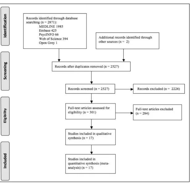
Different research methodologies were used in this thesis. A systematic review and meta-analysis of observational studies was performed to evaluate both the frequency of post-stroke (ictal and interictal) epileptiform activity in the EEG, and the quality of studies about this
IX
subject. Furthermore, different types of observational studies (including clinical case report, case series and cohort studies) were completed to answer relevant clinical questions.
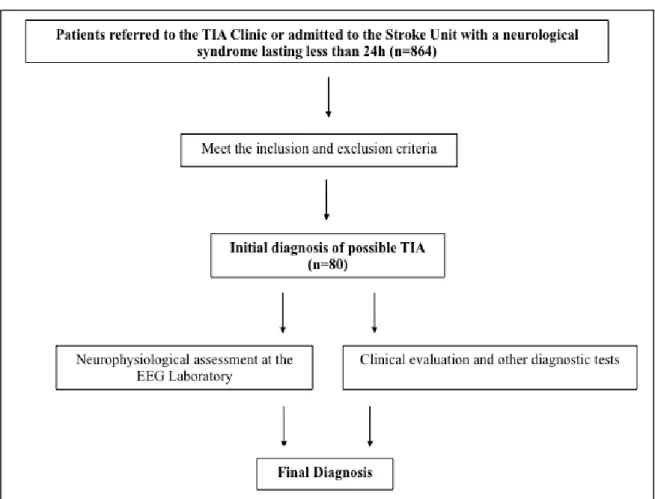
We performed a prospective longitudinal study of possible transient ischaemic attacks (TIA) patients evaluated at a tertiary centre during 36 months, with 1-3 months follow-up and also of acute anterior circulation ischaemic stroke patients, consecutively admitted to a Stroke Unit over 24 months and followed-up for one year. In both studies, patients underwent standardized clinical, diagnostic and neurophysiological assessment.
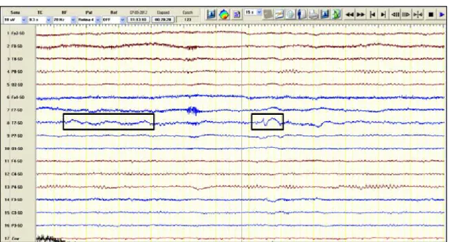
A short duration (≤60 minutes) video-EEG protocol with an extended montage including 64 EEG, two electrooculogram, one electrocardiogram and at least one electromyogram channel was established. Different electroencephalographic investigation technics including visual, back-average and quantitative analysis were used in the clinical workup of patients with possible and definite, transient and established, cerebrovascular disease as tools for the differential diagnosis and for brain functional assessment, concerning not only epileptic manifestations detection and prediction but also to search for predictors of ischaemic stroke functional outcome and vital prognosis.
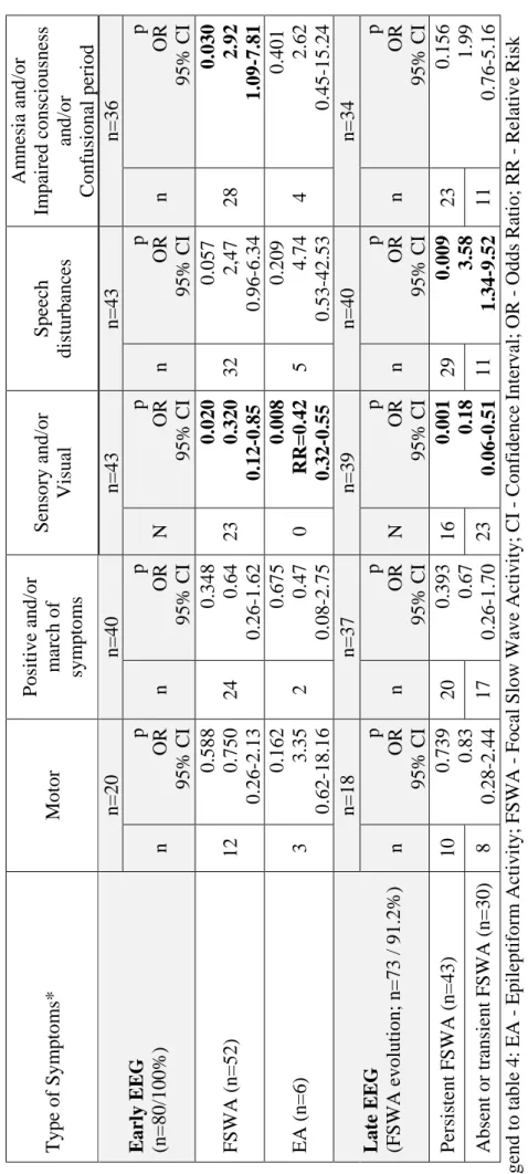
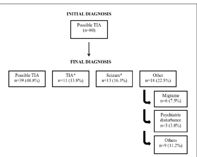
Although epileptic seizures were the most frequent defined final diagnosis (n=13; 16.3%) in our series of 80 patients with difficult-to-diagnose transient neurological symptoms, visual inspection of EEG supported this diagnosis only in 7.5% (n=6) of patients with possible TIA. Moreover, the majority (n=6; 53.8%) of patients with the final diagnosis of epileptic seizures did not have interictal epileptiform activity in an early EEG. Furthermore, early focal slow wave activity, the most frequent EEG abnormality in this patient’s series, did not distinguished between TIA and seizure patients.
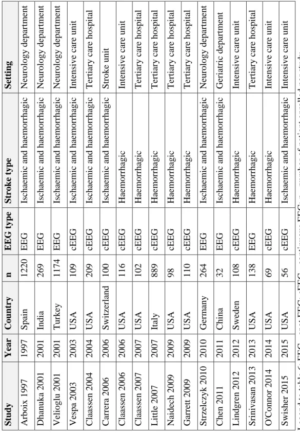
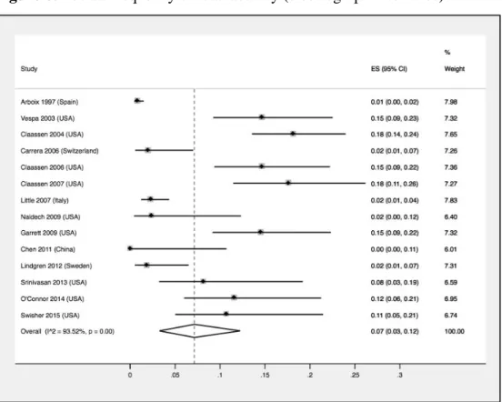
Our systematic review and random-effects meta-analysis showed that the pooled frequency of post-stroke ictal and interictal epileptiform activity was 7% (95%CI: 3%-12%) and 8% (95%CI: 4%-13%) respectively. Only 2 out of 17 included studies (11.7%) attained the maximum quality score. Moreover, no study exclusively enrolled ischaemic stroke patients, highlighting the need for higher quality studies in the evaluation of epileptiform activity frequency in this type of cerebrovascular disease. Furthermore, due to detection bias, it was not possible to correlate clinical and electrographic seizures.
In our prospective cohort of 151 anterior circulation acute stroke patients, we identified different post-stroke, clinical and electroencephalographic, epileptic manifestations including 22.7% (5/22) of acute symptomatic seizures that were exclusively electrographic and therefore could not otherwise be recognised. Furthermore, only EEG back-average
X
analysis allowed the diagnosis of cortical myoclonus during intravenous alteplase perfusion in one clinical vignette included in this work and the recognition of epilepsia partialis continua as a chronic complication of this stroke type in 1.7% of patients. This original work also showed that studied clinical and EEG epileptic manifestations were not significantly different between intravenous alteplase treated and non-treated patients.
This thesis work established which abnormalities of an early EEG after acute stroke (background activity asymmetry and the presence of interictal epileptiform activity) are independent predictors of epilepsy in the year after (even when adjusted for clinical and imaging stroke severity). Besides this, early (within the first 72h) post-stroke EEG features, extracted from visual (background activity diffuse slowing and asymmetry) and quantitative (such as delta-theta to alpha-beta ratio and alfa relative power) analysis were recognized as independent predictors of death or functional dependency, at hospital discharge and at 12 months after stroke. Furthermore, outcome models that incorporate delta-theta to alpha-beta ratio or alpha relative power were better than models based exclusively on clinical and imaging-related ischaemic stroke severity at hospital admission. Additionally, post-stroke acute symptomatic seizures and epilepsy were independently associated to death and to an unfavourable outcome 1 year after an acute anterior circulation ischaemic stroke, respectively.
Globally, these research projects have shown the value of EEG in the current paradigm of stroke patient’s care. Furthermore, they expand the knowledge both about the EEG role as a complementary neurophysiological tool in general Neurology and about different aspects of the diagnosis and outcome of two of the most prevalent neurological disorders, Cerebrovascular Diseases and Epilepsy, in particular.
Beyond the value of specific results, with this work several other research questions about EEG and seizures in ischaemic cerebrovascular disease emerge. Therefore, new possibilities of future research, ideally multicentric, clinical or translational arise.
Key-words: EEG, cerebrovascular disease, transient ischaemic attacks, ischaemic stroke, alteplase, intravenous thrombolysis, acute symptomatic seizures, unprovoked seizures, epilepsy, diagnosis, prediction, outcome.
XI
O EEG na Doença Vascular Cerebral Isquémica Aguda
O eletroencefalograma (EEG) é uma técnica neurofisiológica com uma elevada resolução temporal e sensibilidade na avaliação da função cerebral em tempo real. Para além disso, o EEG é o gold standard para a identificação de biomarcadores da epileptogénese e ictogénese.
As crises epilépticas e a doença vascular cerebral são duas das doenças neurológicas mais frequentes e que impõem mutuamente desafios importantes. Além disso, nos últimos anos, os cuidados ao doente com AVC evoluíram de uma forma notável e num novo paradigma de cuidados agudos (centrados em equipas multidisciplinares), as crises epilépticas (como complicações pós-AVC) merecem ser re-estudadas. O EEG é um exame neurofisiológico essencial na avaliação de doentes com crises epilépticas, estado de mal epilético e/ou epilepsia, tanto para o diagnóstico e classificação, como para o estabelecimento de um tratamento adequado e predição prognóstica. Para além disso, o EEG foi usado previamente na doença vascular cerebral com diferentes objectivos. Contudo, é ainda incerta a sua utilidade clínica no diagnóstico diferencial dos sintomas neurológicos transitórios, nomeadamente na diferenciação entre um acidente isquémico transitório e alguns tipos de crises epilépticas, e também no diagnóstico ou predição de crises epilépticas após um acidente vascular cerebral (AVC) ou na predição do prognóstico pós-AVC.
Neste trabalho, tivemos como objectivo usar o modelo clínico da doença vascular cerebral para estudar o valor do EEG no diagnóstico diferencial dos sintomas neurológicos transitórios, no diagnóstico e predição das crises pós-AVC, assim como para analisar se as alterações eletroencefalográficas e/ou as crises epilépticas são preditoras independentes do prognóstico num doente com um acidente vascular cerebral isquémico da circulação anterior. Ademais, porque o tratamento atual padrão dos doentes com acidente vascular cerebral isquémico agudo (alteplase endovenosa) está associado a redução da mortalidade e da incapacidade, com possíveis consequências na frequência de crises pós-AVC, mas um efeito pró-convulsivo e epileptogénico da alteplase também foi descrito, quisemos estudar a hipótese dos doentes tratados com alteplase endovenosa terem uma frequência diferente de manifestações epilépticas (clínicas e/ou eletroencefalográficas) comparativamente com os doentes não tratados.
XII
Diferentes metodologias de investigação foram usadas neste trabalho. Foi efetuada uma revisão sistemática e meta-análise de estudos observacionais para avaliar a frequência de atividade epileptiforme (crítica e intercrítica) e a qualidade dos estudos existentes sobre este assunto. Adicionalmente, foram completados diferentes estudos observacionais (incluindo descrição de caso clínico, série de casos e estudos de coorte) respondendo a perguntas clínicas relevantes.
Efetuámos um estudo longitudinal prospectivo de doentes com acidentes isquémicos transitórios possíveis, avaliados num centro terciário durante 36 meses e com 1-3 meses de seguimento. Estudámos ainda doentes com um AVC isquémico agudo da circulação anterior, admitidos consecutivamente numa Unidade de AVC durante 24 meses e seguidos durante 1 ano. Em ambos os estudos os doentes foram submetidos a uma avaliação clínica, diagnóstica e neurofisiológica estandardizada.
Foi estabelecido um protocolo de vídeo-EEG de curta duração (≤60 minutos) com uma montagem que incluía pelo menos 64 canais de EEG, dois de eletrooculograma, um de eletrocardiograma e pelo menos um de eletromiograma. Diferentes técnicas eletroencefalográficas foram usadas na avaliação de doentes com doença vascular cerebral possível ou definitiva, transitória ou estabelecida. Estas ferramentas foram utilizadas para o diagnóstico diferencial e para a avaliação da função cerebral, não só no que diz respeito à detecção e previsão de manifestações epilépticas, mas também à identificação de preditores do prognóstico funcional e vital de doentes com um acidente vascular cerebral isquémico.
Embora as crises epilépticas tenham sido o diagnóstico final definido mais frequente (n=13/16.3%) na nossa série de 80 doentes com sintomas neurológicos transitórios de difícil diagnóstico, a análise visual do EEG suportou este diagnóstico somente em 7.5% (n=6) dos doentes com AIT possível. Aliás, a maioria dos doentes (n=6; 53,8%) com o diagnóstico final de crises epilépticas não tinha atividade epileptiforme no EEG precoce. Por outro lado, a atividade lenta focal precoce, alteração mais frequente no EEG nesta série de doentes, não permitiu o diagnóstico diferencial entre um AIT e uma crise epiléptica.
A revisão sistemática e a meta-análise de efeitos aleatórios efetuadas mostrou uma frequência estimada de atividade epileptiforme crítica e intercrítica de 7% (95%CI: 3%-12%) e 8% (95%CI: 4%-13%), respectivamente. Somente 2 dos 17 estudos incluídos (11,7%) obtiveram uma pontuação máxima na escala de avaliação de qualidade utilizada. Para além disso, nenhum estudo envolveu doentes exclusivamente com AVC isquémico, mostrando a necessidade de trabalhos de melhor qualidade na avaliação da frequência da
XIII
atividade epileptiforme neste tipo de doença vascular cerebral. Além disso, devido a um viés de detecção, não foi possível correlacionar as crises clínicas e eletrográficas.
No estudo prospectivo de 151 doentes com AVC isquémicos da circulação anterior, identificámos diferentes manifestações epilépticas pós-AVC, clínicas e eletroencefalográficas, incluindo 22.7% (5/22) de crises sintomáticas agudas que foram exclusivamente eletrográficas e que, portanto, não poderiam ter sido reconhecidas de outra maneira. Da mesma forma, somente a análise de “back-average” permitiu o diagnóstico de mioclonias corticais durante a perfusão endovenosa de alteplase numa vinheta clínica incluída neste trabalho e o reconhecimento de epilepsia partialis continua como uma complicação crónica deste tipo de AVC em 1.7% dos doentes. Este trabalho original também mostrou que as manifestações epilépticas estudadas, clínicas e eletroencefalográficas, não foram significativamente diferentes entre doentes tratados e não tratados com alteplase endovenosa, após ajustamento para a idade e gravidade clínica e imagiológica do enfarte. Esta tese identificou ainda alterações eletroencefalográficas precoces após um acidente vascular cerebral agudo do território anterior (assimetria da eletrogénese de base e atividade epileptiforme intercrítica no EEG) como preditores independentes de epilepsia pós-AVC no ano seguinte (mesmo após ajuste para a gravidade clínica e imagiológica do enfarte). Além disso, características eletroencefalográficas precoces (das primeiras 72h) pós-AVC, extraídas não só da análise visual (lentificação e assimetria da eletrogénese de base) mas também quantitativa do EEG (como o rácio delta-teta / alfa-beta e a potência relativa do alfa) foram reconhecidas como preditores independentes de morte ou dependência funcional, não só na alta como 12 meses depois de um AVC isquémico da circulação anterior. Para além disso, os modelos de prognóstico que incorporaram o rácio delta-theta / alfa-beta ou a potência realtiva do alfa tiveram uma melhor capacidade discriminativa do que o modelo baseado exclusivamente na gravidade clínica e imagiológica do enfarte aquando da admissão hospitalar do doente com um AVC agudo do território anterior. Adicionalmente, as crises sintomáticas agudas e a epilepsia pós-AVC associaram-se a morte e a um prognóstico desfavorável, respetivamente, no ano seguinte a um AVC agudo da circulação anterior.
Na sua globalidade, os projetos de investigação incluídos nesta tese mostraram o valor do EEG no paradigma atual dos cuidados médicos ao doente com AVC isquémico. Ademais, ampliam o conhecimento sobre o valor do EEG como uma técnica neurofisiológica complementar em Neurologia e sobre o diagnóstico e prognóstico de duas das mais prevalentes doenças neurológicas, as Doenças Vasculares Cerebrais e a Epilepsia.
XIV
Para além do valor dos resultados específicos, surgem com este trabalho várias outras perguntas de investigação sobre o EEG e as crises epilépticas pós AVC. Emergem assim novas possibilidades de investigação futura, idealmente multicêntrica, clínica e translacional.
Palavras-Chave: EEG, doença vascular cerebral, acidente isquémico transitório, AVC isquémico, alteplase, trombólise endovenosa, crises sintomáticas agudas, crises não provocadas, epilepsia, diagnóstico, predição, prognóstico.
XV
ACKNOWLEDGMENTS
“If you want to go fast, go alone. If you want to go far, go together” African Proverb
XVI
To all Patients included in this project, my openhearted and never-ending gratitude. This work was made because of them, with them and for them.
I am obliged to all EEG/Sleep Laboratory staff members for their outstanding team work and friendship, especially to the Clinical Neurophysiology Technicians Joana Pires, Lígia Ferreira e Rosa Santos, the Secretary Isabel Morais de Sousa, and the Assistant Benvinda Ribeiro. Without any one of them this work would not be possible.
To my dear colleague and friend Ana Rita Peralta for her sharp intelligence and precious help in all stages of this thesis. Also, for being a good listener.
My thankfulness to Dr. Teresa Pinho e Melo, Head of the Hospital de Santa Maria Stroke Unit, and Professora Doutora Patricia Canhão, Head of the TIA outpatient clinic at our department, for their mentorship, pertinent criticism and facilitating role during all this teamwork.
My acknowledgement to all Neurological Residents in training at the EEG/Sleep Laboratory during the time period of this thesis, namely Dr. Ana Catarina Franco, Dr. Pedro Viana, Dr. Cristiana Silva and Dr. Isabel Amorim for their supporting attitude and hard work at our laboratory.
I am grateful to all Nurses and Medical Residents working in the Stroke Unit of Hospital de Santa Maria between 2011 and 2013 for their great support for this project.
To my different expert Co-authors, many thanks for their precious time and exceptional work. It will be a pleasure to work with you again.
My gratitude to my thesis committee, Professor Doutor José Manuel Lopes Lima, Professora Doutora Patrícia Canhão and Professor Doutor João Costa for all their valuable time, support, advice and recommendations.
I would like to thank all my department colleagues and friends for the daily challenge of being surrounded by brilliant brains in a stimulating environment. A special mention to recent PhD or other PhD students in the Neurology Department, namely Dr. Ana Verdelho,
XVII
Dr. Miguel Coelho, Dr. Leonor Correia Guedes, Dr. Diana Aguiar de Sousa, Dr. Lara Caeiro and Dr. Ana Catarina Santos who, in their unique manner, always had an encouraging and supporting attitude.
Finally, my profound gratitude to my PhD supervisors Professor Doutor José Manuel Ferro and Professora Doutora Teresa Paiva for their intelligent leadership and coaching during all these years, for their outstanding scientific and clinical knowledge and for being (each in their own way) a role model in Medicine, Science and Life.
XIX
SCIENTIFIC BOARD and ETHICS COMMITTEE APPROVALS
“Science is a way of thinking much more than it is a body of knowledge” Carl Sagan
“Ethics is nothing else than reverence for life” Albert Schweitzer
XX
The Scientific Board of Faculty of Medicine - University of Lisbon on September 20th 2011, approved this CAML PhD project
The “Comissão de Ética para a Saúde” of the HSM-CHLN, on July 27th 2011, approved this CAML PhD project.
XXI
FUNDING
“Money is not the only answer, but it makes a difference” Barack Obama
XXII
The 2012 Research Grant in Cerebrovascular Diseases supported this work.
The scientific promoter of this grant was Sociedade Portuguesa do Acidente Vascular Cerebral and the sponsor Tecnifar. The study sponsor had no involvement in the collection, analysis and interpretation of data.
XXIII
INSTITUTIONS WHERE PROJECTS WERE PERFORMED
“Be sure you put your feet in the right place, then stand firm” Abraham Lincoln
XXIV
1. Department of Neurosciences and Mental Health. Hospital Santa Maria - Centro Hospitalar Lisboa Norte. Lisboa, Portugal
1.1. EEG/Sleep Laboratory (all projects)
1.2. TIA Clinic (project 1)
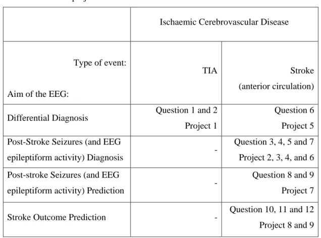
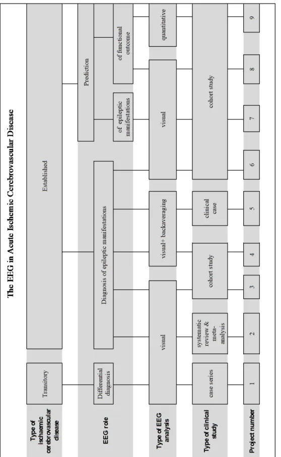
1.3. Stroke Unit (project 1, 3, 4, 5, 6, 7, 8 and 9)
2. Faculty of Medicine, University of Lisbon. Lisboa, Portugal
2.1. Neurology University Clinic (all projects)
XXV
CO-AUTHORS, AFILLIATIONS and CONTRIBUTIONS
“A nossa vida que parece uma linha recta, não o é. Construímos a nossa vida só uns 5%, o resto é feito pelos outros, porque vivemos com os outros”
XXVI
The following co-authors contributed to the projects and publications contained in this PhD thesis:
Professor Doutor José M. Ferro, affiliated to Department of Neurosciences and Mental Health (Neurology) at Hospital de Santa Maria-CHLN and Faculty of Medicine-University of Lisbon in Lisboa, Portugal, contributed fully in all projects included in this thesis, in particular in study conception and design, intellectual input to analysis and interpretations of data and critical revision of all manuscripts. Professor Doutor José Ferro made a critical revision of this thesis.
Professora Doutora Teresa Paiva, affiliated to Centro de Electroencefalografia e Neurofisiologia Clínica in Lisboa, Portugal, contributed in projects 1, 3, 4 and 6 to 9 included in this thesis, in particular in study conception and design, intellectual input to interpretations of data and critical revision of manuscripts and this thesis.
Professora Doutora Patrícia Canhão, affiliated to Department of Neurosciences and Mental Health (Neurology) at Hospital de Santa Maria-CHLN and Faculty of Medicine-University of Lisbon in Lisboa, Portugal, contributed fully in project 1 included in this thesis, in particular in study conception and design, clinical data acquisition and collection, intellectual input to the analysis and interpretation of data and critical revision of the manuscript text. Professora Doutora Patrícia Canhão also contributed to projects 3, 4 and 6 to 9 included in this thesis, in particular in intellectual input to interpretations of data and critical revision of manuscripts.
Professor Doutor João Costa, affiliated to Laboratory of Clinical Pharmacology and Therapeutics, Centre for Evidence-Based Medicine and Portuguese Collaborating Center of the IberoAmerican Cochrane Network at Faculty of Medicine-University of Lisbon, and Clinical Pharmacology Unit at Instituto de Medicina Molecular in Lisboa, Portugal, contributed in project 2 included in this thesis, in particular in study conception and design, intellectual input to interpretations of data and critical revision of the manuscript.
Dr. Teresa Pinho e Melo, affiliated to Department of Neurosciences and Mental Health (Neurology) at Hospital de Santa Maria-CHLN and Faculty of Medicine-University of Lisbon in Lisboa, Portugal, contributed in projects 3 to 9 included in this thesis in particular
XXVII
in study conception and design, intellectual input to interpretation of data and critical revision of manuscripts. Dr. Teresa Pinho e Melo also contributed to projects 1 included in this thesis, in particular in data acquisition, intellectual input to interpretations of data and critical revision of manuscripts.
Dr. Ana Rita Peralta, affiliated to Department of Neurosciences and Mental Health (Neurology) at Hospital de Santa Maria-CHLN and Faculty of Medicine-University of Lisbon in Lisboa, Portugal, contributed fully in all projects included in this thesis, in particular in study conception and design, data collection and analysis, interpretation of data and critical revision of all manuscripts.
Dr. Hugo Martins, affiliated to Department of Medicine 1.2. Hospital de São José-Centro Hospitalar Lisboa Central in Lisboa, Portugal, contributed in projects 3, 4 and 6 to 9 included in this thesis, in particular in clinical data collection and analysis and critical revision of manuscripts.
Dr. Carlos Morgado and Dr. Carlos Casimiro, affiliated toDepartment of Neuroradiology Hospital de Santa Maria-CHLN and Faculty of Medicine-University of Lisbon in Lisboa, Portugal, contributed in projects 3, 4 and 6 to 9 included in this thesis, in particular in data acquisition and analysis, interpretation of data and critical revision of all manuscripts. Dr. Carlos Morgado also contributed in project 5 in data analysis.
Professora Doutora Ana Catarina Fonseca and Dr. Ruth Geraldes, affiliated to Department of Neurosciences and Mental Health (Neurology) at Hospital de Santa Maria-CHLN and Faculty of Medicine-University of Lisbon in Lisboa, Portugal, contributed in projects 1, 3, 4 and 6 to 9 included in this thesis, in particular in data acquisition, interpretation of data and critical revision of all manuscripts. Dr. Ruth Geraldes is currently affiliated to Nuffield Department of Clinical Neurosciences at John Radcliffe Hospital in Oxford, United Kingdom and Anatomy Institute, Faculty of Medicine-University of Lisbon, Portugal.
Dr. Ana Catarina Franco, affiliated to Department of Neurosciences and Mental Health (Neurology) at Hospital de Santa Maria-CHLN in Lisboa, Portugal, contributed in projects 2 to 9 included in this thesis, in particular in data collection and analysis, interpretation of data and critical revision of all manuscripts.
XXVIII
Dr. Pedro Viana, affiliated to Department of Neurosciences and Mental Health (Neurology) at Hospital de Santa Maria-CHLN and Faculty of Medicine-University of Lisbon in Lisboa, Portugal, contributed in projects 1, 5, 8 and 9 included in this thesis, in particular in data collection and analysis, interpretation of data and critical revision of all manuscripts.
Dr. Filipe B. Rodrigues, affiliated to Laboratory of Clinical Pharmacology and Therapeutics at Faculty of Medicine, University of Lisbon, Clinical Pharmacology Unit at Instituto de Medicina Molecular in Lisboa, Portugal and Huntington’s Disease Centre, University College London, in London, United Kingdom, contributed fully in project 2 included in this thesis, in particular in study conception and design, analysis and interpretations of data, manuscript draft and critical revision.
Dr. Diana Aguiar de Sousa, affiliated to Department of Neurosciences and Mental Health (Neurology) at Hospital de Santa Maria-CHLN and Faculty of Medicine-University of Lisbon in Lisboa, Portugal, contributed in project 2 included in this thesis, in particular in data collection and analysis and interpretations of data.
Dr. Gonçalo S. Duarte and Dr. Raquel Marques, affiliated to Laboratory of Clinical Pharmacology and Therapeutics at Faculty of Medicine-University of Lisbon and Clinical Pharmacology Unit at Instituto de Medicina Molecular in Lisboa, Portugal, contributed in project 2 included in this thesis, in particular in data collection and analysis and interpretations of data. Dr. Gonçalo S. Duarte also contributed to the manuscript draft.
Dr. Hipólito Nzwalo, affiliated to the department of Biomedical Sciences and Medicine, University of Faro in Faro, Portugal, contributed in project 2 included in this thesis, in particular in data collection and analysis and interpretations of data.
XXIX
LIST OF PUBLICATIONS
“Not everything that can be counted counts, and not everything that counts can be counted”
XXX
ORCID: 0000-0003-2399-7678
1. *Bentes, C. et al. Usefulness of EEG for the differential diagnosis of possible transient ischaemic attack. Clin. Neurophysiol. Pract. 3,11-19 (2018).
2. Rodrigues, F. B. et al. Post-stroke epilepsy frequency/incidence – a protocol of a systematic review and meta-analysis of observational studies. PROSPERO 2015:CRD42015029362. (2015).
3. Bentes, C. et al. Frequency of post-stroke electroencephalographic epileptiform activity – a systematic review and meta-analysis of observational studies. Eur. Stroke J. 2, 361–368 (2017).
4. Bentes, C. et al. Post-stroke seizures are clinically underestimated. J. Neurol. 264, 1978–1985 (2017).
5. Bentes, C. et al. Epilepsia partialis continua after an anterior circulation ischaemic stroke. Eur. J. Neurol. 24, 929–934 (2017).
6. Bentes, C. et al. Cortical myoclonus during IV thrombolysis for ischaemic stroke. Epilepsy Behav. Case Reports 2, 186–188 (2014).
7. Bentes, C. et al. Epileptic manifestations in stroke patients treated with intravenous alteplase. Eur. J. Neurol. 24, 755–761 (2017).*
8. Bentes, C. et al. Early EEG predicts poststroke epilepsia. Epilepsia Open 3 (2), 441-452 (2018).
9. Bentes, C. et al. Seizures, electroencephalographic abnormalities, and outcome of ischaemic stroke patients. Epilepsia Open 2, 441–452 (2017).**
10. Bentes , C. et al. Quantitative EEG and functional outcome following acute ischaemic stroke. Clinical Neurophysiology 129, 1680-1687 (2018).
* This article was selected as the Continuing Medical Education (CME) article of the month (June 2017) by the European Academy of Neurology education committee **This article won the Epilepsy Open Prize 2018
XXXI
INITIALS, ACRONYMS and ABBREVIATIONS
“KISS principle” Keely Johnson
XXXII
A
AAS – Acute Symptomatic Seizure(s) ACA – Anterior Cerebral Artery
ACNS– American Clinical Neurophysiology Society ADCI – Acute Delta Change Index
AED – Antiepileptic Drug(s) AP – Absolute Power
ARP – Ana Rita Peralta
ASCI – Acute Symmetry Change Index
ASPECTS – Alberta Stroke Program Early CT Score AVC – Acidente Vascular Cerebral
B
BA – Background Activity
BAA – Back-Average Analysis or jerk-lock back-average analysis (JLBA) BSI – Brain Symmetry Index
C
CAML – Centro Académico de Medicina de Lisboa CB – Carla Bentes
CC – Carlos Casimiro cEEG – continuous EEG
CHLN – Centro Hospitalar Lisboa Norte CI – Confidence Interval
CM – Carlos Morgado
CNS – Central Nervous System CT – Computer Tomography
D
DAR – Delta to Alpha Ratio DS – Diana Sousa
DTABR – Delta-Theta to Alpha-Beta Ratio
DWI – Diffusion-Weighted brain magnetic resonance Imaging
E
EA – Epileptiform Activity ED – Emergency Department
XXXIII EPC – Epilepsia Partialis Continua
ESO – European Stroke Organization
F
FBR – Filipe Brogueira Rodrigues FFT – Fast Fourier Transform
FIRDA – Frontal Intermittent Delta Rhythmic Activity FSWA – Focal Slow Wave Activity
G
GSD – Gonçalo Silva Duarte
H
HFF – High Frequency Filter HN – Hipólito Nzwalo
HSM – Hospital de Santa Maria
HSM-CHLN – Hospital de Santa Maria - Centro Hospitalar Lisboa Norte Hz – Hertz
I
ICU – Intensive Care Unit i.e. – id est ("that is.")
IEA – Interictal Epileptiform Activity
ILAE – International League Against Epilepsy IQR – InterQuartile Range
J
JLBA –Jerk-Lock Back-Average analysis or Back-Average Analysis
K
KISS – keep it simple and straightforward
L
LFF – Low Frequency Filter Ln – natural Logarithm
LPCE – Liga Portuguesa Contra a Epilepsia
M
MAC - Macintosh
MCA – Middle Cerebral Artery
MOOSE – Meta-analyses Of Observational Studies in Epidemiology mRS – Modified Rankin Scale
XXXIV
N
NA – Not Applicable
NCSE – Non-Convulsive Status Epilepticus
NIHSS – National Institutes of Health Stroke Scale ns– non-significant O OR - Odds Ratio P PC – Patrícia Canhão PD – Periodic Discharges
PRISMA – Preferred Reporting Items for Systematic review and Meta-Analysis PRISMA-P – PRISMA Protocols
pTIA – Possible TIA
PROSPERO – International prospective register of systematic reviews
Q
qEEG – quantitative EEG
R
RAWOD – Regional attenuation without delta activity
rtPA – recombinant tissue-type plasminogen activator or alteplase ROC – Receiver Operating Characteristic Curve
RP – Relative Power
RSWA – Rhythmic Slow Wave Activity RR – Relative Risk
S
SAH – SubArachnoid Haemorrhage
SAMPL – Statistical Analyses and Methods in the Published Literature SD – Standard Deviation
Secs – Seconds
SPECT – Single Photon Emission Computed Tomography SPSS – Statistical Package for the Social Sciences
SWI – susceptibility-weighted brain magnetic resonance imaging
T
TIA – Transient Ischaemic Attack
XXXV
U
US – Unprovoked Seizure(s) USA – United States of America
V
XXXVI
CONTENTS
Preamble - I
Title / Abstract / Key-Words -VII 1. In English - VIII 2. Em Português - XI Acknowledgments - XV
Scientific Board and Ethics Committee approvals - XIX Funding - XXI
Institutions where projects were performed - XXIII Co-authors, Affiliations and Contributions - XXV List of Publications - XXIX
Initials, acronyms and abbreviations - XXXI Contents - XXXVI
I. Introduction - 1
1. Research topic importance and knowledge gaps synopsis- 3 2. The EEG in ischaemic cerebrovascular disease - 6
3. Interrelations between seizures and ischaemic cerebrovascular disease - 11
II. Aim of the study - 23
III. Research Questions and Research Outline - 27
IV. Usefulness of EEG for the differential diagnosis of possible TIA (Project 1) - 33
V. Frequency of post-stroke EEG epileptiform activity - a systematic review and meta-analysis of observational studies (Project 2) - 59
VI. Post-stroke seizures are clinically underestimated (Project 3) - 77
XXXVII
VIII. Cortical myoclonus during intravenous thrombolysis for stroke (Project 5) - 111
IX. Epileptic manifestations in patients treated and not-treated with rtPA (Project 6) - 121
X. Early EEG predicts post-stroke epilepsy (Project 7) - 139
XI. Seizures, EEG abnormalities and outcome of ischaemic stroke patients (Project 8) - 165
XII. Quantitative EEG and outcome of ischaemic stroke patients (Project 9) - 201
XIII. Summary of projects main findings - 227
XIV. Discussion - 233 1. General discussion - 235 2. Clinical implications - 247 3. Research perspectives - 248 XV. References - 251 XVI. Appendices - 271 XVII. Facsimile - 303
1
I. INTRODUCTION
“Start by doing what's necessary; then do what's possible; and suddenly you are doing the impossible”
3
1. RESEARCH TOPIC IMPORTANCE AND KNOWLEDGE GAPS SYNOPSIS
The electroencephalogram (EEG) is a neurophysiological technique with high temporal resolution and sensibility in the evaluation of brain function in real time. Besides, EEG is the gold standard for identifying different epileptogenesis and ictogenesis biomarkers4,5. Moreover, EEG does not have relevant contraindications, and is a painless exam that can be done at the patient's bedside, repeatedly or continuously, according to the clinical evolution without increased risks. It is also a low-cost exam, available in most hospitals. First used in humans in 1929 by Hans Berger6, EEG survived rapid technological advances of brain imaging technics arising mainly in the 70s7, and evolved including different technical modalities of recording and analysis of bioelectrical cerebral activity. Therefore, it progressively increased its importance in the diagnosis, treatment programing and evaluation and also in functional prognostication of specific neurological disorders. Currently, it is frequently used in Neurology clinical practice as a complementary technique to the essential clinical assessment of brain function, either alone or in a multimodal perspective7. In fact, EEG is an essential neurophysiological exam in the evaluation of patients with epileptic seizures, status epilepticus and/or epilepsy, both for diagnosis and classification, as well as for the establishment of a correct treatment3,8. Furthermore, EEG has been used in cerebrovascular disease with different clinical and research purposes (described in detail in introduction point 2). However, the clinical usefulness of this neurophysiological technique in the differential diagnosis of transient neurological symptoms (namely in the differentiation between a transient ischaemic attack and some types of epileptic seizures) and also in the diagnosis or prediction of post-stroke seizures or even in stroke prognostication remains uncertain.
Cerebrovascular disease and epileptic seizures are two of the most frequent neurological disorders9 being associated to different mutual challenges and relationships in clinical practice (expanded topic of introduction point 3).
The first clinical challenge is the differential diagnosis10–16. Although expert clinical assessment and cerebral imaging are undoubtedly useful, there is still a need for complementary diagnosis tools in helping this distinction17. However, there are few studies18 about the EEG’s value in the discrimination of these neurological entities.
4
Secondly, cerebrovascular disease is a frequent risk factor for epileptic seizures19, accounting for more than half of all cases of epilepsy in elderly patients20. However, reported frequency of post-stroke seizures after an ischaemic stroke is variable (2-67%)21–26 possibly due to different study methodologies including the lack of an electroencephalographic record27. Furthermore, in recent years, stroke care has evolved remarkably and facing a new paradigm of acute standard care (centred on multidisciplinary Stroke Units), epileptic seizures (as stroke complications) deserve to be rethought. As a matter of fact, intravenous alteplase (rtPA), the gold standard treatment for acute ischaemic stroke, has been associated with clinical seizures and the occurrence of epileptiform activity in the EEG28,29. It has even been documented that seizures during rtPA perfusion can occur even in the absence of a cerebral lesion, as described in 2 patients submitted to thrombolysis for acute myocardial infarction30. In fact, neurotoxic and epileptogenic properties31 of rtPA are known. Other postulated mechanisms for seizures during thrombolysis for ischaemic stroke include secondary cortical infarct from distal embolization or reperfusion/hyperperfusion syndrome32. A different imaging pattern of the infarct in patients treated with rtPA might also predispose to seizure occurrence. Despite the clinical relevance of these observations, rtPA related seizures30,32,33 and the frequency of seizures in patients treated with rtPA34,35 has been mostly described in a few retrospective and non-controlled case series. Furthermore, it is known that clinical stroke severity and infarct dimension are risk factors for post-stroke epileptic seizures and vascular epilepsy27 and that EEG is a sensitive neurophysiological technique in the detection of acute cerebral ischaemia36 and a robust one in the functional assessment of the brain37. However, it is unclear whether electroencephalographic markers of acute vascular injury severity are independently associated with an increased risk of post-stroke seizures or useful for their prediction. The third interaction between cerebrovascular disease and epileptic seizures is a possible outcome influence. Stroke is a leading cause of disability and mortality worldwide and global stroke burden is expected to rise in the future38. Despite the existence of demographic, clinical and imaging27,39–42 predictors of stroke functional outcome, there is large interindividual variability in short and long-term outcome of ischaemic stroke43 and still the need to identify reliable, inexpensive biomarkers that can add prognostic information in these patients. Post-stroke seizures44,45 (and also electrographic seizures and interictal epileptiform discharges in critically ill patients46–48) have been associated to an unfavourable functional outcome of stroke patients. However, it is not known if this association is independent from stroke severity. Furthermore, due to accumulating evidence regarding
5
neuro-vascular uncoupling in acute ischaemic stroke, neurophysiological biomarkers seem increasingly relevant for predicting outcome49.
We thought this project could expand the knowledge both about the EEG’s value as a complementary neurophysiological tool and regarding two of the most prevalent neurological disorders, specifically regarding their differential diagnosis, the frequency and prediction of post-stroke epileptic seizures and epilepsy, and the influence of seizures and EEG abnormalities in stroke outcome.
6
2. THE EEG IN ISCHAEMIC CEREBROVASCULAR DISEASE
The recording and monitoring of EEG in patients with acute ischaemic cerebrovascular disease has been done in different contexts and is based on various types of evidence, which are described in the following paragraphs.
2.1. The EEG in the differential diagnosis of a Transient Ischaemic Attack (TIA)
The use of EEG in clinical models of TIA has been poorly explored. However, De Reuck & Van Maele50 showed the importance of the EEG in the differential diagnosis of a TIA versus an inhibitory epileptic seizure. Comparing clinical and EEG features of patients with TIA and patients with inhibitory epileptic seizures, they found differences in age, sex and cerebrovascular risk factors between the two groups. In addition, most patients with seizures had EEG abnormalities (specific and nonspecific), while 90% of patients with TIA had a normal EEG. Furthermore, in a subgroup analysis (TIA with symptoms lasting less than 24 hours versus less than 1 hour) the clinical differences previously found were not significant, but (regardless of TIA definition) EEG findings remain different between TIA and epileptic seizures patients. The authors conclude that an urgent EEG is essential in the investigation of a transient episode of neurological dysfunction. In fact, EEG differences appeared to be larger than those found when comparing patients with and without unprovoked seizures after stroke51. This evidence increased the number of EEG requests for patients admitted to the TIA Clinic of our Neurology Department, especially in patients with difficult-to-diagnose transient neurological symptoms or possible TIA. However, the type and frequency of EEG abnormalities in possible TIA and their value in the distinction between an epileptic seizure and a TIA is not exactly known.
2.2. The EEG in the detection of acute cerebral ischaemia
EEG abnormalities are early markers of cerebral ischaemia and reflects the disturbance of oxygen metabolism52 and the reduction of cerebral blood flow53,54. The first EEG changes occur when cerebral blood flow reaches 25-30 ml/100g/minute. At this level, therapeutic interventions may prevent brain damage which is established for flows lower than 18 ml/100g/minute55–57 and reversible until a flow of 12ml/100g/minute36. Mild hypoxia (cerebral blood flow of 25-30 ml/100g/min) determines a subtle reduction of the amplitude
7
of rapid activity (>13 Hz) in the EEG. The worsening of ischaemia results in the appearance of polymorphic delta activity and more marked attenuation of rapid activities, including alpha and sleep spindles58,59. The use of quantitative EEG (qEEG) also showed a relationship between focal ischaemia and some indices such as the absolute and relative power of different spectral bands58,60,61 and brain symmetry index62.
Given the availability and characteristics of current cerebral imaging techniques, EEG is not currently used for the diagnosis or for the topographical location of an ischaemic stroke. However, some EEG patterns such as regional attenuation without delta (RAWOD)63 have been suggested as possible support for patient selection in the emergency department for thrombolysis or treatment of cerebral oedema64. In fact, some authors agree that EEG can be used in an acute stroke setting as a continuous monitor of cerebral perfusion to detect progressive or recurrent ischaemia59.
2.3. The EEG in the detection of revascularization and ischaemic stroke treatments effects
The EEG is sensitive to cerebral reperfusion and can demonstrate recovery earlier than the clinical examination65. This observation makes it a potentially very useful tool for continuously monitoring cerebral perfusion and stroke revascularization therapy. In fact, EEG monitoring was classically performed during endarterectomy36,66. This technique demonstrated that cerebral reperfusion is associated with neurophysiological abnormalities including changes in the EEG background activity frequency and amplitude and even epileptiform activity. EEG changes are not exclusive to endarterectomy. In fact, several other studies showed EEG changes associated with different ischaemic stroke treatments:
- Focal EEG slowing normalized when regional cerebral blood flow was elevated above the ischaemic threshold after hypervolaemic and hypertensive therapy67.
- There was a correlation between the percentage of theta and delta activity in relation to total activity (qEEG) and cerebral blood flow measured by single photon emission computed tomography (SPECT), before and after isovolaemic haemodilution in patients with middle cerebral artery stroke68
- There was a good correlation between topographic maps based on qEEG parameters and control of blood pressure in atherosclerotic acute stroke patients69.
- An improvement in acute delta change index (ADCI) and National Institutes of Health Stroke Scale70 (NIHSS) score was observed in the only patient of the Finnigan et al.71 study
8
treated with intravenous thrombolysis. According to the authors, this favourable clinical evolution would not be suspected based on acute MRI.
- A relationship was demonstrated between NIHSS score and brain symmetry index (BSI)62, in 16 patients undergoing intravenous thrombolysis. Van Putten and collaborators suggested that abnormalities of this qEEG index could alert the clinician to re-examine the patient, detecting early clinical abnormalities.
Despite these evidences, and the fact that intravenous thrombolysis72 (sometimes followed by mechanical thrombectomy) is now the gold standard treatment for acute ischaemic stroke, there is no large prospective study, using EEG monitoring during or after thrombolysis. Claassen & Hirsch73 further suggested that monitoring may also be used in hospitalized patients at high risk of stroke after TIA74 for early detection of ischaemia and maximize the benefit of thrombolysis.
2.4. The EEG in the detection of epileptiform activity after ischaemic stroke
Seizures and status epilepticus often occur after acute brain injury. In an intensive care environment, seizures are mainly electrographic and therefore cannot be detected merely by clinical observation. Claassen and colleagues75, in a series of neurological patients admitted to an Intensive Care Unit (ICU) and submitted to continuous EEG monitoring showed that 19% had seizures, mostly (92%) without obvious clinical manifestations. EEG monitoring is also essential in the diagnosis and treatment of status epilepticus in a neurological ICU46. One type of acute brain injury is ischaemic stroke and it is known that among these patients, those with clinical indication for continuous EEG monitoring, 11% will have seizures36,76, 9% exclusively non-convulsive seizures and 7% criteria for the diagnosis of non-convulsive status epilepticus75. Furthermore, a prospective study 29 using EEG monitoring showed that 17% of acute ischaemic and haemorrhagic stroke patients have epileptiform activity but the time interval between stroke and EEG and the record duration time (between 1 and 37 hours) were not predictors of that occurrence. Therefore, the optimal duration of an EEG record to detect epileptiform activity after stroke is still to be defined. Moreover, in the study of Carrera and collaborators29, factors that significantly increased the likelihood of EEG epileptiform activity were: NIHSS score on admission, cortical involvement and intravenous thrombolysis. However, in multivariate regression analysis, only the NIHSS score was an independent predictor. In fact, this study included 100 patients but only 11 were treated with intravenous thrombolysis (4 of whom had epileptiform activity in the EEG). Therefore, a
9
conclusion about the epileptogenic effect of rtPA could not be inferred. However, it is suggested that patients undergoing this treatment, particularly those with secondary clinical worsening, should have a tight EEG follow-up to exclude electrographic seizures. In animal models tissue plasminogen activator may facilitate seizures77 and has been implicated in epileptogenesis78. Nevertheless, the relationship between intravenous alteplase and clinical seizures is not established. There are studies suggesting an association of acute symptomatic seizures and intravenous thrombolysis28 but De Reuck and Van Maele32, comparing the occurrence of seizures in patients treated and or not treated, showed that early seizures were related to stroke severity and late seizures tended to be lower in the treated group. Furthermore, the influence of seizures in the outcome of ischaemic stroke patients submitted to intravenous thrombolysis is also a matter of discussion. Some studies suggest that seizures during thrombolysis represent reperfusion and neurological recovery32 whereas others indicate a potential negative influence of seizures on clinical outcome of thrombolysed stroke patients28.
2.5. The EEG in the prediction of ischaemic stroke outcome
Seizures, status epilepticus and EEG abnormalities have been associated with an unfavourable stroke outcome44,45,64,71,79–89. Nevertheless, it is questionable whether these features are markers of the extent of brain damage or even if they cause further brain injury. Regarding ischaemic stroke, several EEG features have been associated with an unfavourable outcome, including: EEG background slowing85,86 and contralateral slowing86; polymorphic delta/theta, slowing of alpha or depressed beta activity, or both87; different qEEG indexes89 including an increase in Acute Delta Change Index (ADCI)71 and regional attenuation without delta activity (RAWOD)64. Sometimes qEEG abnormalities were better than clinical (using Canadian Neurological Scale)89 or imaging investigation for the outcome prediction71. However, it is unknown whether the association between EEG abnormalities and stroke functional outcome is independent from already known cerebral infarct outcome predictors, namely age and stroke (clinical and imaging) severity39–42.
10
In short, in ischaemic cerebrovascular disease, the EEG can potentially be used for the differential diagnosis of transient ischaemic events and, in acute stroke, as a cerebral perfusion monitor to detect progressive or recurrent ischaemia59, to detect the presence of epileptiform activity, to monitor its therapy or even to predict its outcome73.
11
3. INTERRELATIONS BETWEEN EPILEPTIC SEIZURES AND ISCHAEMIC CEREBROVASCULAR DISEASE
Epileptic seizures and cerebrovascular disease are two of the most frequent neurological disorders9. In addition to their high prevalence, these disorders share different biunivocal and bidirectional associations and relationships with some consequences for their own diagnosis, outcome and therapy. The following paragraphs describe these interrelations including differential diagnosis challenges, the causal relationship, mutual treatment interference, and outcome effects.
3.1. Differential diagnosis between an epileptic seizure and a TIA
One of multiple biunivocal relationships between epileptic seizures and cerebrovascular disease is the difficulty that can exist in their differential diagnosis. In both disorders transient focal neurological symptoms that can appear to be clinically similar are physiopathologically distinct.
3.1.1. Possible TIA and possible epilepsy
About a quarter of transient ischaemic attacks patients have "probable" or "possible" TIA90,91, defined as a clinical syndrome lasting less than 24 hours that do not fulfil accepted criteria for TIA nor for another diagnosis, although a vascular origin cannot be excluded91,92. The main reasons for classification difficulties of possible TIA include odd accompanying symptoms (such as prickles/itching sensations, rigid postures or movement of the limbs), march of symptoms, consciousness disturbance or amnesia with focal symptoms, multiple stereotyped episodes, positive visual phenomena, isolated speech disturbance/confusion and focal symptoms plus panic or anxiety and paucity of details in the symptoms description91. Similarly, in the absence of a seizure documented by video-EEG recording and typical for the patient´s usual seizures, there will be situations where a diagnosis of epilepsy remains uncertain93. One approach to this difficulty is to consider uncertain cases as “probable” or “possible epilepsy”. The ILAE task force, aimed to formulate an operational definition of epilepsy for clinical diagnosis purposes93, has indeed left that possibility open for the future.
12
3.1.2. Akinetic seizures and Todd´s phenomena as TIA mimics
It is true that epileptic seizure semiology is most frequently associated with positive symptoms. Nevertheless, negative ones (when occurring) may bring difficulties to the differential diagnosis with cerebrovascular events. In fact, electrical stimulation of the human cortex typically elicits positive sensorimotor effects although several cortical stimulation studies, since Penfield and Jasper94 (1954), have also reported cortical areas in which stimulation produces inhibition or arrest of on-going movement95. These cortical regions are located laterally in the inferior frontal gyrus and premotor cortex and medially in the supplementary motor area (SMA) and pre-SMA95,96. In the dominant hemisphere, the dorsal negative motor area coincides with Broca´s area. Lüders and collaborators97 named these cortical zones as “negative motor areas", and speculate their involvement in motor planning. Typical negative motor responses by electrical stimulation of these areas include speech arrest and arrest of movements of the hand, leg and foot without loss of awareness. Ikeda et al.96 suggested that negative motor areas are indeed responsible for negative motor seizures, a rare epileptic condition that consists of motor arrest, inability to conduct voluntary movements or praxis, due to epileptic interference in a similar way to how it occurs in higher cortical areas (such as the language areas) where epileptic activation always produces inability of the functions (such as aphasia, but never creates spontaneous words or speech). Negative motor seizures should be distinguished from focal ictal paresis (or ictal atonia), linked to primary sensorimotor cortex inactivation96. Nevertheless this distinction is not always evident in literature 10,98–101 and is often difficult in clinical practice. It seems puzzling that the same cortical area (primary motor cortex) can produce positive and negative symptoms, that is, is capable of generating clonic and also atonic seizures. This phenomenon may be explained by the amount of cortex involved in the epileptic discharge102, a phenomena that was named vertical restriction of focal seizure activity. Engel and Speckmann103 showed that superficial epileptic foci and foci involving all layers of the cortex produce indistinguishable epileptiform discharge. However, only the focus involving all cortical layers was able to produce a spinal discharge and only spikes localized in the superficial layer produced sustained inhibitory activity in the deep layer. This may explain negative motor events when an epileptiform potential is recorded over the motor cortex. Despite having different cortical generator mechanisms, clinically negative motor seizures and focal ictal paresis are rarely distinguished, since simultaneous EMG recording is often not performed. The diagnosis relies on either clinical testing for a movement task or detailed history after the event to determine whether the patient intended to perform movements. To