Outcome and characteristics of infants with
acute viral bronchiolitis submitted to mechanical
ventilation in a Brazilian pediatric intensive care
Evolução e característica de lactantes com bronquiolite viral
aguda submetidos à ventilação mecânica em uma unidade de
terapia intensiva pediátrica brasileira
INTRODUCTION
Acute viral bronchiolitis (AVB) is the most frequent lower airway infection in the irst two years of life. he disease is much more severe in young infants, premature newborns and in children with underlying diseases such as
bron-chopulmonary dysplasia, immunodeiciencies and congenital heart defects.(1-8)
AVB has a seasonal pattern, predominating in the autumn and winter seasons.(5-7,9) he respiratory syncytial virus (RSV) is the main etiological agent
being followed by adenovirus, parainluenza, inluenza, rhinovirus,
metapneu-movirus, coronavirus and mycoplasma.(1,9-13)
Fernanda Umpierre Bueno1,
Jeferson Pedro Piva2, Pedro Celiny
Ramos Garcia3, Patrícia Miranda
Lago4, Paulo Roberto Einloft5
1. Physician at Pediatric Intensive Care Unit at Hospital São Lucas – Pontiicia Universidade Católica do Rio Grande do Sul PUCRS – Porto Alegre (RS), Brazil. 2. Associate Professor of Pediatrics at Pontiicia Universidade Católica do Rio Grande do Sul PUCRS – Porto Alegre, RS and Universidade Federal do Rio Grande do Sul UFRGS – Porto Alegre (RS), Brazil. 3. Associate Professor of Pediatrics at Pontiicia Universidade Católica do Rio Grande do Sul PUCRS – Porto Alegre (RS), Brazil.
4. Physician of Pediatric Intensive Care Researching Group and Pediatric Post Graduation Program at Pontiicia Universidade Católica do Rio Grande do Sul PUCRS – Porto Alegre (RS), Brazil. 5. Associate Professor of Pediatrics at Pontiicia Universidade Católica do Rio Grande do Sul PUCRS – Porto Alegre (RS), Brazil.
ABSTRACT
Objective: To describe the charac-teristics and the outcome of infants with acute viral bronchiolitis submitted to mechanical ventilation.
Methods: We performed a retro-spective study enrolling all infants (less than 12 months old) admitted with the diagnosis of acute viral bronchiolitis and submitted to mechanical ventilation in an university ailiated Brazilian pediat-ric intensive care unit between March, 2004 and September, 2006 (3 consecu-tives winters). he mechanical ventila-tion parameters’ employed on 1st, 2nd, 3rd, 7th day and before extubation were evaluated as well as the evolution (mor-tality rate, presence of acute respiratory distress syndrome and the prevalence of complications). he groups were com-pared using the Student t test, the Mann-Whitney U test and the Chi-square test.
Results: Fifty-nine infants were includ-ed (3.8 ± 2.7 months old, 59% male), with 9.0 ± 9.4 days on mechanical ventilation. Prior mechanical ventilation, non invasive ventilation was instituted in 71% of
chil-dren. Anemia was observed in 78% of the sample. In 51 infants (86.5%) the lower airway obstructive pattern was maintained up to tracheal extubation with a nil mor-tality and low prevalence of pneumotho-rax (7.8%). Acute respiratory distress syn-drome occurred in 8 infants (13.5%), with higher mortality and a higher prevalence of pneumothorax (62.5%).
Conclusions: he declining mortal-ity in acute viral bronchiolitis is observed even in non developed regions, involv-ing children with high rates of anemia and premature labor. he low mortality is associated with the maintenance of the lower airway obstructive pattern during the period on mechanical ventilation. he development of acute respiratory distress syndrome is associated with in-creased mortality and higher prevalence of complications, representing the actual challenge in the management of children with severe acute viral bronchiolitis.
Keywords: Bronchiolitis, viral/ therapy; Respiration, artiicial; Intuba-tion, intratracheal; Acute respiratory distress syndrome
Received from: Pediatric Department, School of Medicine at Pontiicia Universidade Católica do Rio Grande do Sul - PUCRS - Porto Alegre (RS), Brazil and Pediatric Intensive Care Unit at Hospital São Lucas, Pontiicia Universidade Católica do Rio Grande do Sul - PUCRS - Porto Alegre (RS), Brazil.
he above authors declare that they do not have any potential conlict of interest in this study. his research was partially supported by the Brazilian Research Council (CNPq).
he present study was approved by the Ethics Research Committee at PUCRS – University (CEP-06/03068).
Submetido em 17 de Abril de 2009 Aceito em 29 de Maio de 2009
Author for correspondence: Jeferson Pedro Piva
UTI pediátrica Hospital São Lucas da PUCRS
Av. Ipiranga 6.690 – 5º andar CEP: 90.610-000 - Porto Alegre (RS), Brazil.
Despite of the medical advances, the rate of hospital-ization in children with AVB has increased during lasts years.(5,6,13) Between 1 and 3% of the infants with AVB
are hospitalized and up to 15% of them are admitted to the pediatric intensive care unit (PICU).(2,5,13,14) In the last
decade the non invasive ventilation (NIV) as well as the continuous positive airway pressure (CPAP) applied either by nasal prongs or through facial mask have been increas-ingly used in young children with AVB.(15-18) Some studies
demonstrated that mechanical ventilation (MV) could be substantially reduced when CPAP or NIV are ofered to
infants with severe AVB.(18) However this possible beneit
is still matter of debate in consequence of the paucity of well designed randomized studies as well as the non uni-form indications for mechanical ventilation in the difer-ent parts of the world.(2,7,9, 13 -21) Nevertheless MV is still
provided to 1% to 15% of the hospitalized children with AVB being higher among infants with cardiac or chronic respiratory diseases.(2,16-28) he mortality rate in children
with AVB submitted to MV ranges between 1 and 7%, as well as is associated with high rate of complications (pneu-mothorax, secondary lung infection, sepsis, progressive re-spiratory failure and multiple organ failure).(9,14,18-20,24,25,28)
Typically, AVB presents as a lower airway obstructive lung disease (hyperinlation, wheezing and respiratory distress). However few patients might develop a restric-tive lung disease pattern compatible with acute respiratory distress syndrome (ARDS), that has higher mortality and a higher prevalence of complications.(13,26,29-31)
Most of studies describing the outcome of children with AVB on mechanical ventilation are based on European and/ or North American reports.(9,18,20,22,23,32,33) To our knowledge
there are no published studies conducted in Latin America describing the outcome and the pattern of MV provided to infants with AVB. We hypothesize that the outcome of Lat-in-American children with AVB submitted to MV would be afected by some typical aspects of this region (e.g.: high prevalence of anemia and low numbers of pediatric inten-sive care beds available, which delays and restricts children
admission to the PICU).(34) he purpose this study was to
evaluate the evolution of infants with AVB and submitted to MV in one referral Brazilian PICU, describing the char-acteristics (parameters) of MV and relating it to the out-come (survival, ARDS, complications) and to the age.
METHODS
A retrospective observational study was conducted including all infants (less than 12 months old), admit-ted to the PICU at Hospital São Lucas from Pontiicia
Universidade Católica do Rio Grande do Sul (PUCRS) - Brazil, with the diagnosis of AVB and submitted to MV between March 1, 2004 and September 30, 2006 (cover-ing three consecutive winters). he study was approved by the Ethics and Research Committee at PUCRS.
his university ailiated PICU is located in Porto Alegre (southern region of Brazil) and has 12 beds, with an average annual admission of 450 patients (0 to 15 years old), 55% of them are submitted to MV and the general mortality rate ranges between 7 and 9%. It is a referral PICU for medical and surgical diseases including advanced programs of kidney transplant, neurosurgery and cardiac surgery.
Children with severe AVB not responding to oxygen delivered by mask or nasal prongs are submitted to nasal
CPAP (pressure up to 10 cmH2O) or bi-level
non-inva-sive ventilation (5 and 10 to 15 cmH2O). MV support is
indicated at the discretion of the PICU medical staf and is based on the lack of response to CPAP or NIV as well as on clinical aspects (apnea, severe respiratory distress, refractory hypoxemia). he patients are ventilated pref-erably in the controlled pressure mode, with synchro-nized intermittent mandatory ventilation plus pressure support using the Siemens Servo I or Siemens Servo 300 ventilators.
Infants (less than one year old) on MV with a clinical diagnosis of AVB deined by the irst episode of wheez-ing associated with the presence of signs and symptoms of acute viral infection and signs of respiratory distress were included. All children included in this study, had samples of nasopharyngeal secretions collected to per-form direct immunoluorescence test to identify the
most common virus (Respiratory Syncytial Virus,
Adeno-virus, Parainfluenza and Influenza).
We excluded children with previous tracheostomy, chronic pulmonary disease (eg.: bronchopulmonar dys-plasia, conirmed or possible diagnosis of cystic ibrosis) oxygen-dependent patients, severely compromised cen-tral nervous system and a prior history of MV beyond the neonatal period.
study anemia was deined as hemoglobin serum levels below than 10 g/dl.
In this PICU every patient submitted to MV has a speciic low sheet where the MV settings are reported. Every change in the MV parameters is reported in this low sheet with the respective time. Some of these MV parameter’s (peak inspiratory pressure (PIP), positive end-expiratory pressure (PEEP), fraction of inspired
oxygen (FiO2) and respiratory rate (RR)) were evaluated
sequentially: at the end of the irst 6 hours on MV, at 2nd,
3rd and 7th days and 6 hours before tracheal extubation.
he higher values reported during the speciic day were included in the study.
Two researchers (FB and JP) evaluated independently the medical charts and the chest x-ray looking for the clinical evolution of the lung disease. It was necessary 100% of agreement to classify the children in two groups: a) Obstructive group - children that maintained the low-er airway obstructive pattlow-ern up to tracheal extubation. he inclusion criteria were: signs of lung hyperinlation in all chest x-ray up to the extubation and MV with a
FiO2 equal or inferior to 60%; b) ARDS group - children
that presented the arterial oxygen partial pressure(PaO2)/
FiO2 ratio below 200 mmHg, with a difuse pulmonary
iniltrate without hyperinlation in the chest x-ray and absence of cardiogenic pulmonary edema (or luid lung overload).(9,13,26,29,33)
Statistical analysis: he continuous variables were ex-pressed by means with the respective standard deviation and were compared using the Student t test (when dem-onstrate normal distribution) or the U Mann-Whitney test for asymmetrical distribution. Categorical variables were expressed as percentage and were compared with the Chi-square test or exact Fisher test.
RESULTS
During the 30 months of the study (covering three consecutive winters), 1204 children were admitted to the PICU, being 59 infants (less than one year old) with AVB submitted to MV (4.9% of all PICU admis-sions and 9.2% of patients on MV). he mean time on MV was 9.0 ± 9.4 days, with a median of 12 days in the PICU. he mean age was 3.8 ± 2.7 months old, the mean weight was 5.5 ± 2.2 kg and 59% of them were male. Prior MV, NIV or nasal CPAP were instituted in 71% of children (Table 1). he causes for indicating MV were: fatigue (73%), hypoxemia (15 %) and apnea (12%). here wasn’t any indication of MV due to car-diorespiratory arrest. RSV was the main etiological agent
(69.5%) followed by parainluenza (20%) and adenovi-rus (5%).
Transfusion with packed red cells was indicated in 46 children (78%) during the MV support, being the hemoglobin levels before transfusion of 7.5 ± 1.0 g/dL. he initial transfusion of packed red cells occurred at 4.0 ± 4.3 days of MV with an average of 16.5 ±10.8 ml/kg per patient.
he mortality rate in this series was 6.8% (4/59). In the survival group (55 patients), the extubation failure was observed in 4 patients (7.3%), being fatigue the main reason for reinstitution the MV in the irst 48 hours after tracheal tube withdrawn. he mean age in the extuba-tion failure group was 3.8 ± 2.8 months (versus 3.7 ± 2.7 months in the “extubation success” group; p= 0.97).
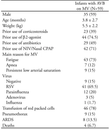
Table 1 - Characteristics of 59 children with acute viral bron-chiolitis submitted to mechanical ventilation
Infants with AVB on MV (N=59)
Male 35 (59)
Age (months) 3.8 ± 2.7
Weight (kg) 5.5 ± 2.2
Prior use of corticosteroids 23 (39) Prior use of β2-agonist 44 (74.5) Prior use of antibiotics 29 (49) Prior use of NIV/Nasal CPAP 42 (71) Main reason for MV
Fatigue Apnea
Persistent low arterial saturation
43 (73) 7 (12) 9 (15) Virus
Negative RSV Parainluenza Adenovirus Inluenza
9 (15) 41 (69.5)
12 (20) 3 (5) 1 (1.7) Transfusion of red packed cells 46 (78)
Pneumothorax 9 (15)
ARDS 8 (13.5)
Deaths 4 (6.7)
AVB - acute viral bronchiolitis; NIV - non invasive ventilation (deli-vered by face mask); Nasal CPAP - continuous positive airway pres-sure (delivered by nasal prongs); ARDS - acute respiratory distress syndrome; RSV - Respiratory Syncytial Virus; MV - mechanical venti-lation. Results are expressed as N (%) or mean ± standard deviation.
syndrome between the 3rd and 7th day of MV (ARDS
group), and the four deaths occurred in this group. In the obstructive group, 42 infants (82.3%) had a positive virology result being the RSV the most fre-quent agent (88%). Children with positive and nega-tive virology had no diferences regarding demographic characteristics, treatment or outcomes; being analyzed as a unique group. Stratifying the 51 infants of the ob-structive group according to their age (older and younger than three months old), we observed that the younger infants stayed longer time in the PICU (p= 0.039), but without diferences regarding the length on MV. Fatigue was the main reason for indicating MV in both groups of age; nevertheless apnea was identiied just in young in-fants (20%). he prevalence of RSV, prematurity, cardiac disease, prior treatment with NIV or nasal CPAP and incidence of pneumothorax or extubation failure was the same in younger and older infants (Table 2).
he peak inspiratory pressure (PIP) in the obstructive
group remained close to 30 cmH2O during days 1,2,3,7
and even on the extubation day, without diference
be-tween the younger and older infants. he only diference occurred on the irst 6 hours of MV, when infants older than 3 months used slight higher PIP (32.3 ± 3.9
cm-H2O versus 30.2 ± 3 cmH2O; p= 0.032). In igure 1 it is
shown that the groups did not present diferences regard-ing the FiO2 levels (which remained constant between 0.3 and 0.4), the respiratory rate, which varied from 16
to 20 rpm, with a mean of 11 rpm at the 6th hour before
extubation, independent of age group analyzed.
he eight infants with AVB who developed ARDS (Table 3) were older than the 51 infants in the
obstruc-tive group (5.4 versus 3.6 months; p=0.01). Although the
initial hemoglobin level was not diferent between the two groups on the day of the irst transfusion, the ARDS group received, on average, a greater volume of packed
red cells during MV (28.8 ± 9.9 ml/kg versus 13.9 ± 9.2
ml/kg; p<0.01). Compared to the obstructive group, the MV parameter’s in the ARDS group were diferent just
on the 7th day of MV, when presented higher PIP, PEEP,
respiratory rate and FiO2, as well as, higher prevalence of
pneumothorax (62.5% versus 7.8%, p=0.001).
Table 2 - Comparison between young and older infants with acute viral bronchiolitis submitted to mechanical ventilation
0 – 3 months (N=30) Older than 3 months (N=21) p Value
Weight (Kg) 4.5 ±1.5 6.9 ± 2.4 < 0.0001a
Age (months) 1.8 ± 1 6.1 ± 2.4 <0.0001a
Male 19 (63) 11 (52) 0.43c
PICU length of stay (days) 23.4 ± 56.4 10.3 ± 6.3 0.039b
Prior treatment with NIV/Nasal CPAP 19 (63.3) 17 (81.0) 0.29d
Cause of MV Fatigue Hypoxemia Apnea
19 (63) 5 (16.6) 6 (20)
20 (95) 1 (4.8)
0
0.009c
0.38d
-MV length of time (days) 7.7 ± 4.4 6.7 ± 4.9 0.16b
Deaths 0 0
-Extubation failure 2 (6.6) 2 (9.5) 1.00d
Virus Negative RSV Parainluenza Adenovirus Inluenza
5 (16) 22 (73)
5 (16) 1 (3.3) 1 (3,3)
4 (19) 15 (71) 2 (9.5) 1 (4.7)
0
1.00d
0.80c
0.68d
1.00d
-Premature infants
Congenital heart disease
10 (33) 2 (6.6)
7 (33) 1(4.8)
1.00c
1.00d
Prior treatment Use of steroids Use of Beta2-agonists
3 (10) 16 (53)
16 (76) 21 (100)
<0.0001d
-Pneumothorax 2 (6.6) 2 (9.5) 1.00d
NIV - non invasive ventilation (delivered by face mask); Nasal CPAP - continuous positive airway pressure (delivered by nasal prongs); MV - mecha-nical ventilation; RSV - Respiratory Syncytial Virus; PICU - pediatric intensive care unit. Results are expressed as N (%) or mean ± standard deviation.
Figure 1 - Comparing old and young infants with acute viral bronchiolitis concerning the parameters of mechanical ventila-tion. he only diference was observed on the 1st day of mechanical ventilation when peak inspiratory pressure was higher in infants older than 3 months old (32.3 ± 3.9 cmH2O vs 30.2 ± 3 cmH2O; p=0.032).
Table 3 - Comparing infants admitted with acute viral bronchiolitis who developed acute respiratory distress syndrome (ARDS) to those infants that maintained the lower airway obstructive pattern (no ARDS)
ARDS (N=8) No ARDS (N=51) p Value
Weight (Kg) 5.9 ± 2.1 5.5 ± 2.2 0.65a
Age (months) 5.4 ± 1.7 3.6 ± 2.8 0.01b
Male sex 5 (62.5) 30 (58.8) 1.00c
PICU Length of stay (days) 29.0 ± 23.7 18.0 ± 43.6 0.067b
Prior treatment with NIV/Nasal CPAP 6 (75) 36 (70.6) 1.00c
Cause of MV Fatigue Hypoxemia Apnea
4 (50) 3 (37.5)
1(12.5)
39 (76.4) 6 (11.8) 6 (11.8)
0.19c
0.09c
1.00c
Length of time on MV (days) 20.2 ± 20.6 7.3 ± 4.6 0.06b
Deaths 4 (50) 0
-Virus Negative RSV Parainluenza Adenovirus Inluenza
0 4 (50) 5(62.5) 1 (12.5)
0
9 (17.6) 37 (72.5)
7 (13.7) 2 (3.9)
1
-0.23c
0.006c
0.36c
-Premature infants
Infants with cardiac diseases
4 (50) 1 (12.5)
17 (33.3) 3 (5.9)
0.43c
0.45c
Transfusion of packed red cells ml/kg
8 (100) 28.8 ± 9.9
38 (74.5) 13.9 ± 9.2
-<0.001b
Pneumothorax 5 (62.5) 4 (7.8) 0.001c
DISCUSSION
In the Southern of Brazil AVB occurs as epidemic out-breaks being one of the main reasons for PICU admission and MV support in infants (respectively 4.9% and 9.4% in this study). he magnitude of these numbers could be even higher, considering that in this study we didn’t in-clude children with AVB older than 12 months.
Aside the known public health problems in this
re-gion,(1,35) this group of infants with AVB on MV have
un-questionable associated risk factors such as: high rate of anemia (78%) requiring blood transfusion, one third were preterm newborns and two thirds didn’t respond to the prior treatment with CPAP or NIV.(3,4,7,13,18,21,22) Even
con-sidering such risks, it is appealing that the observed mor-tality during MV (6.7%) was similar what has been re-ported in developed countries.(13,20,22-25,27) More intriguing
is that the mortality in infants with AVB on MV seems be strongly associated with the pulmonary pattern response. Our results demonstrate that a benign evolution with a nil mortality and low rate of complications is expected when the lower airway obstructive pattern is maintained during all course of MV. On the other hand, higher mortality and elevated rate of complications are concentrated on those cases that develop ARDS.
In the last few years several studies have demonstrated that CPAP/NIV could avoid MV in infants with severe AVB.(15-18,36-38) Unfortunately, the response to this
treat-ment is unpredictable and a substantial number of infants with AVB still need be submitted to invasive MV. he ventilatory strategy in this group is based on the respirato-ry physiologic changes.(2,13,32,39) AVB afects preferentially
lower airways with variable intensity. he inlammatory process cause non homogeneous obstruction, leading air trapping and lung hyperinlation. he high airway resis-tance prolongs the time constant, enlarging the length of time to empty and inlate the alveoli. he PIP should be suicient to overcome the high airway resistance and be accomplished with a low respiratory rate to respect the prolonged inspiratory and expiratory times.(2,13,20,32,39,40)
Similarly to our results, several studies have reported
PIP between 25 and 35 cmH2O and respiratory rate
low-er than 30 rpm for ventilating infants with AVB.(20,36,39,40)
In AVB, PIP could opening the obstructed lower airways (recruiting collapsed bronchioles), decreasing the FiO2 needs.(2,36,39,40) On the other hand, as we demonstrated,
levels of PIP close to 30 cmH2O weren’t associated with
higher incidence of pneumothorax, even when ventilating young infants with AVB. As shown in other pulmonary obstructive diseases, the use of high PEEP doesn’t avoid
the lower airway obstruction neither the progressive alveo-lar hyperinlation.(41) Maintaining PIP close to these
val-ues, even in the weaning phase, possibly overcome both the thoracic wall resistance and airway resistance, deliver-ing an adequate tidal volume.(2,13,20,36,39,40) We presume that
the low rate of extubation failure in our study might be explained on the beneicial of this weaning strategy.
A small group of patients with AVB changes the natu-ral course and progress to ARDS manifested by reduced pulmonary compliancy due to alveolar and interstitial involvement.(26) Similarly what was reported previously,
we observed that 13.5% of children with AVB developed
ARDS between the 3rd and 7th day of MV, with high rate
of complications and elevated mortality. In such circum-stances, the ventilatory strategy is based on high PEEP
(proportional to the FiO2 requirements), low PIP and low
tidal volumes (to avoid alveolar hyperdistension). Never-theless the mortality rate is still high oscillating between 30 and 50%.(26,30,31) Aside the protective ventilatory
strate-gy, some other adjunct therapy should be better evaluated in this condition as: exogenous surfactant administration, support ventilation with high frequency oscillation, corti-costeroids and other therapies.(13,14,29,38,42,43)
he ARDS pattern in children with AVB submitted to MV could be primarily associated with: a) he speciic virus agent Inluenza virus, rhinovirus and adenovirus have been associated with worst evolution and poor out-come;(1,7,10,11,13,19,21,23,24) b) Factors involved with the
indi-vidual immune response have been associated with poor outcome (genetic predisposition, pro/anti-inlammatory imbalance response); (13,19,21,23,26) c) Ventilator-induced
lung injury can´t be ruled as an isolated or associated fac-tor with ARDS pattern in the course of MV in this group of children with AVB.(9,20,31,33) While we do not identify
the exact mechanism causing ARDS in children with AVB we will not be able to ofering the efective strategies to decrease the crude mortality in this challenge situation.
We are aware that we could have some bias in this retro-spective study based on data extraction from single referral center and without the strict standardization to manipu-late the respirator parameters. Another aspect that is inher-ent to the vast majority of studies evaluating MV relays on the subjective basis for indicating and withdrawing the MV.(2,9,13-18,26,33,36) In the present study, this aspect could be
methodological aspects, we strongly believe that our results relects the local reality as well as are quite comparable what has been reported in other regions of the planet.
CONCLUSION
We demonstrate that the declining mortality rate in AVB (between 1 and 7%)(13,20,22-25,27) is observed even
in non developed regions, involving children with high rates of anemia and premature labor. Another important inding in this study is that the low mortality in children with AVB is associated with the maintenance of the lower airway obstructive pattern during the period on MV. On the contrary, those children that developed ARDS had an increased mortality rate and higher prevalence of compli-cations, representing the actual challenge in the manage-ment of children admitted to the PICU with AVB.
RESUMO
Objetivo: Descrever as características e a evolução de lactan-tes com bronquiolite aguda submetidos à ventilação mecânica.
Métodos: Estudo retrospectivo desenvolvido entre março 2004 e setembro 2006 (três invernos consecutivos), recrutando todos os lactantes (menos de 12 meses de idade) com diagnóstico de bronquiolite viral aguda e submetidos à ventilação mecânica em uma unidade de terapia intensiva, brasileira, ligada a uma
universidade. Os parâmetros de ventilação mecânica adotados no 1°, 2° 3° e 7° dia e antes da extubação foram avaliados, assim como a evolução (taxa de mortalidade, presença da síndrome de desconforto respiratório agudo) e prevalência de complicações. Os grupos foram comparados usando o teste t de Student, o teste U de Mann-Whitney e o teste Qui-Quadrado.
Resultados: Foram incluídos 59 lactantes ((3,8 ± 2,7 me-ses de idade, 59% de sexo masculino) com 9,0 ± 9,4 dias em ventilação mecânica. Antes da ventilação mecânica, ventilação não-invasiva foi instituída em 71% dos lactantes. Foi observada anemia em 78% da amostra. Em 51 lactantes (86,5%), o padrão obstrutivo de vias aéreas inferiores foi mantido até extubação in-tratraqueal, com mortalidade nula e baixa prevalência de pneu-motórax (7,8%). A síndrome de desconforto respiratório agudo, ocorreu em 8 lactantes (13,5%) com mortalidade mais elevada e alta prevalência de pneumotórax (62,5%).
Conclusões: O declínio na mortalidade em crianças com bronquiolite viral aguda tem sido observado mesmo em regiões não desenvolvidas, com altas taxas de anemia e partos prematu-ros. A baixa mortalidade está associada à manutenção o padrão obstrutivo de vias aéreas inferiores durante o tempo em ventilação mecânica. O desenvolvimento da síndrome de desconforto res-piratório agudo está associado a uma mortalidade mais elevada e maior porcentagem de complicações representando o desaia atu-al para o tratamento de crianças com bronquiolite viratu-al aguda.
Descritores: Bronquiolite viral/terapia; Respiração artiicial; Intubação intratraqueal; Síndrome do desconforto respiratório do adulto
REFERENCES
1. Calegari T, Queiroz DA, Yokosawa J, Silveira HL, Costa LF, Oliveira TF, et al. Clinical-epidemiological evaluation of respiratory syncytial virus infection in children attended in a public hospital in midwestern Brazil. Braz J Infect Dis. 2005;9(3):156-61.
2. Rodríguez Núñez A, Martinón Torres F, Martinón Sanchez JM; Sociedad Española de Cuidados Intensivos Pediátricos. [Ventilation in special situations. Mechanical ventilation in bronchiolitis]. An Pediatr (Barc). 2003;59(4):363-6. Spanish. 3. Meissner HC. Selected populations at increased risk from
respiratory syncytial virus infection. Pediatr Infect Dis J. 2003;22(2 Suppl):S40-4; discussion S44-5.
4. Weisman LE. Populations at risk for developing respiratory syncytial virus and risk factors for respiratory syncytial vi-rus severity: infants with predisposing conditions. Pediatr Infect Dis J. 2003;22(2 Suppl):S33-7; discussion S37-9. 5. Shay DK, Holman RC, Newman RD, Liu LL, Stout JW,
An-derson LJ. Bronchiolitis-associated hospitalizations among US children, 1980-1996. JAMA. 1999;282(15):1440-6. 6. Fjaerli HO, Farstad T, Bratlid D. Hospitalisations for
res-piratory syncytial virus bronchiolitis in Akershus, Norway,
1993-2000: a population-based retrospective study. BMC Pediatr. 2004;4(1):25.
7. Welliver RC. Review of epidemiology and clinical risk fac-tors for severe respiratory syncytial virus (RSV) infection. J Pediatr. 2003;143(5 Suppl):S112-7.
8. Papadopoulos NG, Moustaki M, Tsolia M, Bossios A, As-tra E, Prezerakou A, et al. Association of rhinovirus infec-tion with increased disease severity in acute bronchiolitis. Am J Respir Crit Care Med. 2002;165(9):1285-9. 9. Tasker RC, Gordon I, Kif K. Time course of severe
respi-ratory syncytial virus infection in mechanically ventilated infants. Acta Paediatr. 2000;89(8):938-41.
10. Straliotto SM, Siqueira MM, Muller RL, Fischer GB, Cunha ML, Nestor SM. Viral etiology of acute respiratory infections among children in Porto Alegre, RS, Brazil. Rev Soc Bras Med Trop. 2002;35(4):283-91.
11. Hall CB. Respiratory syncytial virus and parainluenza virus. N Engl J Med. 2001;344(25):1917-28. Comment in: N Engl J Med. 2001;345(15):1132-3. N Engl J Med. 2001;345(15):1132; author reply 1133.
13. Frankel LR, Derish MT. Respiratory syncytial virus-indu-ced respiratory failure in the pediatric patient. New Horiz. 1999;7:335-46.
14. Davison C, Ventre KM, Luchetti M, Randolph AG. Ei-cacy of interventions for bronchiolitis in critically ill in-fants: a systematic review and meta-analysis. Pediatr Crit Care Med. 2004;5(5):482-9. Comment in: Pediatr Crit Care Med. 2004;5(5):498-500.
15. Campion A, Huvenne H, Leteurtre S, Noizet O, Binoche A, Diependaele JF, et al. [Non-invasive ventilation in in-fant with severe infection presumably due to respiratory syncytial virus: feasibility and failure criteria]. Arch Pedia-tr. 2006;13(11):1404-9. French.
16. Larrar S, Essouri S, Durand P, Chevret L, Haas V, Chaber-naud JL, et al. [Efects of nasal continuous positive airway pressure ventilation in infants with severe acute bronchio-litis]. Arch Pediatr. 2006;13(11):1397-403. French. 17. hia LP, McKenzie SA, Blyth TP, Minasian CC,
Kozlo-wska WJ, Carr SB. Randomised controlled trial of nasal continuous positive airways pressure (CPAP) in bronchio-litis. Arch Dis Child. 2008;93(1):45-7. Comment in: Arch Dis Child. 2008;93(7):637-8.
18. Javouhey E, Barats A, Richard N, Stamm D, Floret D. Non-invasive ventilation as primary ventilatory support for infants with severe bronchiolitis. Intensive Care Med. 2008;34(9):1608-14. Comment in: Intensive Care Med. 2008;34(9):1560-1.
19. Willson DF, Landrigan CP, Horn SD, Smout RJ. Com-plications in infants hospitalized for bronchiolitis or res-piratory syncytial virus pneumonia. J Pediatr. 2003;143(5 Suppl):S142-9.
20. Chevret L, Mbieleu B, Essouri S, Durand P, Chevret S, Devictor D. [Bronchiolitis treated with mechanical ven-tilation: prognosis factors and outcome in a series of 135 children]. Arch Pediatr. 2005;12(4):385-90. French. 21. Buckingham SC, Quasney MW, Bush AJ, DeVincenzo JP.
Respiratory syncytial virus infections in the pediatric in-tensive care unit: clinical characteristics and risk factors for adverse outcomes. Pediatr Crit Care Med. 2001;2(4):318-23.
22. Wang EE, Law BJ, Stephens D. Pediatric Investigators Collaborative Network on Infections in Canada (PICNIC) prospective study of risk factors and outcomes in patients hospitalized with respiratory syncytial viral lower respira-tory tract infection. J Pediatr. 1995;126(2):212-9. 23. Shay DK, Holman RC, Roosevelt GE, Clarke MJ,
Ander-son LJ. Bronchiolitis-associated mortality and estimates of respiratory syncytial virus-associated deaths among US children, 1979-1997. J Infect Dis. 2001;183(1):16-22. 24. hompson WW, Shay DK, Weintraub E, Brammer L, Cox
N, Anderson LJ, Fukuda K. Mortality associated with in-luenza and respiratory syncytial virus in the United Sta-tes. JAMA. 2003;289(2):179-86. Comment in: JAMA. 2003;289(2):227-9. JAMA. 2003;289(19):2499-500;
author reply 2500-2. JAMA. 2003;289(19):2499; author reply 2500-2. JAMA. 2003;289(19):2500; author reply 2500-2.
25. Holman RC, Shay DK, Curns AT, Lingappa JR, Ander-son LJ. Risk factors for bronchiolitis-associated deaths among infants in the United States. Pediatr Infect Dis J. 2003;22(6):483-90.
26. Hammer J, Numa A, Newth CJ. Acute respiratory distress syndrome caused by respiratory syncytial virus. Pediatr Pulmonol. 1997;23(3):176-83.
27. Leader S, Kohlhase K. Recent trends in severe respiratory syncytial virus (RSV) among US infants, 1997 to 2000. J Pediatr. 2003;143(5 Suppl):S127-32.
28. horburn K, Harigopal S, Reddy V, Taylor N, van Saene HK. High incidence of pulmonary bacterial co-infection in children with severe respiratory syncytial virus (RSV) bronchiolitis. horax. 2006;61(7):611-5. Comment in: horax. 2006;61(12):1098. horax. 2007;62(3):278. 29. Cepkova M, Matthay MA. Pharmacotherapy of acute lung
injury and the acute respiratory distress syndrome. J Inten-sive Care Med. 2006;21(3):119-43. Review.
30. Ventilation with lower tidal volumes as compared with traditional tidal volumes for acute lung injury and the acute respiratory distress syndrome. he Acute Res-piratory Distress Syndrome Network. N Engl J Med. 2000;342(18):1301-8. Comment in: ACP J Club. 2001;134(1):16. N Engl J Med. 2000;342(18):1360-1. N Engl J Med. 2000;343(11):812-3; author reply 813-4. N Engl J Med. 2000;343(11):812; author reply 813-4. N Engl J Med. 2000;343(11):813; author reply 813-4. 31. Moloney ED, Griiths MJ. Protective ventilation of
pa-tients with acute respiratory distress syndrome. Br J Anaes-th. 2004;92(2):261-70. Review.
32. Kneyber MC, Blussé van Oud-Alblas H, van Vliet M, Ui-terwaal CS, Kimpen JL, van Vught AJ. Concurrent bac-terial infection and prolonged mechanical ventilation in infants with respiratory syncytial virus lower respiratory tract disease. Intensive Care Med. 2005;31(5):680-5. 33. Lebel MH, Gauthier M, Lacroix J, Rousseau E, Buithieu
M. Respiratory failure and mechanical ventilation in severe bronchiolitis. Arch Dis Child. 1989;64(10):1431-7. Com-ment in: Arch Dis Child. 1990;65(3):332.
34. Piva JP, Schnitzler E, Garcia PC, Branco RG. he burden of paediatric intensive care: a South American perspective. Paediatr Respir Rev. 2005;6(3):160-5.
35. Victora CG, Vaughan JP, Barros FC, Silva AC, Tomasi E. Explaining trends in inequities: evidence from Brazilian child health studies. Lancet. 2000;356(9235): 1093-8. 36. Bernet V, Hug MI, Frey B. Predictive factors for the
suc-cess of noninvasive mask ventilation in infants and chil-dren with acute respiratory failure. Pediatr Crit Care Med. 2005;6(6): 660-4.
years experience in a pediatric intensive care unit. Pediatr Crit Care Med. 2006;7(4): 329-34.
38. Tasker RC. CPAP and HFOV: diferent guises of the same underlying intensive care strategy for supporting RSV bronchiolitis. Intensive Care Med. 2008;34(9):1560-1. Comment on: Intensive Care Med. 2008;34(9):1608-14. Intensive Care Med. 2008;34(9):1698-702.
39. Leclerc F, Scalfaro P, Noizet O, humerelle C, Dorkenoo A, Fourier C. Mechanical ventilatory support in infants with respiratory syncytial virus infection. Pediatr Crit Care Med. 2001;2(3):197-204.
40. Levin DL, Garg A, Hall LJ, Slogic S, Javis JD, Leiter JC. A prospective randomized controlled blinded study of three bronchodilators in infants with respiratory sincytial virus bronchiolitis on mechanical ventilation. Pediatr Crit Care Med. 2008;9(6):598-604. Comment in: Pediatr Crit Care Med. 2008;9(6):659-61.
41. Guerin C, LeMasson S, de Varax R, Milic-Emili J, Four-nier G. Small airway closure and positive end-expiratory pressure in mechanically ventilated patients with chronic obstructive pulmonary disease. Am J Respir Crit Care Med. 1997;155(6):1949-56.
42. Luchetti M, Ferrero F, Gallini C, Natale A, Pigna A, Tor-torolo L, Marraro G. Multicenter, randomized, controlled study of porcine surfactant in severe respiratory syncytial virus-induced respiratory failure. Pediatr Crit Care Med. 2002;3(3):261-8.