MASTER THESIS PRESENTED TO INSTITUTE OF
BIOMEDICAL SCIENCE ABEL SALAZAR OF THE
UNIVERSITY OF PORTO IN TOXICOLOGY AND
ENVIRONMENTAL CONTAMINATION
Anna Alfeus
Anna Alfeus
Dissertation for the master degree in Environmental Toxicology and Contamination submitted to the Institute of Biomedical Sciences of Abel Salazar from the University of Porto.
Supervisor – Professor Maria do Rosário Martins. Category – Full professor.
Affiliation – School of Health Technology of Porto- Polytechnic of Porto. Category - Researcher.
Affiliation – Interdisciplinary Center of Marine and Environmental Research (CIIMAR).
Co-supervisor – Ralph Urbatzka. Category – Postdoctoral researcher.
Affiliation - Interdisciplinary Center of Marine and Environmental Research (CIIMAR).
Co-supervisor- Prof Vitor Vasconcelos Category: Full Professor
Affiliation: Faculty of Science, University of Porto Category: President of the Board
Affiliation-Interdisciplinary Centre of Marine and Environmental Research (CIIMAR)
i Glory be to God Almighty who in his intimate mercy has allowed me to take this step in academics.
My profound appreciations go to my research supervisor Dr Rosario Martins whose wonderful contributions to the quality success of this work cannot be over emphasized. I must also acknowledge the contribution and support of my second supervisor Dr Ralph Urbatzka and Prof Vitor Vasconcelos for the wonderful support of this work. A great appreciation goes to Dr. Aldo Barreiro for his great contribution and devotion of your time particularly in the statistical analysis of this thesis result. My sincere appreciation goes to Dr. Lucia Guilhermino (Director of Master of Toxicology and Environmental contamination) and the entire staff involved in the deliverance of the coursework. Thank you very much for the profound foundation you have installed me and the level of intelligence that I have acquired.
My special thanks go to the whole staff of the LEGE (CIIMAR) for providing me with all the support I needed to finish my research. Thank you for being accommodative and for the conducive environment you have provided for me to work. Your contribution and support is extremely heavy to weigh. I feel God is on my side to send me such good people in my life. Once again, thank you very much Prof Vitor Vasconcelos for allowing to work in the lab and complete this part of work
I am indebted to my parent Ms Selma Iindombo and Mr Matheus Alfeus for their support till this moment. To my grandparents & family, my siblings Johannes, Saima & Emma, I thank them for their understanding and cooperation during my studies and I pray to God to give them wisdom to achieve their dreams in life.
ii Furthermore I would love to thank the colleagues of the Master of Toxicology and Environmental Contamination. It was a long and interesting bumpy road but with you I have found courage and confidence to proceed until I reached this stage. Thank you for assistance for without you I would not have made it.
Last but not least I would like to thank my friend who have made their support in all means. Thank you for your support and understanding and your most responsibility of my social life. You have been a back-up who enlightened my soul and spirit when I am down until I completed this work.
The studies were funded by the Kite partnership within the framework of the Erasmus Mundus Action 2 represented by the coordinating institution Masaryk University, Czech Republic to stay for the duration of 22 months at the University of Porto for the full Master degree mobility.
This research was partially supported by FCT – Foundation for Science and Technology under the project UID/Multi/04423/2013 and by the Structured Program of R&D&I INNOVMAR - Innovation and Sustainability in the Management and Exploitation of Marine Resources (reference NORTE-01-0145-FEDER-000035, Research Line NOVELMAR), funded by the Northern Regional Operational Programme (NORTE2020) through the European Regional Development Fund (ERDF).
iii Cyanobacteria are known to synthesize secondary metabolites of high diversity, which emerged with recent research of bioactivity potential. Research has explored and found interesting compounds of cyanobacteria with potential of anticancer, antiparasitic, antibacterial, antiviral, antifungal. At the Laboratory of Ecotoxicology Genomics and Evolution, LEGE – CIIMAR, studies have been performed concerning the bioactive potential of marine cyanobacteria isolated from the Portuguese coast. Recent results revealed interesting bioactivities such cytotoxicity in cancer cell lines, bacteria and fungi and toxicity in marine invertebrates. Against this background, we aimed with this work to study cyanobacteria strains from the LEGE Cyanobacteria Culture Collection in terms of its potential in skin care. Eight strains of the picoplanktonic and filamentous genera Cyanobium, Leptolyngbya, Nodosilinea, Pseudoanabaena and Synechocystis, were selected. These include strains LEGE06113, LEGE07175, LEGE06099, LEGE07167, LEGE06102, LEGE06194 LEGE06108 and LEGE06173. Cyanobacterial cultures were performed and a crude extract obtained from lyophilized biomass. Cytotoxicity of the crude extracts was assayed against keratinocytes, fibroblasts and endothelial cells. The antioxidant potential was screened by the DPPH assay and the superoxide radical scavenging assay. Effects on melanin production were assessed by the inhibition of the mushroom tyrosinase enzyme.
In vitro, the keratinocyte cell line HaCat, the fibroblast cell line 3T3L1 and the endothelial cell line hCMEC/D3 were exposed to the cyanobacteria extracts at concentrations of 100 µg/mL, 10 µg/mL and 1 µg/mL. Cellular viability was evaluated by the 3-(4.5-dimethylthiazol-2-yl)-2.5-diphenyltetrazolium bromide (MTT) assay after 24, 48 and 72 hours. The MTT results confirmed an absence of toxic effects of the selected cyanobacteria strains, in most of the cases, with an increase in cell viability at 1 µg/mL and at the 72hours incubation. From all the eight cyanobacteria strains, LEGE06102, LEGE06113 and LEGE06099 revealed to be more active, since an induction on cell viability occurred in all cell lines. Concerning the antioxidant potential, a weak antioxidant activity was registered with the DPPH assay, with strains LEGE 06113, LEGE06108 and LEGE06102 exhibiting the highest scavenging activities. In the superoxide radical scavenging activity assay, a higher antioxidant potential was registered from strains LEGE06108, LEGE06099, LEGE 07167 and LEGE07175, with
iv 49.7%, 49.57, 42.77% and 47.39% respectively at 10 µg/mL. The results of the effects of extracts on the inhibition of the mushroom tyrosinase enzyme, showed inhibition for strains LEGE06102; LEGE06099 and LEGE06113. Taking all the results together strains LEGE06102, LEGE06113 and LEGE06099 revealed a higher potential in terms of application in skin products. These results are emphasized by the fact that these strains already demonstrated bioactive potential in previous studies. Far from being completed this study represent a first step in the study of the bioactive potential of marine cyanobacteria in skin care. Additional methodologies must be performed in order to confirm the results, alongside with the quantification and identification of compounds such as phenolic compounds.
v As cianobactérias são conhecidas por produzirem compostos secundários com potencial bioactivo, nomeadamente potencial anticancerígeno, antiparasítico, antibacteriano, antivírico e, antifúngico. No Laboratório de Ecotoxicologia Genómica e Evolução, LEGE - CIIMAR, têm sido realizados estudos sobre o potencial de bioativos de cianobactérias marinhas isoladas da costa portuguesa. Resultados recentes revelaram bioactividades interessantes, tais como citotoxicidade em linhas celulares de cancro, bactérias e fungos, e toxicidade em invertebrados marinhos. Neste contexto, pretendeu-se com este trabalho estudar estirpes de cianobactérias da coleção de isolados do LEGE em termos de seu potencial em cuidados da pele. Oito estirpes dos géneros picoplanctónicos e filamentosas Cyanobium, Leptolyngbya, Nodosilinea, Pseudoanabaena e Synechocystis, foram selecionados. Estas incluíram as estirpes LEGE06113, LEGE07175, LEGE06099, LEGE07167, LEGE06102, LEGE06194 LEGE06108 e LEGE06173. As cianobactérias foram cultivadas em laboratório para obtenção de biomassa, tendo sido preparado um extracto bruto a partir de biomassa liofilizada. A citotoxicidade dos extractos brutos foi avaliada em queratinócitos, fibroblastos e células endoteliais. O potencial antioxidante foi rastreado pelo ensaio de DPPH e o ensaio de sequestro de radicais superóxido. Efeitos na produção de melanina foram avaliados pela inibição da enzima tirosinase de cogumelo.
In vitro, a linhagem celular de queratinócitos HaCat, a linhagem celular de fibroblastos 3T3L1 e a linhagem de células endoteliais hCMEC/D3 foram expostas aos extractos de cianobactérias, em concentrações de 100 μg/ml, 10 μg/mL e 1 μg/mL. A viabilidade celular foi avaliada pelo ácido 3- (4,5-dimetiltiazol-2-il) -2,5-difeniltetrazolio (MTT) após 24, 48 e 72 horas de incubação. Os resultados de MTT confirmaram na maioria dos casos, uma ausência de efeitos tóxicos das estirpes de cianobactérias seleccionadas, com um aumento na viabilidade celular na concentração de 1 μg/mL e 72 horas de incubação. Das oito estirpes de cianobactérias, as estirpes LEGE06102, LEGE06113 e LEGE06099 revelaram maior interesse, uma vez que uma induziram um aumento na viabilidade celular em todas as linhas celulares. Em relação ao potencial antioxidante, registou-se uma fraca atividade com o ensaio DPPH, tendo as estirpes LEGE06113, LEGE06108 e LEGE06102 exibido as maiores atividades. No ensaio de atividade de sequestro de radicais superóxido, foi registado um maior potencial antioxidante, tendo
vi as estirpes LEGE06108, LEGE06099, LEGE 07167 e LEGE07175, registado valores de 49,7%, 49.57, 42.77% e 47.39%, respectivamente, a 10 μg/mL . Os resultados dos efeitos dos extractos sobre a inibição da enzima tirosinase de cogumelo, mostraram inibição pelas estirpes LEGE06102; LEGE06099 e LEGE06113. Considerando todos os resultados em conjunto, as estirpes LEGE06102, LEGE06113 e LEGE06099 revelaram um maior potencial em termos de aplicação em produtos para a pele. Estes resultados são apoiados pelo fato de já terem sido descritas bioactividades em linhagens celulares em estudos anteriores. Longe de estar concluído este estudo representa um primeiro passo no estudo do potencial bioativo de cianobactérias marinhas no cuidado da pele. Metodologias adicionais devem ser realizadas a fim de confirmar os resultados obtidos assim como a quantificação e identificação de compostos, tais como compostos fenólicos.
vii
1. Introduction ... 1
1.1. Cyanobacteria general characteristics ... 1
1. 2. Bioactive compounds from marine cyanobacteria ... 2
1.3. Cyanobacteria and UV light ... 4
1.4. Cyanobacteria in cosmetics ... 8
1.4.1 Effect of cyanobacteria on keratinocytes ………..…….……..11
1.4.2 Effect of cyanobacteria on fibroblasts ………...………...……11
1.4.3 Effect of cyanobacteria on melanin production ……….…….……12
1.4.4. Effects of cyanobacteria as antioxidants ……….12
2. Objectives ... 14
3. Materials and Methods………16
3.1. Cyanobacteria strains ... 16
3.2. Cyanobacteria culture and biomass ... 16
3. 3 Preparation of a crude extract ... 17
3.4 Cell cultures and Cytotoxicity assays ... 17
3.4.1 Cell culture ……….….17
3.4.2 Viability assay - MTT assay ………..………..18
3.5 Antioxidant activity ... 19
3.5.1 DPPH radical scavenging assay ……….19
viii
3.6. Assay of tyrosinase inhibitory activity ... 20
3.7. Statistical analysis ... 20
4. Results and discussion ... 21
4.1. Cytotoxicity assays ... 21
4.2. DPPH radical scavenging activity ... 28
4.3. Superoxide radical scavenging activity ... 30
4.4. Tyrosinase inhibitory assay ... 31
5. Final considerations and future perspectives ... 36
ix Figure 1- Production of reactive oxygen species (ROS) induced by UVA radiation. ……10 Figure 2 - The homogenous subsets of the cyanobacteria strains with respect to their effect in keratinocytes. ………..………22 Figure 3 - Cell viability of the keratinocytes cell line HaCat when exposed to cyanobacteria crude extract. Extracts were tested at a concentration of 100 µg/mL, 10 µg/mL and 1 µg/mL. Solvent control consisted in 1% DMSO in cell culture medium and positive control consisted in 20 % DMSO. ………...……….……….23 Figure 4 - The homogenous subsets of the cyanobacteria strains with respect to their effect in fibroblasts. ………24 Figure 5 - Cell viability of the fibroblasts cell line 3T3L1 when exposed to cyanobacteria crude extract. Extracts were tested at a concentration of 100 µg/mL, 10 µg/mL and 1 µg/mL. Solvent control consisted in 1% DMSO in cell culture medium and positive control consisted in 20 % DMSO. ……….……….25 Figure 6 - The homogenous subsets of the cyanobacteria strain with respect to their effect in endothelial cells. ……….26 Figure 7 - Cell viability of the endothelial cell line hCMEC/D3 when exposed to cyanobacteria crude extract. Extracts were tested at a concentration of 100 µg/mL, 10 µg/mL and 1 µg/mL. Solvent control consisted in 1% DMSO in cell culture medium and positive control consisted in 20 % DMSO. ……….………..27 Figure 8 - Inhibitory effects of the cyanobacteria strains on mushroom tyrosinase. Extracts were tested at a concentration of 100 µg/mL, 10 µg/mL and 1 µg/mL. Solvent control consisted in 1% DMSO. Positive control consisted in ascorbic acid at 50 μg/mL, 25 μg/mL and 12.5 μg/mL. ………..………...33
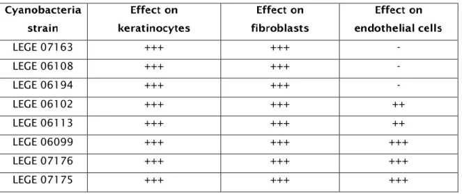
x Table 1- Botanical classification of Cyanobacteria, main structural characteristic and main genera. ……….………...2 Table 2. Cyanobacteria species with photo protective compounds. ….…………...………7 Table 3 – Bioactivities of marine cyanobacteria from the LEGE cyanobacteria culture collection. ………..…….15 Table 4 – Cyanobacteria strains included in this study and bioactivities already described. ………..………..16 Table 5 – Cell lines included in the study. ……….….….………18 Table 6 – Effect of the cyanobacteria strain with respect to extract, concentration and time on the cell lines. ………..……….……….22 Table 7 - Antioxidant activity of the cyanobacteria extracts as determined by the DPPH assay. ………...………….……..29 Table 8 - Antioxidant activity of the cyanobacteria extracts as determined by the superoxide radical assay. ……….……….…………31 Table 9 – Inhibitory activity of cyanobacteria as determined by mushroom tyrosinase enzyme against L-DOPA. ……….…..32 Table 10 – Summary of cell viability data from the MTT (3-(4,5-dimethylthiazol-2-yl)-2,5-di phenyl tetrazolium bromide) assays, after exposure to the 1μg/mL cyanobacterial crude extract at 72 hrs. +++ indicates viability higher than 100%; ++ indicates viability 90-100%; + indicates viability 90-80%; - indicates viability lower than 80%. ……….……….…………..34 Table 11: Summary of the DPPH scavenging activity, superoxide scavenging activity and
xi tyrosinase inhibitory activity after exposure to the cyanobacteria crude extract 1μg/mL……….35
xii 3T3L1 – Mouse fibroblasts
BBE - Blue Biotechnology and Ecotoxicology research group
CIIMAR – Interdisciplinary Center of Marine and Environment Research DMEM - Dulbecco's Modified Eagle Medium
DMSO – Dimethyl sulfoxide DNA - Deoxyribonucleic acid
DPPH - 2, 2-diphenyl-1-picrylhydrazyl HaCaT - Human normal keratinocytes
hCMEC/D3 – Human normal endothelial cells L- DOPA - L-3,4-dihydroxyphenylalanine
LEGE – Laboratory of Ecotoxicology, Genomics and Evolution MAAs - Mycosporine-like amino acids
MTT - 3-(4.5-dimethylthiazol-2-yl)-2.5-diphenyltetrazolium bromide NADH - Reduced nicotinamide adenine dinucleotide
NBT - Nitro blue Tetrazolium Chloride PBS - Phosphate buffered saline
PMS - Phenazine methosulphate PSI – Photosystem I
PSII – Photosystem II
ROS - Reactive oxygen species SCY - Scytonemin
UV – Ultraviolet
UVR – Ultraviolet radiation UVA – Ultraviolet A
UVB- Ultraviolet B UVC – Ultraviolet C
1 Cyanobacteria are gram-negative prokaryotes, which according to fossil records evolved about 2.7 billion years ago (Battistuzzi et al. 2004; Castenholz, 2015). According to the Botanical code of classification, cyanobacteria are formerly known as blue-green algae, which are derived from the presence of phycocyanin, a blue-green colored pigment used for photosynthesis (Whitton and Potts, 2007). Later their bacteriological similarities were discovered and changed their designation from algae to blue-green bacteria or cyanobacteria. Today both the International code of Nomenclature of Bacteria and the Botanical code of Nomenclature are valid and being utilized in the classification of cyanobacteria. In the classic botanical taxonomic scheme of cyanobacteria, five orders are recognized (Table 1) whereas in the bacterial classification scheme these orders are described as subsections. Structurally, cyanobacteria are identified as unicellular or colonial, filamentous without differentiated cells or as filamentous with differentiated cells with the absence of a nucleus and other intracellular membrane surrounded organelles such as chloroplast and mitochondria (Castenholz, 2015).
Like plants, cyanobacteria have oxygenic photosynthesis by two photosystems (PSII and PSI), using CO2 as a carbon source and H2O as the photo reductant in photosynthesis providing energy to most living organisms as well as releasing oxygen into the atmosphere (Cohen & Gurevitz, 2006; Castenholz et al., 2001). In addition to their ability to synthesize food, many cyanobacteria contain heterocysts, a thick-walled cell with a nodule of cyanophycean, which enable them to fix atmospheric nitrogen as well as anaerobic respiration (Stal & Krumbein, 1984). Based on their genotypic and phenotypic differences, cyanobacteria occur in wide and broad geographical distribution (Flombaum et al., 2013). Due to their adaptation features they are found in diverse ecological habitats of limnic, estuarine and marine environment where they play an important role in nutrient cycling, as well as terrestrial habitants (Bergman et al., 2008; Khetkorn et al., 2012). Depending on the genetic influence on species, some can cope in extreme environmental stress, such as the extreme thermal springs, or even surviving in different environmental influences associated with global warming (Papke et al., 2003; Paul, 2008). While anthropogenic activities and climate change are causing significant
2 degradation of ecosystems such as coral reefs, cyanobacteria have successfully adapted to this new altered environment dominating other organisms, due to their ability to tolerate extreme conditions (Hallock, 2005).
Table 1 - Botanical classification of Cyanobacteria, main structural characteristic and main genera.
1.Chroccocales Coccoid cells that reproduce by binary fusion or budding
Aphanocapsa,Aphanothece,Gloeocapsa,M erismopedia,Microcystis,Synechococcus,S ynechocystis
2.Pleurocapsales Coccoid cells, aggregates or pseudo-filaments that reproduce by baeocytes
Chroococcidiopsis, Pleurocapsa
3.Oscillatoriales Uniseriate filaments, without heterocytes or akinetes
Lyngbya,Leptolyngbya,Microcoleus,Oscill atoria,Phormidium,Planktothrix
4.Nostocales Filamentous cyanobacteria that divide in only one plane, with heterocytes; false branching in genera such as scytonema
Anabaena,Aphanizomenon,Calothrix,Cyli ndrospermopsis,Nostoc,Scytonema,Tolyp othrix
5.Stigonematales Division in more than one plane, true branching and multiseriate forms, heterocytes
Mastigocladus (Fishcherella),Stigonema
Historically, cyanobacteria are known to produce toxic secondary metabolites - toxins that are potentially harmful to both vertebrates and invertebrates (Codd, 1995). In humans, these toxins are known to affect the liver, the nervous system, skin as well as the gastro-intestinal tract (Drobac et al., 2013). As a result, cyanobacteria are extensively studied as an environmental nuisance or a source of toxins, hazardous to the ecosystem. However due to the high diversity in its secondary metabolites spectrum, recent discovery emerged with different bioactivities potentials (Singh et al., 2011). Research have focused more on filamentous marine cyanobacteria to produce secondary
3 metabolites with promising natural compounds because they grow in large density forming colonies, are easy to collect and analyzed directly from environmental samples (Martins et al., 2008). However, recent research works based on marine species of less studied genera such as the picoplanktonic Cyanobium, Synechocystis, Synechococcus and also other filamentous such as Nodosilinea, Leptolyngbya, Pseudanabaena and Romeria revealed several strains as promising for isolation of bioactive compounds (Costa et al., 2013). From the picocyanobacterium group, Cyanobium sp (LEGE 06113) was found to synthesize a natural compound, hierridin B, whose biological activities were tested and showed selective cytotoxicity towards HT-29 colon adenocarcinoma cells (Leão et al., 2013). Overall cyanobacteria offer interesting advantage in studies as producer of natural bioactive compounds because they can be easily cultivated in laboratory on a large scale in less efforts comparing to other aquatic microalgae, macroalgae and invertebrates (Pratt et al., 1945). Another added advantage with cyanobacteria is that, a strain of cyanobacteria is capable of producing an array of secondary metabolites with different chemical properties and bioactivities. For example, a strain of Lyngbya majuscula was found to produce around 236 compounds (Gerwick et al., 2008) and strains of Synechocystis and Synechococcus species produced compounds with both antimicrobial and anti-cancer potency (Martins et al., 2008). Although many researchers have focused on the exploration of cyanobacteria for anticancer bioactive compounds (Costa et al., 2013, Nagle et al., 1995; Bai et al., 1990; Mooberry, et al., 2003), many compounds revealed also antiparasitic potential, antibacterial, antiviral, antifungal; antiprotozoal; protease inhibitors as well as immunodulatory activities (Martins et al., 2008; Kreitlow et al., 1999; Hol, 2015). In an ecological point cyanobacteria have been identified to synthesize UV filter compounds that protect them against photochemical damage (Sinha & Häder, 2008). They also play a role in nitrogen fixing, provinding a constant fertilization of the soil ecosystem in rice fields leading to a higher yield (Sinha & Häder, 1996; Vaishampayan et al., 1998; Vaishampayan et al., 2001). They also constitute 50% biomass of the phytoplankton community in the marine environment, which are primary producer in the food chain (Whitton & Potts, 2007). During the food production process, cyanobacteria produce oxygen as a by-product, which quench in catalysing with an anaerobic enzyme the presence of molecular oxygen, which could inhibit the nitrogenase enzyme responsible for reducing the N2 into the reactive form, which is being used in the nitrogen cycle
4 (Berman-Frank et al., 2003). Irrespective of the historical growth, habitant and taxonomies, cyanobacteria have proven the ability to photo assimilate carbon monoxide in facultative anoxygenic photosynthesis (Garlick, et al., 1977).
Cyanobacteria are originated in the Precambrian era when ozone shield was absent and they faced high fluxes of ultraviolet radiation (UVR), which presumed an evolutionary pressure to adapt mechanisms against photochemical damage (Sinha & Häder, 2008). Since cyanobacteria need to harvest solar energy to drive the processes of photosynthesis and nitrogen fixation, they are exposed to UVR, which is detrimental to living organisms (Singh et al., 2010; Ehling-Schulz & Scherer, 1999). Moreover, the depletion of the stratospheric ozone layer due to anthropogenic activities enhanced the ultraviolet B radiation (UVB - 290-320nm), creating a life threatening situation for these organisms (Ehling-Schulz & Scherer, 1999; Shick & Dunlap, 2002). The decline of the atmospheric ozone content due to chlorinated chlorofluorocarbon pollutants released into the atmosphere through anthropogenic activities led to increased UVB radiation penetrating into the oceans making phytoplankton community susceptible to photo damage resulting in the altered dynamic of the marine environment (Solomon et al., 1986; Smith et al., 1992; Kerr & McElroy, 1993). The ultraviolet A radiation (UVA) has a long wavelength (320nm-400nm), thus penetrate deep into the water column causing DNA damage in cyanobacteria indirectly through the production of reactive oxygen species (ROS) or by the UVA stimulated chromophores to the DNA target or photosensitization (Cadet et al., 2005; Ravanat, et al., 2001; He & Häder 2002). Meanwhile UVB radiation being the highly energetic wavelength are absorbed directly by the targeted DNA bases inducing DNA strand breaks, mutagenic and cytotoxic DNA lesions manipulating several physiology and alteration of the genomic functions and integrity (Ravanat, et al., 2001). Studies confirmed the change in proteins and proteomes quantities of different cyanobacteria due to UVB radiations ultimately affecting key metabolic activities such as photosynthesis, N2 fixation, CO2 uptake, ribulose 1,5-bisphosphate carboxylase/oxygenase (RuBisCO) activity, cellular morphology, growth, survival, and buoyancy (Sinha et al., 1995; Gao et al., 2009). In a brief summary, both
5 UVA and UVB can negatively affect cell morphology, differentiation, orientation, growth, mobility, pigmentation, nitrogen metabolism, DNA, RNA, and proteins in cyanobacteria (Singh et al., 2010). In response, cyanobacteria has adapted mechanisms to counteract the photo damage such as migration into the mat community to avoid strikes of UV radiation, scavenging and repair of DNA and protein damage by detoxifying enzymes and antioxidants, synthesis of UV protection compounds to screen and absorb UVR as well as elimination of damaged cells (Singh et al., 2010; Sinha & Häder, 2002; Kobayashi et al., 2004; Mittler & Tel-or, 1991). Mycosporine-like amino acids (MAAs) and scytonemin (SCY) are photo protectants (Table 2) that have shown putative role in absorption of the UV region in the spectrum (Proteau et al., 1993; Sinha et al., 1999; Sinha & Hader, 2008). Most of the cyanobacteria reported with photo protective compounds have been isolated from a diverse environment inclusive of the hypersaline, benthic, freshwater, terrestrial environment, extreme environment such as desert rocks, hot springs, tree barks as well as rice field environments (Oren, 1997; Kedar et al., 2002; Sinha et al., 1999 Ehling-Schulz et al., 1997), due to the cosmopolitan distribution of cyanobacteria and their ability to synthesize a diversity of secondary metabolites, UV compounds from the less studied picoplanktonic cyanobacteria of the marine environment are explored for dermatological protection.
Favre-Bovin et al. (1987) described MAAs as water-soluble, substituted cyclohexenones, which are linked to amino acids and amino alcohols through the shikimate pathway, and maximally absorbed between 310 nm and 360 nm (Ehling-Schulz & Scherer, 1999). In addition to their screening ability and high absorption maxima, antioxidant capability and photo stability are other photo protective features (De la Coba et al., 2009; Conde et al., 2000). Besides the structural variations among mycosporine, there is Glycine, a common amino acid in many MAAs (Bandaranayake, 1998). Since the photo protective efficiency depends on the position of MAAs within the cell, significant protection limits have been reported in the cytoplasm (Böhm et al., 1995). Although the sun screening capabilities of cyanobacteria have not yet been tested, three MAAs compounds: shinorine, porphyra-334, and mycosporine–glycine from scallop ovaries have shown protective effects on human cells against UV light, making them a potential ingredients for cosmetics as a UV protectors and activators of cell proliferations (Oyamada et al, 2008). In addition to plant extracts, there is a successful tale of MAAs compound from a marine algae utilized in cosmetics as a photo protector against UVA radiations (Daniel
6
et al., 2004). Albeit MAAs being common and diverse among cyanobacteria, not all of them are induced by UVR, therefore significant protection is reliable from the presence of both MAAs and scytonemin (Bandaranayake, 1998; Garcia-Pichel & Castenholz, 1993). Scytonemin (SCY) is another photo protective compound established with a sunscreen role found in the extracellular sheath of cyanobacteria cluster or colonies and exclusively produced by cyanobacteria (Mishra et al., 2014). It is a yellow–brown, lipid-soluble, dimeric compound composed of indolic and phenolic subunits, with a molecular mass of 544 Da (Proteau et al., 1993). The chemical structure of SCY is preceded by the condensation of tyrosine and tryptophan allowing the absorption of 370 nm within in vivo studies (Jones et al., 2011). The organic extract of Scytonema sp., collected on the Mitaraka inselberg, French Guyana, yielded three new pigments, tetramethoxyscytonemin, dimethoxyscytonemin, and scytonine, derived from the scytoneman skeleton of scytonemin (Bultel-Poncé et al., 2004). SCY is described as a passive UVA sunscreen with the ability to reduce 90% of UVA radiation encroaching on the cell even in its low concentration (Garcia‐Pichel et al., 1992). Although SCY shown high absorbing strength within a spectral region of 325-425nm UVA-violet-blue, significant absorption were also noted in the UVC (λmax=250nm) and UVB (280-320 nm) (Proteau et al., 1993).
7 Table 2 - Cyanobacteria species with photo protective compounds
Lyngbya sp.; Nostoc punctiforme Scytonemin 325-425nm (UV A) 280-320 nm (UVB) 250 nm (UVC) Proteau et al.,(1993) Soule et al, (2007) Euhalothece sp. Mycosporine Glycine 331nm Kedar et al., (2002) Nodularia sp; Microcystis aeruginosa
Porphyra 334 334nm Liu et al.,
(2004). Anabaena Sp ; Nostoc commune ; Sytonema sp ; Nodularia sp ; Microcystis aeruginosa
Shinorine 334 nm Sinha et al.,
(2001). Sinha et al., (1999). Liu et al., (2004). Euhalothece sp. Euhalothece-362 362nm Volkmann et al., (2006).
8 SCY have gained appraisal by dermatologists due to its protection mechanisms in cyanobacteria. In this sense this compound can be used to replace other chemical UV absorbing compounds that were proved to cause dermatological allergies (Karlsson, 2011). Despite its great potential against skin allergies and reactivity, it lacks solubility and therefore presents an extra job of finding a suitable solvent (Gao & Garcia-Pichel, 2011). In addition to MAAs and SCY, other UV absorbing compounds have been isolated from cyanobacteria. An example is the prenostodione isolated from a natural bloom of the cyanobacterium Nostoc sp. has maximum UV absorption at 217, 230, 287, and 318 nm. However this compound has not been chemically characterized and therefore requires further work (Ploutno & Carmeli, 2001). Another instance is an unidentified water soluble brownish compound from the Scytonema sp. which has shown absorption affinity in the UVB region of the spectrum and produced as wavelength absorbed maximally at 315nm (Sinha et al., 2001).
The integumentary system is the largest organ of the body with a surface area of about 1.5-2.0 m2, which protect the internal organs against environmental factors such as excessive exposure to sunlight (Wlaschek et al., 2001). The epidermis is a superficial layer, which is mainly dominated by the keratinocytes and melanocytes providing the first line of defense against UVR (Brenner & Hearing, 2008). The dermis is another skin layer consisting in connective tissue composed by collagen, elastin and hyaluronic acid and by the typical connective tissue cells the fibroblasts (Djavaheri-Mergny et al., 1999). Being the external organ, it is highly exposed to UVR reaching the earth. The UVR reaching the earth's surface consists of approximately 5% UVB (290-320nm) and 95% UVA (320nm-400nm). While the stratospheric ozone layer absorbs the UVC (280 nm), the skin does a great job by absorbing the highly energetic UVB (280-315) and deeper penetrative UVA (315-400nm) consequently triggering biological effects on the skin (Rass & Reichrath, 2008). The DNA is the main absorbing chromophore but specifically the superficial layer of the skin (epidermis) absorbs UVB radiation with maximum absorption obtained at 260-290nm and the absorption decreased beyond 320nm. It’s for this reason that the UVB effects, such as skin cancer development, occurs in the
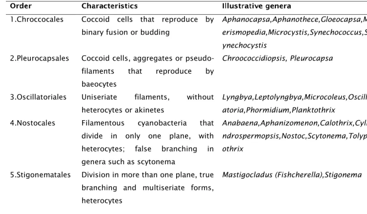
9 epidermis (De Fabo & Noonan, 1983). Whereas, the UVA penetrates deeper into the dermis and deposit 30-50 % of its energy into the dermal stratum papillare, thus why UVA effects such as skin aging, solar elastosis occurs in the dermis (Wlaschek et al., 2001). On the contrary, acute exposure to UV radiation within the solar spectrum leads to sunburn inflammation (erythema) and tanning while chronic exposure is the key factor in the initiation of premature aging of the skin, basal cell carcinoma, squamous cell carcinoma, malignant melanoma as well as an immunosuppressant of the immune system facilitating cell proliferation into abnormal tumour growth (Ullrich, 2002). The ozone hole in the stratospheric atmosphere results in the increased UVB penetrating the human skin which will affect the susceptible biomolecules and modify the physiology of human life (De Fabo, 2005). UVB is more cytotoxic and mutagenic, which induces the formation of pyrimidine dimers, a typical UV signature mutation. During the repair of cyclobutane pyrimidine dimers by P53 gene, the DNA photoproducts may mutate the gene leading to its loss of function causing especially non-melanocytes carcinoma (Rass & Reichrath, 2008). On the other hand, UVA sensitizes trans-urocanic acid causing it to react with oxygen which lead to the formation of reactive oxygen species (ROS) such as superoxide anion (O
-2) and singlet oxygen (1O2). The superoxide dismutase converts the superoxide anion radicals into hydrogen peroxide (H2O2) which can easily cross the cell membrane into the dermal layer. In conjunction with the transitional iron (FeII) is highly reactive hydroxyl radicals (HO ) can occur which react with lipids to form lipid peroxidation leading to the regulation of matrix degrading metalloproteases responsible for the degradation of connective tissues, a hallmark in carcinogenesis and skin aging (Brenneisen et al., 1998) (Figure1). Studies have proven that ROS such as singlet oxygen and H2O2 leads to the UVA-induction of mRNAs and synthesis of 1, 2 and MMP-3 with specificities for dermal (collagen I, II, III) and basement membranes compounds (collagen IV, VII, proteoglycan, laminin). In contrast MMPT-1 remained unaltered and this imbalance plays a role in tissue breakdown leading to the loss of interstitial collagen, the main structural protein in the dermal connective tissue and an overgrowth of elastin fibers (Scharffetter-Kochanek et al.,1993; Wlaschek et al., 1995; Herrmann et al.,1993; Brenneisen et al., 1997).
10
Transurocanic acid + O2 O-2 H2O2
Figure 1- Production of reactive oxygen species (ROS) induced by UVA radiation (Adapted from Brenneisen et al., 1998).
Despite of the negative effects of the sunlight on the skin, positive effects occur such as the production of vitamin D by the skin, which induce the maturity of proliferative cells leading to the formation and development of tissues, organs and bones (Holick, 2003) and regulate calcium homeostasis in the body (Heaney, 2008). In an attempt to attenuate the harmful effect of sunlight exposure, UV filter are developed for dermal application in a form of sunscreen cosmetics aiming to absorb the UV radiation striking the human skin. Sunscreens are applied on the superficial layer of the skin ensuring primary defense of the internal organs against UV radiation (Benson, 2000). The UV filters absorb photons and rapidly emit the energy thermally to cool down the excited molecule back to the ground state avoiding photo degradation of the filter (Stavros, 2014). Evidence has shown that the daily application of sunscreen reduce skin aging (Hughes et al., 2013). However, the efficacy of the UV filters was found to be reduced due to photo induced decomposition, the absorptions by the skin and into the environment (Giokas et al., 2007). Due to this reduced efficiency of synthetic UV filters, there is a need to invent new ways to protect the skin against huge doses of solar radiation that continue to engulf the earth. Plant extracts strongly proved to play evident role as antioxidant defense, hence the interest in using biological compound as photo protective ingredients in cosmetics (Almeida et al., 2015). In addition to plant antioxidants, cyanobacteria had been screened to synthesize compounds that are photo protective against UV rays (Rastogi et al., 2010). Due to its ability to synthesize UVR filter compounds and survive in extreme heat environments, cyanobacteria are indeed a good candidate of exploration as UV active ingredients in cosmetics. As an example, the porphyra 334 from the cyanobacterium Aphanizomenon flos-aquae was found with the highest mean critical
Superoxide dismutase
2H+
O
2Fe
2+11 wavelength against the two commercial products, Nivea (Nivea Moisturising Sun Lotion) and Boots (Boots Soltan Extra Moisturising Sun Lotion, batch 1Z) making it more efficient as a potential sunscreen cosmetics for UVR protection (Torres et al., 2006). Another cyanobacteria compound that was found with sunscreen potential is the syctonemin (Karlsson, 2011).
Human Keratinocytes are the primary target cells of the UVR and they respond to UVR by expression of anti-inflammatory proteins (Grewe et al., 1995). In addition, recent studies also found that UVA radiation-induced generation of singlet oxygen plays a role in mitochondria DNA mutations induced by increased 4977 base pair deletion in photoaging of human skin (Berneburg et al., 1999). Plant extracts have shown positive effect with keratinocytes such as improved membrane integrity, inhibited lipid peroxidation, stimulated elastin expression and inhibited MMP-1 expression after UV radiation (Philips et al., 2003). Another study has also indicated the use of the Plukenetia volubilis, a domesticated vine, which has shown no cytotoxicity effect of human keratinocytes and proven to be a good cosmetic candidate (Gonzalez-Aspajo et al., 2015). To the best knowledge, only a few studies were performed that include the effect of cyanobacteria on keratinocytes proliferation. These include a study in which an extract from Spirulina platensis was found to inhibit the herpes simplex virus (HSV) without affecting keratinocytes (Mader et al., 2016).
Cell proliferation is a frequently chosen marker to study the effects of UVR in the cell viability because UVR has demonstrated ability to damage the cytoskeleton of the cell (Alonso-Lebrero et al., 2003). While focusing on fibroblast proliferation, a Spirulina extract was found to reduce apoptotic cell death on mouse fibroblast (Chu et al., 2010). Other study found that MAAs such as shinorine, porphyra-334 (P-334) and
mycosporine-12 glycine (MG) protect the human fibroblast cells from UV-induced cell death, it was also found that these MAAs stimulated the cell proliferation of the human fibroblast skin cells which is very important for skin cell renewal (Oyamada et al., 2008). A mixture of two MAAs, P-334 and shinorine from red algae was found to induce cell viability against UV-induced aging of human fibroblast cells making it a potential candidate for use in cosmetics as UV protectors and activators of cell proliferation (Daniel et al., 2004). A
Spirulina extract was found to have potential in regeneration of dermal fibroblast layers by proven to positively affect viability and proliferation of mouse fibroblasts in extract-imbedded electrospun nanofibers (Jung et al., 2013).
Melanin serve as a natural antioxidant defense which filter and scatter the UV radiation preventing further reactions with cells (Brenner & Hearing, 2008). However studies have shown that melanin also causes deleterious effect by reacting with DNA resulting in UV induced melanogenesis or tanning (Gilchrest & Eller, 1999). However it is still an argument of which UV radiation is responsible for melanogenesis (Gilchrest et al., 1996). Daniel et al., 2004 clearly demonstrates that a cream with MAAs could provide protection equally to a cream containing a mixture of both UV filter and this can reduce the production of melanin which may cause further adverse effect to the cell. Although MAAs were already described from cyanobacteria (Table 2), no evidence is provided about their mechanisms of action.
Many physiological processes in the biological system results in the production of ROS and oxygen centered free radicals, which are responsible for the onset of degenerative diseases such as cancer, heart disease, cataracts, congestive disorders and aging. Therefore antioxidant compounds play an important role on scavenging the free radical and blocking the oxidation process which could lead to the onset of diseases and aging
13 (Ani et al., 2006). As it was mentioned earlier, UV radiation damage the cell’s DNA directly or indirectly, during absorption through the skin chromophores like urocanic acid or DNA. Especially when the light is absorbed by trans-urocanic acid, singlet oxygen is generated which has a high affinity to lipids or proteins (Scharffetter-Kochanek et al., 1993). The production of the singlet oxygen by transuronic acid react with the lipids causing lipid peroxidation and loss of cellular function leading to finally loss of skin resilience and formation of wrinkles. The products of lipid peroxidation or protein carbonylation are ROS themselves, which leads to loss of cellular function. Transcription factors as NF-κB and AP-1 gets activated, resulting in the increased production of matrix metalloproteinases, a group of protease enzymes responsible for the degradation of collagen and elastin fibers (Djavaheri-Mergny et al., 1999). A reduction in collagen contents and fiber fragmentation are typical signs of photo aging. Antioxidant compounds from plants have indicated to be photostable, hence absorbing the UV radiation and not producing photoproducts preventing DNA damage (Almeida et al., 2015). MAAs from a red algae were found to reduce lipid peroxidation by encapsulation in the liposomes reducing firmness and smoothness in the skin of human fibroblast cells as a result of early photo aging (Daniel et al., 2004). Other studies of plant UV protective compounds such as anthocyanin have shown inhibitory effect of UV-induced lipid peroxidation on animal cell in vitro. Cyanobacteria studies related to the antioxidant potential are scarce and publications are quite recent. Hossain et al. (2016) found that freshwater cyanobacteria species of the genera Oscillatoria, Lyngbya ,Microcystis., and Spirulina exhibited antioxidant properties through widely used antioxidant assays. Also, Pumas et al., 2011, described antioxidant properties of cyanobacteria of the genera Cyanosarcina, Phormidium, Scytonema and Leptolyngbya; Aydas et al. (2013) found also antioxidant properties in extracts of cyanobacteria of the genus Schynechocystis and Huang et al. (2007) from Spirulina. Chauhan et al. (2014) described also antioxidant properties in Nostoc species.
14 The Blue Biotechnology and Ecotoxicology research group (BBE) at the Interdisciplinary Centre of Marine and Environmental Research (CIIMAR) hosts a cyanobacteria culture collection that comprises strains from the different cyanobacteria families (LEGE Culture Collection). From this collection several strains have been screened for diverse bioactivities such as anticancer, antimicrobial and antifouling, and several strains have been considered interesting. In the last years, several papers have been published reflecting the potential of strains of the collection as natural compounds producers (Leão et al., 2013; Martins et al., 2013; Brito et al., 2015) and different bioactivities have also been reported (Table 3).
Considering the potential of the collection, as demonstrated in these previous studies, we aimed with this work to infer about the potential of cyanobacteria as producers of compounds with cosmetic application in skin protection. We intended to study the effect of extracts on different skin cell types, on the antioxidant potential and on melanin production.
For this work the following specific tasks were defined:
1. To culture cyanobacteria strains in order to get biomass 2. To prepare cyanobacteria extracts
3. To study the effects of cyanobacteria extract on proliferation of keratinocytes and fibroblasts
4. To study the effects of the cyanobacteria extracts on other skin cells namely on endothelial cells
5. To study the potential of cyanobacteria extracts as antioxidants 6. To study the effects of cyanobacteria extracts on melanin production
15 Table 3 – Bioactivities of marine cyanobacteria from the LEGE cyanobacteria culture collection
Bioactivity Model organism/cells Reference
Histopathology Mice Martins et al., 2005
Antimicrobial Gram + and Gram- bacteria Martins et al 2008; Costa et al., 2015;
Toxic to marine
invertebrates
Paracentrotus lividus, Artemia salina
Martins et al., 2007; Lopes et al., 2010; Costa et al., 2015
Anticancer (cytotoxic)
Human Cancer cell lines Selheim et al., 2005; Costa et al., 2013; Leão et al., 2014; Leão et al., 2015; Costa et al., 2015
Antiparasitic Plasmodium falciparum Leão et al., 2014
Platelet Blood platelets Selheim et al., 2005;
16 For this work, eight cyanobacteria strains were selected (Table 4). These strains were isolated from the water samples and solid materials from the Portuguese coast and are part of the LEGE cyanobacteria culture collection. Strains were selected considering interesting bioactivities already described for extracts and compounds.
Table 4 – Cyanobacteria strains included in this study and bioactivities already described.
Cyanobium sp. LEGE 06113
Anticancer Costa et al., 2013; Leão et al., 2013 Freitas et al., 2016 Cyanobium sp. LEGE 07175 Anticancer Antibacterial Cytotoxic Costa et al., 2013 Costa et al., 2015 Costa et al., 2015 Synechocystis salina LEGE 06099 Anticancer Costa et al., 2013 Leptolyngbya fragilis LEGE 07167 Anticancer Costa et al., 2015
Nodosilinea sp. LEGE 06102 Anticancer Costa et al., 2013
Pseudonabaena sp. LEGE 06194 Anticancer Costa et al., 2015
Leptolyngbya sp. LEGE 06108 Anticancer Costa et al., 2013
Pseudonabaena sp. LEGE 06173 Anticancer Costa et al., 2015
The cyanobacteria strains were cultured in Z8 medium supplemented with 20% NaCl at 25 °C, with a light intensity of 10 μmol photons m−2·s−1 and with a light/dark cycle of 14:10. Cultures were performed in 6 L flask filled with 4L medium. Cyanobacteria grow for approximately one month with constant aeration. At the exponential growth phase, cells were harvested by centrifugation at a rotational speed of 4600rpm and 4 ºC. The
17 collected biomass was frozen and freeze-dried. Then the lyophilized biomass was stored at −20 ºC.
A crude extract of each cyanobacteria strain was obtained by extraction of cyanobacterial biomass (dry weigh) with a dichloromethane: methanol (2:1, v/v) solution. 1g of lyophilized material of each strain was extracted for 10 minutes with 50mL of a dichloromethane: methanol (2:1) solution by stirring periodically. The solution was placed into a Bücher funnel, under vacuum. At the end of the filtration the residual mass was collected for new extraction, for another 10 minutes. In the end of the extraction the solvents were evaporated in the rotary evaporator and at the dichloromethane pressure. The flask was then washed with a mixture of isooctane: ethanol (1:1, v/v) to dissolve the pellet, using ultrasounds, and transferred to a previously weighted 22 mL clear glass vial. The mixture was evaporated using N2. The glass vial was weighted and the total mass of the crude extract calculated. The dry extract used in all assays was dissolved in DMSO.
Cytotoxic assays with the cyanobacteria crude extract were performed with keratinocytes, which are the main cells in epidermis, and fibroblasts, which are the main cells in the connective tissue of the dermis. Since the skin is a much irrigated organ we also study the effect of the extracts on normal endothelial cells. Information on cell lines is presented in Table 5.
18 Table 5
–
Cell lines included in the study.Keratinocytes HaCaT American Type Culture Collection (ATCC) Fibroblasts 3T3L1 American Type Culture Collection (ATCC) Endothelial cells hCMEC/D3 Donated by Dr. PO Couraud (INSERM, France)
Cells were cultured in Dulbecco’s Modified Eagle Medium (DMEM Glutamax-Gibco Invitrogen) supplemented with 10% fetal bovine serum (FBS-Gibco Invitrogen), 2.5 μg/ml fungizone (Gibco Invitrogen) and 2.5μg/ml penicillin-streptomycin. Cells were incubated in a humidified atmosphere with 5% of CO2, at 37ºC. Culture medium were renewed every two days. At 80-90% cell confluence, adherent cells were enzymatically released with a solution of 0.25% trypsin or using triplex (Gibco).
The cellular viability was evaluated by the reduction of the 3-(4,5-dimethylthiazole-2-yl)-2,5-diphenyltetrazolium bromide (MTT), a yellow tetrazole soluble in water. In this assay, cell survival was estimated by measuring the mitochondrial-dependent conversion of the tetrazolium salt MTT, to a purple colored formazan product, which is insoluble in water. Keratinocytes, fibroblasts and endothelial cells, were seeded in 96 well plates at a density of 2.5×10⁴ cell/mL, 3.3×10⁴ cell/mL and 1.0 ×10⁵cel/mL, respectively. After 24 hours of cell adhesion, new medium containing the cyanobacteria extracts at 100 μg/mL, 10 μg/mL and 1 μg/mL concentration was provided. Cells were then incubated for 24, 48 and 72 hours. After each incubation time, cells were treated with 0.05 mg/mL MTT final concentration in medium and incubated for 4h. After incubation with MTT, the medium was carefully aspirated, and formazan crystals solubilized with 100% DMSO. Absorbance was read at 550 nm in a “GEN5TM – Multi – detection Microplate Reader (Biotek) (Alonso-Lebrero et al., 2003). All tests were run in triplicate and averaged. Cytotoxicity was expressed as a percentage of cell viability considering 100% viability in the solvent control (cells treated with 1% DMSO). For reproducibility of the results each assay was independently repeated three times.
19 The antioxidant activity of the extracts was determined by the 2, 2-diphenyl-1-picrylhydrazyl (DPPH) assay which infers about the radical scavenging activity of different extracts. The DPPH assay was performed according to Sanches-Moreno et al., (1998). The extracts were dissolved in DMSO in order to test the concentrations of 100 μg/mL; 10
μ
g/mL and 1 μg/mL. The reaction mixture consisted in 19.4 μl from each cyanobacteria concentration and 175 μl DPPH in ethanol at a final concentration of 90 μM. Plates were incubated at room temperature for 30 minutes. After incubation time, the decrease in the absorption of the DPPH solution was measured at 515nm. Ascorbic acid was used as a positive control at the concentration of 50 μg/mL, 25 μg/mL and 12.5 μg/mL. All assays were run in triplicates. The % radical scavenging activity (% RSB) of the extracts was calculated using the following formula, % RSB = [A0-A1]/A0×100, where A0 is the absorbance of control and A1 is the absorbance of the test.Superoxide anion radical (O2–•) scavenging activity was determined by the nitro blue tertazolium chloride (NBT) method, with the reduced nicotinamide adenine dinucleotide/phenazine methosulphate (NADH/PMS) system as generator of superoxide radicals. The reaction mixture consisted in 75 μl NADH (166 μM), 150 μl NBT (43 μM) and 10 μl PMS (2.7 μM) all prepared in 19 mM phosphate buffer pH 7.4. 26.1 μl of extracts dissolved in DMSO were tested at concentrations of 200 µg/mL, 100µg/mL and 10 µg/mL. Ascorbic acid at the concentration of 50 μg/mL, 25 μg/mL and 12.5 μg/mL was tested as positive control. The reaction mixture was incubated at 37°C for 5 min and the absorbance was measured at 560nm. Percent inhibition of O2–•was calculated by the formula % Inhibition= [A0-A1]/A0×100, where A0 is the absorbance of control and A1 is the absorbance of the test. All assays were run in triplicate.
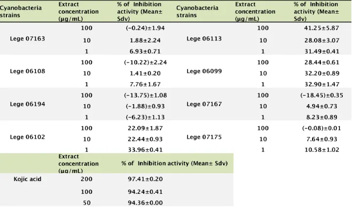
20 To study the effect on melanin production the effect of the extracts on the enzyme tyrosinase was assayed. Commercial mushroom tyrosinase at a concentration of 25U/mL was exposed to the cyanobacteria extracts at 100 µg/mL, 10 µg/mL and 1 µg/mL and then mixed with the substract L-DOPA at a concentration of 2.5mM. After 10 min incubation at 25 ºC, the enzymatic activity was measured by spectrophotometric analysis at 475 nm. Kojic acid at a concentration of 200 µg/mL, 100 µg/mL and 50 µg/mL was used as positive control. All solutions were prepared in 50 mM phosphate buffer at pH 6.5. The percentage of inhibition of tyrosinase activity was calculated by the formula % Inhibition= [A0-A1]/A0×100, where A0 is the absorbance of control and A1 is the absorbance of the test. All assays were run in triplicate.
The software employed was R version 2.15.2 (R Foundation for Statistical Computing, Vienna, Austria). In order to test the effect of the experimental factors, with the inclusion of experiment as a random factor, we fitted generalized linear models with Gamma distribution for the residuals. This was implemented with gmler function from lme4 package. Then we tested the significance of the fixed factors (Cyanobacterial strain as cathegorical factor and time and extract concentration as covariates) with analysis of deviance implemented with the Anova function from car package. Post hoc pairwise comparisons among factor levels were performed with the Tukey test implemented with the glht function from the multcomp package. Homogeneous subsets of levels of the factor “cyanobacterial strain” were obtained based on the Tukey pairwise comparisons, with the function multcompBoxplot from the multcompView package.
21 In order to study the effect of the cyanobacteria extract on cells that are present on the skin, the MTT assay was performed with keratinocytes, fibroblasts and endothelial cells. The MTT assay is a cell viability assay widely used, since it allows the evaluation of the cell viability, and indirectly cell proliferation. Although the objective was to study the effects on human skin, due to logistic issues, mice fibroblasts and brain barrier endothelial cells were used.
For the cytotoxicity assays cells were exposed to a crude extract of eight different cyanobacteria strains: LEGE07163; LEGE06108; LEGE06194; LEGE06102; LEGE06113; LEGE06099; LEGE07167 and LEGE07175, at three different concentration 100 µg/mL; 10 µg/mL and 1 µg/mL and exposure times of 24 hours, 48 hours and 72 hours.
For all the three cell lines, the effects of the different cyanobacteria strains were significant. Concentration had a positive significant effect in fibroblasts and a negative significant effect in endothelial cells. Time was only significant for the keratinocytes with a positive effect (Table 6).
In Figures 2, 3, 4, 5, 6 and 7, results concerning cell viability are presented. The values presented are averages of the three assays performed, since assay was a significant random factor in the statistical model implemented to analyze cell viability (Table 6).
22 Table 6 – effect of the cyanobacteria strain with respect to
extract, concentration and time on the cell lines.
CELL LINE FACTOR Χ2 DF P
FIBROBLASTS Extract 852.0615 8 <0.001 Concentration 56.0806 1 <0.001 Time 0.0164 1 0.90 KERATINOCYTES Extract 265.102 8 <0.001 Concentration 8.3239 1 0.051 Time 34.6712 1 <0.001 ENDOTHERIAL Extract 105.5931 8 <0.001 Concentration 4.8447 1 <0.05 Time 0.4884 1 0.49
Results of the effect of cyanobacteria in the keratinocytes HaCat are presented in Figures 2 and 3.
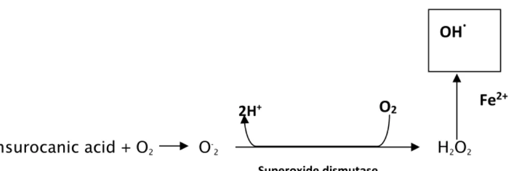
Figure 2 - The homogenous subsets of the cyanobacteria strains with respect to their effect in keratinocytes.
23 Figure 3 - Cell viability of the keratinocytes cell line HaCat when exposed to cyanobacteria crude extract. Extracts were tested at a concentration of 100 µg/mL, 10 µg/mL and 1 µg/mL. Solvent control consisted in 1% DMSO in cell culture medium and positive control consisted in 20 % DMSO.
0 20 40 60 80 100 120 140 CE LL V IA BI LIT Y (% ) EXTRACT CONCENTRATION 24 hrs 48 hrs 72 hrs 0 20 40 60 80 100 120 140 CE LL V IA BI LIT Y (% ) EXTRACT CONCETRATION 24 hrs 48 hrs 72 hrs
24 Results showed both a reduction and increase of the keratinocytes viability. The strains that induced a reduction in cell viability were LEGE 06108, LEGE 06194 and LEGE 07163. This reduction in cell viability was particularly registered for the extract concentration of 100 µg/mL and 10 µg/mL. At these concentrations, a reduction in cell viability was registered at 48 hours when compared to the results of 24 hours, followed by an increase in cell viability at 72 hours. At 1 µg/mL the extract also induced a slight reduction in cell viability at 24 and 48 hours but a more evident recovery on cell viability at 72 hours occurred. The highest percentage of cell viability was observed for strains LEGE06102; LEGE06113; LEGE06099; LEGE07167 and LEGE07175, which had a chronological increase in cell viability with the increase of the exposure time.
The keratinocytes dominate the epidermis, the superficial layer of the integumentary system responsible for the primary defence against the UV radiation (Brenner & Hearing, 2008). Physiological regenerative events in skin include proliferation of keratinocytes leading to epidermal thickening (Iglesias-de la Cruz et al., 2012). Therefore, cyanobacteria strains included in this study may be interesting for the isolation of compounds inducing keratinocytes proliferation.
Figure 4 - The homogenous subsets of the cyanobacteria strains with respect to their effect in fibroblasts.
25 Figure 5 - Cell viability of the fibroblasts cell line 3T3L1 when exposed to cyanobacteria crude extract. Extracts were tested at a concentration of 100 µg/mL, 10 µg/mL and 1 µg/mL. Solvent control consisted in 1% DMSO in cell culture medium and positive control consisted in 20 % DMSO.
0 20 40 60 80 100 120 140 CE LL V IA BI LIT Y (% ) EXTRACTS CONCENTRATION 24 Hrs 48 Hrs 72 Hrs 0 20 40 60 80 100 120 140 CE LL V IA BI LT Y (% ) EXTRACTS CONCENTRATIONS 24 Hrs 48 Hrs 72 Hrs
26 For fibroblasts (Figure 4 and 5) only for strain LEGE07167 a reduction in cell viability was registered at 100 µg/mL concentration. Also in most of the cases, after a reduction in cell viability after the 48 hours, an increase in cell viability was registered after 72 hours incubation. For strains LEGE06102, LEGE06113, LEGE07167 and LEGE07175, the higher viability values were obtained at the extract concentration of 1 µg/mL.
Fibroblasts produce major skin components such as collagen and hyaluronic acid important in skin texture, regeneration and hydration. These cells play also major functions in the interactions between dermis and epidermis (Tanaka et al., 2015), and thus are targets for cosmetics compounds.
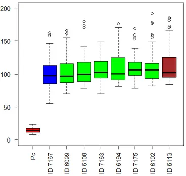
For endothelial cells, strains in which a significant reduction in cell viability occurred were LEGE6108, LEGE6194, LEGE7163 and LEGE7167 (Figures 6 and 7). For strains LEGE 06102, LEGE06113, LEGE 06099 and LEGE07175 a more evident increase in cell viability occurred at 72 hours. Also in this case, and except for strain LEGE06099, the higher viability values were registered for the extract concentration of 1 µg/mL.
Figure 6 - The homogenous subsets of the cyanobacteria strain with respect to their effect in endothelial cells.
27 Figure 7 - Cell viability of the endothelial cell line hCMEC/D3 when exposed to cyanobacteria crude extract. Extracts were tested at a concentration of 100 µg/mL, 10 µg/mL and 1 µg/mL. Solvent control consisted in 1% DMSO in cell culture medium and positive control consisted in 20 % DMSO.
0 20 40 60 80 100 120 140 CE LL V IA BI LIT Y (% ) EXTRACT CONCENTRATION 24 hrs 48 hrs 72 hrs 0 20 40 60 80 100 120 140 CE LL V IA BI LIT Y (% ) EXTRACT CONCENTRATION 24 hrs 48 hrs 72 hrs
28 Skin vascularization regulates the blood supply required for skin physiology and regeneration being its deregulations the basis of several human diseases (Bassino et al., 2016). It is therefore important that compounds used in skin treatments don’t induce injury in endothelial cells.
It was an overall finding that a recovery in cell viability occurred at 72 hours incubation and that higher cell viability was registered at the lower concentration of 1 µg/mL. One explanation for these results could be a hormetic response. Hormesis is a dose-response effect relying on low-dose stimulation and high-dose inhibition (Stebbing, 1982 in Li et al., 2015). In biological models low-dose stimulation were already described and are interpreted as a consequence of a homeostasis shift which leads to a moderate overcompensation response.
Free radicals are oxidants with one or more unpaired electron. Free radicals are formed from molecules via the breakage of a chemical bond, by cleavage of a radical to give another radical and, also via redox reactions (Pham-Huy, 2008). Free radicals include hydroxyl (OH•), superoxide (O2•ˉ), nitric oxide (NO•), nitrogen dioxide (NO2•), peroxyl (ROO•) and lipid peroxyl (LOO•) (Pham-Huy et al., 2008). When in excess, free radicals generate oxidative stress, which is a deleterious process that can damage cells through alteration on proteins, lipids, lipoproteins, and DNA. Assays based on radical scavenging have been used to evaluate the antioxidant activity of compounds. The DPPH radical scavenging assay is a simple and high sensitive assay widely used to evaluate free radical scavenging activity of compounds and thus to evaluate their potential antioxidant activity. This is a colorimetric assay in which the color of DPPH solution changes from purple to yellow due to the acceptance by DPPH of electron or hydrogen atoms donated by the antioxidants compounds (Alam et al., 2013). The reaction is measured as a decrease in absorbance at 515 -517nm.
In this work the antioxidant potential of cyanobacteria extracts was determined by its ability to decrease the optical density of the DPPH solution. The concentration of
29 antioxidant needed to decrease by 50% the initial substrate concentration (IC50) is used to measure the antioxidant power (Sanches-Moreno et al., 1998).
The results of the antioxidant activity of extracts determined by the DPPH assay are shown on Table 7.Extracts from LEGE 06113, 06108 and 06102 have shown the highest antioxidant power with 19.53%, 14.27% and 12.22% respectively. The extracts demonstrated however a weak antioxidant power when compared to ascorbic acid. Also compared to phytochemical compounds such as flavanoids and polyphenolics, these antioxidant activities are considered very low. In a work by Sethyia et al. (2014), by using the DPPH assay, the IC50 from 17 phytochemical from dietary plant sources used globally as functional food varied between 13.62 ± 2.03 μg/mL and 58.54 ± 1.72 μg/mL for vitamin E and stigmasterol respectively. In this work, the highest DPPHradical scavenging activity was only 19.53%, recorded at the concentration of 100 μg/mL. Table 7 - Antioxidant activity of the cyanobacteria extracts as determined by the DPPH assay
μ μ
30 Living cells generate free radicals and other reactive oxygen species as products of physiological and biochemical processes. ROS are highly reactive molecules, which include free radicals such as, superoxide ions (O2-). In this study, the NADH-PMS-NBT system was used to determine the superoxide anion scavenging activities of the crude extracts of the cyanobacteria strains. In this system superoxide radicals are generated by the NADH/PMS system reducing the nitroblue tetrazolium (NTB) to a blue chromogen. Therefore the production of superoxide radicals can be followed spectrophotometrically at 560nm. An extract present in the assay with scavenging activity against superoxide radicals will compete with NBT for superoxide radicals inhibiting, therefore the production of the blue chromogen. The concentration of antioxidant needs to decrease by 50% the control’s blue chromogen production (IC50) is a parameter that can be used to measure the antioxidant power (Alam et al., 2013)
The results concerning the superoxide radical scavenging activity of the cyanobacteria tested are present in Table 8. The general interpretation is that at lower concentration (10 μg/ml) the extracts showed the highest scavenging activities than at higher concentrations (200 and 100 μg/ml). Extracts from the cyanobacteria strains LEGE 06108 and 06099 registered the highest activity with 49.70% and 49.57% respectively at 10 μg/ml. Other strains, which deserve recognition, are LEGE 07167 and LEGE07175, which has shown superoxide scavenging activity at all concentrations studied in this work. In this assay the concentration of 1μg/ml was not assayed due logistic issues. The tendency observed in most of the strains is to an increase in the scavenging activity in lower extract concentrations. In this case it will important to test concentrations below 10 μg/ml. Although the IC50 was not calculated, for strains LEGE 06108 and 06099 this value is around 10 μg/ml. This IC50 value is in the range of values obtained for other phytochemicals using the same methodology (Pratap Chandran et al., 2013).
It is interesting in this assay that the highest percentage of scavenging activity was observed in the lowest concentrations. These results could also be due to a hormetic response, which are described for antioxidant compounds. Li et al. (2015), described a hormetic response of caffeine in cells treated with hydrogen peroxide.
31 Table 8 - Antioxidant activity of the cyanobacteria extracts as determined by the superoxide radical assay
Although melanin is described as being essential for protecting human skin against UV radiation it is also well known that its abnormal accumulation induces pigmentation disorders, such as melasma, freckles, ephelides, and senile lentigines (Slominski et al., 2004). Tyrosinase is the enzyme involved in the first two steps of the melanin biosynthesis, in which L-tyrosine is hydroxylated to 3,4-dihydroxyphenylalanine (L-DOPA) and subsequently oxidated to dopaquinone (Hearing, 2011). This enzyme is common in fungi, higher plants and animals (Parveem et al., 2010). In this work, the screen for cyanobacteria strains as tyrosinase inhibitors was based in a wide used tyrosinase inhibition assay in which the activity of a mushroom tyrosinase against DOPA is evaluated. The results of this assay are presented in Table 9 and figure 8.
Concerning the tyrosinase assay, the results shown that the highest inhibition of the enzyme was observed at 1μg/ml. Also in this case a hormetic response seems to be
μ μ