Ablation and Laparoscopic Reflux Operative
Results in More Effective and Efficient Treatment
of Barrett Esophagus
Trudie A Goers,
MD, Pedro Leão,
MD, Maria A Cassera,
BS, Christy M Dunst,
MD,
FACS,
Lee L Swanström,
MD,
FACSBACKGROUND: Barrett esophagus (BE) caused by gastroesophageal reflux disease can lead to esophageal cancer. The success of endoscopic treatments with BE eradication depends on esophageal anatomy and post-treatment acid exposure.
STUDY DESIGN: Between January 2008 and December 2009, 10 patients were selected for combination treat-ment of BE using laparoscopic anti-reflux surgery and endoscopic radiofrequency ablation. Retrospective review of preoperative, procedural, and postoperative data was performed. RESULTS: Seven study patients had a pathologic diagnosis of nondysplastic BE and 3 patients had a
diagnosis of low-grade dysplasia. Average length of BE lesions was 6.4⫾ 4.8 cm. Procedure time averaged 154.4⫾ 46.4 minutes. At the time of surgery, the mean number of ablations per-formed was 4.39⫾ 1.99. Six patients were noted to have major hiatal hernias requiring reduction. Five patients (80%) had 100% resolution of their BE at their first postoperative endoscopy. The remaining 3 patients had aⱖ50% resolution and underwent subsequent endoscopic ablation. Symptomatic results revealed that 4 patients had substantial dysphagia to solids and other symptoms were minimal. Two patients were noted to have complications related to the ablative treatments. One stricture and 1 perforation were observed.
CONCLUSIONS: Endoscopic radiofrequency ablation of BE at the time of laparoscopic fundoplication is feasible and can effectively treat BE lesions. A single combined treatment can result in fewer overall procedures performed to obtain BE eradication. ( J Am Coll Surg 2011;213:486–492. © 2011 by the American College of Surgeons)
Esophageal cancer has the fastest growing incidence rate of all cancers in the United States and Western Europe, in-creasing 400% during the past 35 years.1-4Barrett esopha-gus (BE) represents a marker for the potential development of esophageal adenocarcinoma. This condition develops when gastroesophageal reflux damages the distal esopha-geal squamous mucosa and the resulting injury heals by a metaplastic process.4,5BE can progress in a stepwise fashion from intestinal metaplasia to low-grade dysplasia to
high-grade dysplasia to carcinoma in situ and, ultimately, inva-sive adenocarcinoma. The mechanisms of this progression are not completely understood.
With current understanding of BE’s relationship to gastroesophageal reflux disease and its risk of cancer, we believe that treatment of dysplastic BE should be 2-fold, ie, attention to the dysplastic mucosa and effective treat-ment of chronic gastric reflux exposure that leads to such changes.
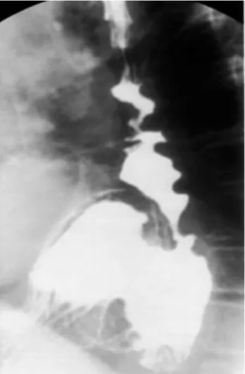
Radiofrequency ablation (RFA) has been safely and ef-fectively applied to BE with and without dysplastic changes.6-9It usually requires 2 to 3 ablation sessions to achieve complete eradication of BE. However, anatomic distortion of the esophagus from large hiatal hernias, stric-tures, esophageal dilation or tortuosity, shortened-esophagus, or, possibly, a previous fundoplication (Fig. 1), can make it more difficult to achieve effective ablation.10-12 In these scenarios, RFA of BE can require multiple at-tempts or be impossible to accomplish.
Disclosure Information: Authors have nothing to disclose. Timothy J Eber-lein, Editor-in-Chief, has nothing to disclose.
Abstract presented at Digestive Disease Week, New Orleans, LA, May 2010. Received November 3, 2010; Revised June 15, 2011; Accepted June 15, 2011. From the GI/Minimally Invasive Surgery Division, The Oregon Clinic, Port-land, OR (Goers, Cassera, Dunst, Swanström) and Department of Surgery, Hospital de Braga, Braga, Portugal (Leão).
Correspondence address: Lee L Swanström, MD, FACS, GI/Minimally In-vasive Surgery Division, Oregon Health Sciences University, 1040 NW 22nd Ave, Suite 560, Portland, OR 97210. email:lswanstrom@orclinic.com
486
© 2011 by the American College of Surgeons ISSN 1072-7515/11/$36.00
As the patient’s underlying genetic predisposition to de-velop BE remains unchanged, prevention of reflux seems mandatory to prevent recurrence. Optimal control of re-flux after ablation, however, has not been defined. Current options include lifetime high-dose acid suppression or anti-reflux surgery (ARS). The role of ARS certainly has theo-retical appeal because it provides an absolute barrier to all gastric contents and there is ample evidence that acid alone is not the causative agent of BE or dysplasia. If one accepts the idea of adding an ARS to BE ablation, the question of the timing of the 2 procedures arises. For example, al-though ARS before ablation can straighten the esophagus and cure esophagitis and strictures, the fundoplication it-self can interfere with effective RFA by obscuring the land-marks of the gastroesophageal junction or making access to the distal-most segments of the metaplasia more difficult. Performing ARS after an ablation can also be more difficult because of transmural inflammatory changes, or complete ablation can never be possible because of the anatomical distortion of the esophagus (angulation, dilatation, short-ening) related to hiatal herniation.13
We hypothesize that performing endoscopic RFA of BE at the time of ARS would be a safe and effective method that can reduce the number of treatments needed to erad-icate the metaplasia by reducing the hernia and straighten-ing the esophagus, as well as providstraighten-ing the best chance of preventing future BE recurrence.
METHODS
This study involved the retrospective review of patient charts and information that was prospectively collected for an IRB-approved data registry containing patients who had endoscopic balloon⫺based esophageal RFA. All proce-dures were performed between January 2008 and Decem-ber 2009.
Patients
Two groups of patients were included. The first included patients who were scheduled for ARS because of either failure or dislike of chronic medical therapy and who had endoscopic and histologic presence of BE with no or low-grade dysplasia. Patients with high-low-grade dysplasia were excluded from selection in this group, as the presence of a
fundoplication might compromise future oncologic resec-tion and reconstrucresec-tion if needed.
The second group of patients included those who orig-inally had biopsy-proven BE with either low- or high-grade dysplasia and who were considered failures to ablate (⬎3 attempts) because of anatomic distortion of the esophagus from dilation, tortuosity, or hiatal hernias; and those who currently had either nondysplastic or low-grade dysplasia on biopsy. Likewise, patients with persistent high-grade dysplasia were excluded and were instead considered for minimally invasive esophagectomy.
Presenting and procedural data
Demographic data were prospectively collected including age, sex, and medical history. Histopathology in the ab-sence of esophagitis, 24-hour pH, and esophageal mano-metric testing were recorded in an Access-based (Mi-Figure 1. Contrast study of patient with long-standing gastroesoph-ageal reflux disease, paraesophgastroesoph-ageal hernia, and chronic esopha-geal mucosal scarring.
Abbreviations and Acronyms ARS⫽ anti-reflux surgery BE ⫽ Barrett esophagus RFA⫽ radiofrequency ablation
crosoft) database. Baseline BE measurements were taken from the first endoscopy. Endoscopy was performed using white light and narrow-band imaging. The endoscopic bi-opsy protocol included 4 quadrant biopsies taken at 2-cm intervals and all nodular areas were removed with endoscopic submucosal resection (EMR). Dysplasia was recorded based only on biopsies taken in the absence of acute inflammation. Presence, type, and size of hiatal hernia were also recorded. At the time of surgical procedure, information about the length of ablation, type of electrode, and details of treatment were recorded from the operative report.
Surgical procedure
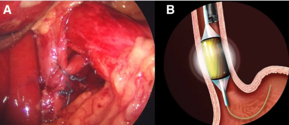
All procedures were done under general anesthesia in the supine position using 5 laparoscopic ports. Mediastinal dis-sections were performed with the end point of the gastro-esophageal junction lying 3 cm below the hiatus (Fig. 2). After mobilization of the distal esophagus and stomach, the esophagus was sized with a sizing balloon and an appropriate-sized RFA ablation balloon (HALO360; Barrx
Medical) was selected by a second surgeon performing en-doscopy. After irrigating the esophagus with 1% N-acetylcysteine to remove mucous, the balloon ablation catheter was inserted over a guidewire and ablation per-formed as the laparoscopic surgeon applied careful traction to straighten and align the esophagus to maximize contact of the ablation balloon. Two cycles of ablations were per-formed at either 10 J (no dysplasia) or 12 J (history of dysplasia). Two patients with more localized and limited disease had ablation with a endoscope-mounted electrode (HALO90; Barrx Medical) instead of the balloon electrode, but using the same ablation protocol. Once the ablation was completed, repair of any hiatal hernia was performed and a tailored fundoplication was performed.
Postoperative care
All patients were admitted overnight and observed. Pa-tients were placed on proton pump inhibitors and were given liquid pain medication as needed. Those who had concomitant performance of a paraesophageal hernia were kept nothing per os overnight and underwent esophago-gastric radiographic study with water-soluble contrast on the first postoperative morning. If no leak was detected, patients were started on a liquid diet. Those with minimal hiatal hernias were started on a liquid diet the night of surgery. All patients were instructed to continue the liquid diet for 2 days, followed by a pureed diet for 2 weeks. The patients were instructed to take a single-dose proton pump inhibitor for the first 3 weeks after surgery to protect the denuded distal esophagus.
Outcome measurements
A standardized gastrointestinal symptom assessment tool was administered at each visit. Patients were followed up in clinic at 2 weeks, 3 months, and 6 months and then yearly. A focused physical examination was performed and any complications or side effects from the surgery were re-corded. Patients who had any severe symptoms at any visit underwent appropriate studies and treatment at that visit. Routine follow-up endoscopy was performed 2 to 3 months after the combination procedure to assess com-pleteness of the ablation. Esophageal mucosa was examined using white light and narrow-band imaging. Subjective mapping of residual BE lesions’ length and circumferential vs. focal nature were recorded in the endoscopic report. Four quadrant biopsies were repeated at 2-cm intervals of both normal-appearing mucosa and columnar mucosa us-ing endoscopic jumbo forceps and were sent in formalin for pathology. Percentage of BE resolution was estimated by the endoscopist based on comparison with preoperative Figure 2. (A) Extensive type II mediastinal dissection allows straightening of the distal
esoph-agus and the gastroesophageal junction rests within the abdomen. (B) The radiofrequency balloon electrode can contact esophageal mucosa.
photos and detailed endoscopy notes. Then, at 6 months, the integrity of the fundoplication was determined per our usual protocol using endoscopy, high-resolution manome-try, and 24-hour pH studies. A competent fundoplication was defined as an intact wrap on endoscopic retroflection (Hill grade 1) and/or normal acid exposure by 24-hour pH tests (DeMeester score⬍14.7). If BE was present at the 3-or 6-month visit, the patient had an additional RFA at that time.
Data analysis
Data that was collected was stored in an IRB-approved database that was developed and maintained by the princi-pal investigator. The means of all of the continuous vari-ables were compared using appropriate parametric or non-parametric tests. Statistical analysis was performed using Predictive Analytics Software (version 18.0; SPSS, Inc).
RESULTS
During the 24-month data collection period, 78 patients with BE underwent treatment, of these, 15 patients met the inclusion criteria to undergo concomitant endoscopic RFA and laparoscopic fundoplication for the simultaneous treatment of BE and gastroesophageal reflux disease. Of these, 1 patient requested esophagectomy, 1 patient re-jected the follow-up protocol, and 3 did not want a fundo-plication. Therefore, 10 patients agreed to the combined procedure.
Patients varied in age from 23 to 80 years old (Table 1). Similarly, American Society of Anesthesiologists scores were all 2, with the exception of a 23-year-old patient who was scored as a 1. The average body mass index of the group
was in the category of obese (34⫾ 9), and no patient was considered to be normal (interquartile range 26.1⫺38.0). At the time of the study, all patients had biopsy-confirmed BE. Seven study patients had a pathologic diag-nosis of nondysplastic BE and 3 patients had a diagdiag-nosis of low-grade dysplasia. All 10 patients had abnormal 24-hour pH testing. All patients had high-resolution manometry and 1 patient had a profound primary esophageal dysmo-tility. Only 1 patient had active esophagitis at the time of diagnosis. Seven of the patients were considered to have failed RFA because of BE persistence despite multiple ab-lation attempts (4 to 7 attempts). The other 3 were de novo patients with long-segment BE (2 with low-grade dyspla-sia, 1 without) who were seeking ARS for symptom control because of failure of medical management.
Procedural data
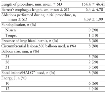
The combined procedure time averaged 154.4 ⫾ 46.4 minutes (Table 2). At the time of surgery, 6 patients were noted to have major hiatal hernias (type III) requiring reduction and crural reconstruction. Nine patients un-derwent 360-degree fundoplication and the patient with poor esophageal motility had a 270-degree posterior fundoplication.
The average length of BE lesions was 6.4 ⫾ 4.8 cm (Table 2). Eighty percent required the 360-degree balloon electrode for circumferential disease. The 90-degree HALO electrode was used in 20% for more focused energy application. One patient had both circumferential and fo-cal islands of disease and required the use of both the 360-degree and 90-360-degree electrodes. Of the 3 patients with dysplasia, all had energy applied at 12 J/cm2 3 with the Table 1. Demographic and Preoperative Data of
Study Patients
Age, y, mean⫾ SD 58⫾ 16.6
Body mass index, mean⫾ SD 34⫾ 9
Male sex, n (%) 7 (70) ASA, n (%) 1 1 (10) 2 9 (90) Histology, n (%) Nondysplastic 7 (70) Low-grade dysplasia 3 (30) GERD 10 (100) Esophagitis 1 (10) Preprocedure ablations, n (%) None 3 (30) Multiple 7 (4 –6)
ASA, American Society of Anesthesiologists; GERD, gastroesophageal reflux disease.
Table 2. Procedural Data of Study Patients
Length of procedure, min, mean⫾ SD 154.4⫾ 46.41 Barrett’s esophagus length, cm, mean⫾ SD 6.4⫾ 4.78 Ablations performed during initial procedure, n,
mean⫾ SD 4.39⫾ 1.99
Fundoplication, n (%)
Nissen 9 (90)
Toupet 1 (10)
Presence of large hiatal hernia, n (%) 6 (60) Circumferential lesions/360 balloon used, n (%) 8 (80) Balloon size, mm, n (%)
25 5 (50)
28 2 (20)
31 3 (30)
Focal lesions/HALO90used, n (%) 3 (30)
Energy, J, n (%)
10 6 (60)
360-degree device. The diameter of the 360-degree balloon electrode used varied. Fifty percent of patients required the 25-mm balloon. However, other patients had larger esoph-ageal diameters and the 28-mm (n⫽ 2) or 31-mm (n ⫽ 3) balloon electrodes were used.
There were no surgical or endoscopic complications and blood loss was⬍50 mL in all cases. Eight patients were discharged home on postoperative day 1 and 2 at 48 hours because of transportation issues. There were no readmis-sions or acute perioperative problems.
Symptomatic evaluation
Patients were seen between 2 and 4 weeks postoperatively for acute recovery data, including pain control, diet pro-gression, and other subjective data. Results of the symptom questionnaire are shown inFigure 3.
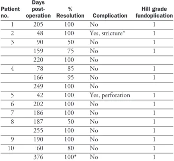
BE resolution
Long-term follow-up ranged from 7 months to 28 months (mean 17 months). All patients completed their 6-month comprehensive evaluation, 8 completed their 1-year evalu-ation, and 4 completed a 24-month follow-up.
All patients were free of BE at time of last follow-up. One had biopsies with columnar epithelium, but no intes-tinal metaplasia. Six patients (60%) had 100% resolution of their BE after 1 intraoperative ablative treatment per-formed at the time of their fundoplication (Table 3). This included 4 patients who had failed previous multiple at-tempts at ablation. None of the remaining BE patients had dysplasia. The remaining 4 patients had aⱖ50% resolu-tion and underwent endoscopic ablaresolu-tion. At their second follow-up endoscopy, 3 patients were found to have resid-ual BE, however, their overall disease burden was less. A third ablation succeeded in complete control, although 1
patient continued to have columnar epithelium with no intestinal metaplasia at 24-month follow-up.
Two patients had major complications related to the ablation treatments. One patient had a soft stricture noted at their first diagnostic endoscopy performed on postoper-ative day 48 for mild solid-food dysphagia. A second pa-tient was evaluated at 6 weeks postoperatively because of a report of a food impaction. This patient was evaluated with upper endoscopy and was found to have a 1.5-cm perfora-tion within the proximal RFA field.
There were no particular postoperative complications attributable to the fundoplication, although patients fre-quently noted common side effects, such as early satiety, bloating, and flatulence. At longest follow-up, no patients had reflux complaints and 1 patient reported heartburn. Three patients were on peptic medications. All fundopli-cations were intact (Hill grade I) on last endoscopy. Eight patients had postoperative manometry and pH studies and all results were within the normal range, including the 1 patient with heartburn and all 3 patients on proton pump inhibitors postoperatively.
DISCUSSION
Treatment of BE with endoscopic RFA is a relatively new concept. The technique so far has been reported to be both effective and well-tolerated.10,11 Exact indications for its use, however, have yet to be worked out completely and the long-term efficacy of endoscopic ablations remains un-known. The cost-effectiveness of this treatment is also a Figure 3. Postoperative patient symptomatic evaluation at 2 to 4
weeks. White bar, absent; black bar, present.
Table 3. Endoscopic Evaluation Results of Barrett Esopha-gus Resolution Status Post Combined Therapy
Patient no. Days post-operation % Resolution Complication Hill grade fundoplication 1 205 100 No 1 2 48 100 Yes, stricture* 1 3 90 50 No 1 159 75 No 1 220 100 No 4 78 85 No 1 166 95 No 1 249 100 No 5 42 100 Yes, perforation 1 6 202 100 No 1 7 186 100 No 1 8 187 50 No 1 255 100 No 1 9 190 100 No 1 10 60 80 No 1 376 100* No 1
controversial subject and both the efficacy and cost-effectiveness of the procedure rely primarily on the success rate of BE eradication; how many treatments it takes to achieve eradication; and how long the BE stays ablated. The literature describes a⬎95% eradication rate after an average of 2 to 6 treatments.6,12,14 Anecdotally, however, most practitioners have encountered a substantial number of patients who seemingly cannot be ablated despite mul-tiple treatment sessions. At our high-volume center (⬎250 RFA procedures), we have had 1 patient referred to us after 16 ablation attempts and another after 12. Even with our experience, we have had occasions where as many as 5 ablations failed to achieve clearance. Considering the stan-dard protocol of treatment with RFA, reassessment 2 to 3 months later and retreatment if there is residual disease, interspersed with occasional endoscopies for biopsy evalu-ation, it is easy to understand the enormous health care burden that multiple treatment sessions impose on the pa-tient, practitioner, and health system.
In our experience, multiple sessions of endoscopic RFA were most often needed for patients who had anatomic distortion of their distal esophagus because of hiatal her-nias, chronic peptic scarring, or esophageal dilation and tortuosity. We therefore theorized that laparoscopically dis-secting and freeing the distal esophagus would allow us to straighten it and make its lumen more uniform. This, in turn, would allow us to better visualize endoscopically the esophageal mucosa, more accurately calibrate balloon sizes, and more effectively deliver radiofrequency energy to the BE lesions. In fact, our study shows that even in patients who had failed multiple treatments, the majority of the study patients had complete resolution of BE after 1 intra-operative ablation session using this intraintra-operative strategy. We also like the idea of leaving a patient with a mechan-ical reflux barrier at the completion of their myotomy. It is presumed, but not known at this time, that the neosqua-mous mucosa after BE ablation will have the same genetics as the original esophagus and, therefore, be at high (per-haps inevitable) risk of reconverting to BE and possible dysplasia unless something additional is done to prevent it. An intact fundoplication is well known to give patients a supraphysiologic reflux barrier that probably would have an impact on the genetic predisposition to metaplastic transformation. Whether medical therapy would have the same effect is controversial. There is much evidence that would support BE progression, even in the face of medical treatment or perhaps because of it. We do plan to follow this patient cohort, as well as our medically treated pa-tients, for the long-term to document the rate of BE recur-rence in both groups.
Although the combined procedure required more
oper-ative time (154 minutes) than an average fundoplication case, the overall cost savings to the patient, physician, and health care system as a whole could be substantial. If BE could be eliminated in 20% of chronic reflux patients using 20% fewer resources and personnel, the longer operative time and initially higher procedure costs would yield large cost savings overall.
Poor understanding of the genetic pathways of BE neo-genesis results in our incomplete knowledge of how to best treat histologic subtypes of BE.15,16In this study, 2 patients (patients 3 and 4) required⬎2 ablations, even with intra-operative mobilization. Both of these patients’ initial pa-thology was nondysplastic BE. There were no significant differences in the endoscopic RFA portion of these 2 cases. Despite no notable difference from the other study patients who achieved complete ablation in 1 session, these 2 pa-tients had persistent BE. Besides technical error, a possible explanation for the different response to treatment could be the genetic profile of the patients. It might be that they possess proto-oncogenes or RNA triggers that make their BE cells more resistant to radiofrequency energy, or make regenerating neomucosa more likely to go down the BE pathway. A better understanding of genetic predisposition and therapeutic sensitivities is certainly needed in BE patients.
The complication rate for our study was 20% (2 pa-tients), indicating that the combined procedure is fairly well tolerated. Individually, laparoscopic ARS and endo-scopic RFA treatment are acceptably safe and have a com-plication rate⬍10%.The patient in whom the minor stric-ture developed was minimally symptomatic, underwent a single dilation, and has not had any residual symptoms. The other complication, necrotic perforation, is more wor-risome. The national BARRX registry had not previously reported any perforations related to its device and no other authors have described this phenomenon.6, 9,17Our patient in whom a perforation developed was an octogenarian who had a 13-cm length of BE. His procedure was uneventful and he recovered without incident. He presented 6 weeks later with a report of progressive dysphagia and food im-paction. Endoscopy revealed a 1.5-cm necrotic perforation 6 cm above the lower esophageal sphincter (LES). A con-trast study confirmed a contained, self-draining perfora-tion. This patient was treated conservatively and dis-charged to home on a liquid diet and oral antibiotics. After 2 months, endoscopy showed complete healing of his esophagus, no BE, and an intact fundoplication. He was advanced to a regular diet and has subsequently done well. We now treat long segments in a staged fashion. The overall tissue quality in these patients with long-standing disease is generally poor, distal esophageal mobilization can be
diffi-cult and result in tissue damage and thinning of the esoph-ageal wall. With this, radiofrequency energy application overall distance can compromise tissue microvasculature and result in easier necrosis. Because of these concerns, we currently limit our ablations to lesionsⱕ5 cm or we will do them in a planned staged program.
Despite these questions, there is little doubt that RFA for BE has dramatically altered the treatment paradigm for dysplastic BE and has resulted in the sparing of many an esophagus that would have otherwise been removed for this problem. The purpose of this study was to use endoscopic ablative technology in conjunction with laparoscopic ARS to improve electrode contact, thereby increasing the success rate of complete ablation. We show that this improves over-all BE ablation efficiency, decreases BE recurrence, and will hopefully impact long-term cancer risk.
CONCLUSIONS
Intraoperative endoscopic RFA of BE at the time of lapa-roscopic fundoplication is feasible and might be a more efficient and cost-effective way to treat BE. We show that a single combined treatment results in the need for fewer overall procedures performed to obtain BE eradication. Al-though the complication rate of this pilot study was not negligible, patients did well with conservative treatment and our procedural approach has been subsequently mod-ified with promising results. We believe that prospective study of this combined treatment modality for patients with BE is warranted.
Author Contributions
Study conception and design: Goers, Swanström. Acquisition of data: Goers, Leão, Cassera.
Analysis and interpretation of data: Goers, Leão, Cassera, Swanström.
Drafting of manuscript: Goers, Cassera, Swanström. Critical revision: Goers, Cassera, Dunst, Swanström.
REFERENCES
1. Sharma P, Dent J, Armstrong D, et al. The development and
validation of an endoscopic grading system for Barrett’s esoph-agus: the Prague C & M criteria. Gastroenterology 2006;131: 1392–1399.
2. Sharma P, McQuaid K, Dent J, et al. A critical review of the
diagnosis and management of Barrett’s esophagus: the AGA Chicago Workshop. Gastroenterology 2004;127:310–330.
3. Wang KK, Sampliner RE. Updated guidelines 2008 for the
di-agnosis, surveillance and therapy of Barrett’s esophagus. Am J Gastroenterol 2008;103:788–797.
4. Shaheen NJ, Sharma P, Overholt BF, et al. Radiofrequency
ab-lation in Barrett’s esophagus with dysplasia. N Engl J Med 2009; 360:2277–2288.
5. Spechler SJ. Clinical practice. Barrett’s esophagus. N Engl J Med
2002;346:836–842.
6. Fleischer DE, Overholt BF, Sharma VK, et al. Endoscopic
abla-tion of Barrett’s esophagus: a multicenter study with 2.5-year follow-up. Gastrointest Endosc 2008;68:867–876.
7. Ganz RA, Utley DS, Stern RA, et al. Complete ablation of
esophageal epithelium with a balloon-based bipolar electrode: a phased evaluation in the porcine and in the human esophagus. Gastrointest Endosc 2004;60:1002–1010.
8. Dunkin BJ, Martinez J, Bejarano PA, et al. Thin-layer ablation
of human esophageal epithelium using a bipolar radiofrequency balloon device. Surg Endosc 2006;20:125–130.
9. Vassiliou MC, von Renteln D, Wiener DC, et al. Treatment of
ultralong-segment Barrett’s using focal and balloon-based radio-frequency ablation. Surg Endosc 2010;24:786–791.
10. Hubbard N, Velanovich V. Endoscopic endoluminal
radiofre-quency ablation of Barrett’s esophagus in patients with fundo-plications. Surg Endosc 2007;21:625–628.
11. dos Santos RS, Bizekis C, Ebright M, et al. Radiofrequency
ablation for Barrett’s esophagus and low-grade dysplasia in com-bination with an antireflux procedure: a new paradigm. J Thorac Cardiovasc Surg 2010;139:713–716.
12. Smith CD, Bejarano PA, Melvin WS, et al. Endoscopic ablation
of intestinal metaplasia containing high-grade dysplasia in esophagectomy patients using a balloon-based ablation system. Surg Endosc 2007;21:560–569.
13. Awad ZT, Mittal SK, Roth TA, et al. Esophageal shortening
during the era of laparoscopic surgery. World J Surg 2001;25: 558–561.
14. Sharma VK, Wang KK, Overholt BF, et al. Balloon-based,
cir-cumferential, endoscopic radiofrequency ablation of Barrett’s esophagus: 1-year follow-up of 100 patients. Gastrointest En-dosc 2007;65:185–195.
15. Nicholson A, Jankowski J. Editorial: one small step for
metapla-sia, but one giant leap for biomarkers is needed. Am J Gastro-enterol 2009;104:2681–2683.
16. Vallbohmer D, Marjoram P, Kuramochi H, et al. Towards the
molecular characterization of disease: comparison of molecular and histological analysis of esophageal epithelia. J Gastrointest Surg 2007;11:1095–1104.
17. Csendes A, Braghetto I, Burdiles P, et al. Late results of the
surgical treatment of 125 patients with short-segment Barrett esophagus. Arch Surg 2009;144:921–927.