DOI: 10.1590/0004-282X20160042
ARTICLE
Surgical outcomes of the endoscopic
endonasal transsphenoidal approach for large
and giant pituitary adenomas: institutional
experience with special attention to
approach-related complications
Resultados cirúrgicos do acesso endonasal endoscópico transesfenoidal para adenomas
hipofisários grandes e gigantes: experiência institucional com ênfase às complicações
relacionadas ao acesso cirúrgico
Edson Rocha Constantino, Rafael Leal, Christian Cândido Ferreira, Marcus André Acioly, José Alberto Landeiro
Universidade Federal Fluminense, Hospital Universitário Antônio Pedro, Divisão de Neurocirurgia, Niterói RJ, Brazil.
Correspondence: Marcus André Acioly; Departamento de Especialidades Cirúrgicas, Divisão de Neurocirurgia; Rua Marquês de Paraná, 303, 3º andar; 24033-900 Niterói RJ, Brasil; E-mail: marcusacioly@yahoo.com.br
Conflict of interest: There is no conflict of interest to declare. Received 5 January 2016; Accepted 17 February 2016.
ABSTRACT
Objective: In this study, we investigate our institutional experience of patients who underwent endoscopic endonasal transsphenoidal approach for treatment of large and giant pituitary adenomas emphasizing the surgical results and approach-related complications.
Method: The authors reviewed 28 consecutive patients who underwent surgery between March, 2010 and March, 2014. Results: The mean preoperative tumor diameter was 4.6 cm. Gross-total resection was achieved in 14.3%, near-total in 10.7%, subtotal in 39.3%, and partial in 35.7%. Nine patients experienced improvement in visual acuity, while one patient worsened. The most common complications were transient diabetes insipidus (53%), new pituitary deficit (35.7%), endonasal adhesions (21.4%), and cerebrospinal fluid leak (17.8%). Surgical mortality was 7.1%. Conclusions: Endoscopic endonasal transsphenoidal surgery is a valuable treatment option for large or giant pituitary adenomas, which results in high rates of surgical decompression of cerebrovascular structures.
Keywords: endoscopic endonasal approach; giant pituitary adenomas; large pituitary adenomas; complication rates.
RESUMO
Objetivo: Neste manuscrito investigamos a experiência institucional com o acesso endonasal endoscópico transesfenoidal no tratamento de adenomas hipofisários grandes e gigantes com ênfase às complicações relacionadas ao acesso cirúrgico. Método: Foram incluídos neste estudo 28 pacientes consecutivos submetidos à cirurgia entre Março de 2010 e Março de 2014. Resultados: O diâmetro médio pré-operatório dos tumores era 4,6 cm. Uma ressecção total foi obtida em 14,3%; quase total, em 10,7%; subtotal, em 39,3% e parcial, em 35,7%. Nove pacientes evoluíram com melhora na acuidade visual, enquanto um paciente apresentou piora da função visual. As complicações mais comuns foram diabetes insipidus transitório (53%), novo défice hipofisário (35,7%), sinéquias endonasais (21,4%) e fistula liquórica (17,8%). A mortalidade cirúrgica foi 7,1%. Conclusões: A cirurgia por via endonasal endoscópica transesfenoidal é uma opção terapêutica extremamente útil para adenomas hipofisários grandes e gigantes, a resultar numa significativa descompressão das estruturas cerebrovasculares.
Palavras-chave: acesso endoscópico endonasal; adenoma hipofisário gigante; adenoma hipofisário grande; índices de complicação.
Introduced initially in the 1960s by Guiot et al.1 as an ad-ditional visualization tool to the microscope for sellar tu-mors, the endoscope has revolutionized the treatment of pituitary disorders because of improved visualization allow-ing ultimately the expansion of the surgical view2. At present, the purely endoscopic endonasal approach is widely used
worldwide for the treatment of pituitary adenomas and oth-er sellar tumors due to its similar safety, resection and com-plication rates, as well as surgical outcome in comparison to the traditional microscopic approach3,4. Large and giant
purely morphological criterion in the way that when the tu-mor achieves a diameter greater than 4 cm in any plane, it is considered giant5,6,7. here is no consensus, however, on
the deinition of large pituitary adenomas8,9. Juraschka et al.10 have considered large tumors as those having > 3 cm in the maximal diameter, while Cusimano et al.11 have introduced a new volumetric assessment (≥ 10 cm3)on the assumption of a better estimation of the tumor size and also for a better
quantiication of the extent of resection.
Traditionally, these tumors have been handled by the transcranial route, alone or in combination with the trans-sphenoidal resection7,9,11,12. Some recent reports have de-scribed the usefulness of the endoscopic endonasal route
in the their management, even though its safety and efec
-tiveness of treating large and giant pituitary adenomas is yet to be established in the literature6,8,10,11,13,14,15,16. Herein, we sought to investigate our institutional experience of pa-tients who underwent endoscopic endonasal transsphe-noidal approach for treatment of large and giant pituitary adenomas emphasizing the surgical results and approach-related complications.
METHOD
his study consists in a retrospective review of 28 consec -utive patients who underwent endoscopic endonasal trans-sphenoidal surgery for treatment of large and giant pituitary adenomas between March, 2010 and March, 2014. Clinical pre-sentation, endocrine status, neuroimaging, histopathological evaluation, extent of resection, clinical outcomes, and
compli-cation rates were analyzed. he institutional review board ap -proved the terms and conditions of the present study.
Inclusion criteria
All patients undergoing endoscopic endonasal transsphe-noidal resection for histologically proven pituitary macroad-enomas with a maximal diameter > 3–4 cm (large) or > 4 cm (giant) were included in this analysis. Patients harboring smaller tumors, distinct histological diagnosis, or those who have been submitted to a previous transcranial resection were excluded.
Patient characteristics
The medical charts were reviewed to collect clinical data. The clinical characteristics included were gender, age, visual acuity (Humphrey visual field [HVF] testing), ophthalmoplegia, pituitary dysfunction (hypo- or hyper-function syndromes), diabetes insipidus (DI), and prior surgery. All patients underwent preoperative magnetic resonance imaging (MRI) and computed tomography (CT) with navigation protocol. Neuroimaging were evaluated to measure the maximal tumoral diameter in any plane, and to assess suprasellar and parasellar extension based
on the classification of Hardy17 and Mohr et al.18, as well as the occurrence of optic nerve compression, hydrocephalus, or haemorrhagic component. All patients were seen by an experienced endocrinological team being tested for se-rum growth hormone (GH), follicle-stimulating hormone (FSH), luteinizing hormone (LH), adrenocorticotrophic hormone (ACTH), cortisol, insulin-like growth factor-I (IGF-1), prolactin, thyroid-stimulating hormone (TSH), T4, testosterone and estrogen levels.
Postoperatively, all patients performed cranial CT within 48 hours and cranial MRI at 2-, 12-months, and yearly there-after. Gross-total resection (GTR) was assigned when the MRI scan showed no residual tumor, while near total resec-tion (NTR) was attributed to the removal of ≥ 90%, subtotal resection (STR) with a tumor reduction from 70% to 89.9% and partial resection (PR) in the case of ≤ 70% of tumor section. Follow-up imaging also addressed regrowth of a
re-sidual tumor or tumor recurrence in the case of GTR. he
occurrence of postoperative complications, including DI (transient/permanent), new hypopituitarism,
cerebrospi-nal luid (CSF) leak, meningitis, ophthalmoplegia, endona -sal synechiae, acute sinusitis, epistaxis, internal carotid ar-tery injury, subarachnoid haemorrhage (SAH) and ischemic stroke were considered.
For hormone-secreting tumors, chemical remission was based on the following criteria: for prolactinomas, a prolac-tin level < 15ng/mL in men and < 20ng/mL in women; and for GH-secreting tumors, normalization of serum levels of IGF-1 matched for age and gender, random serum GH level of < 1ng/mL or a serum GH nadir of < 0.4 ng/mL following oral glucose tolerance test19,20.
Surgical approach
The operative technique used was the endoscopic endonasal transsphenoidal approach. All surgeries were performed primarily by the senior neurosurgeon ( J.A.L.) during this time period. The primary objective was maxi-mum decompression of the optic apparatus, the pituitary gland and surrounding brain structures12. Patients were positioned with their heads secured by a Mayfield head holder. Then, the patients were registered to a frame-less stereotactic navigation system (Stealth; Medtronic, Jacksonville, Florida, USA) for intraoperative guidance and anatomical verification. Lumbar drains were not used routinely.
he nasal cavity was prepared with adrenalin (1:1000)
soaked cottonoids for at least 5 minutes in order to
de-crease bleeding. he surgical procedure started with the
inventory of nasal cavity with a 0-degree 4-mm endoscope (Karl Storz GmbH & Co.KG, Tuttlingen, Germany). A right middle turbinate luxation or middle turbinectomy was de-veloped before harvesting mucoperiosteal nasoseptal
vas-cularized lap for skull base reconstruction after tumor re
nostril bimanual technique. An anterior sphenoidotomy was then carried out. Inside the sphenoid sinus, the sellae borders were demarcated by the aid of image guidance. Its
front wall was opened with a ine chisel and expanded with
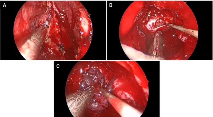
a Kerrison forceps or drilling. Dural exposure was limited by the tuberculum sellae superiorly, clival recess inferiorly, and laterally by the medial walls of the cavernous sinus. Tumor was debulked by piecemeal resection and suction and
ulti-mately the tumor border was deined for bimanual prepara
-tion of the neurovascular structures (Figure 1). he recon
-struction of the skull base was performed with autologous fat patch, which was placed in the resection cavity with a special care not to overpack, followed by placement of the
nasoseptal mucous lap. Tissue glue (Beriplast™, Behring) was applied to the lap edges and covered with Surgicel
(Ethicon). A nasal Foley catheter was introduced for
pack-ing of the sphenoidal cavity in order to prevent lap migra
-tion only in the case of intraoperative CSF leakage.
RESULTS
Preoperative characteristics
Patients included 17 men (60%) and 11 women (40%). he
mean age was 46 years (range, 15–62 years). he average tu
-mor size was 4.6 cm (range, 3.9–9.7 cm). he most common
presenting symptoms were endocrinopathy (57%), visual
acu-ity deicits (42%), and headache (35%). here were 5 patients
(17.8%) with preoperative hypogonadism, 4 patients (14.3%)
had hypothyroidism, one patient (3.6%) had acromegaly and
one patient (3.6%) was afected by galactorrhea. Gait apraxia
and cognitive impairment were found in 7.1% of patients (2 pa-tients) due to obstructive hydrocephalus (grade D of Hardy).
In 2 patients (7.1%), there was acute worsening of vi-sual impairment and headache, which together with the imaging findings confirmed the diagnosis of tumor apo-plexy. Prior to surgery, the imaging morphological char-acteristics of tumors are illustrated on Table 1. The ma-jority of tumors were nonfunctioning pituitary adenomas (82%) (Figure 2).
Extent of resection and Clinical outcomes
he tumor resection rates were estimated by analy
-sis of postoperative MRI exams, which were performed two months after surgery. Gross-total resection (GTR) was achieved in 14.3% (4 patients), near-total (NTR) in 10.7% (3 patients), subtotal (STR) in 39.3% (11 patients), and par-tial (PR) in 35.7% (10 patients). Nine (32.1%) patients expe-rienced improvement in visual acuity, while only one pa-tient (3.6%) worsened. Surgical resection alone achieved
endocrinological remission in four of the ive patients with
hormone-secreting tumors (the remaining patient died af-ter 30 days postoperatively).
Complications
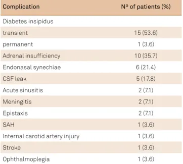
he authors identiied 11 diferent immediate/
short-term and late complications related to the endoscop-ic transsphenoidal resection of giant pituitary adenomas
Figure 1. Intraoperative imaging demonstrating the surgical steps involved in the resection of a giant pituitary adenoma. (A) Anterior sphenoidotomy. (B) Dural opening and initial preparation of the tumor capsule. (C) Intracapsular resection.
A
B
(Table 2). he most common complications were transient
diabetes insipidus (53%), new pituitary deicit (35.7%), en
-donasal adhesions (21.4%), and CSF leak (17.8%). From the immediate/short-term group, nasosinusal disorders, such as acute sinusitis and epistaxis occurred in 7.1%, each. Intraoperatively, CSF leak was detected in 21% and correct-ed at the end of tumor resection, even though 17.8% of the patients experienced postoperative CSF leak. All patients were placed on a conservative regimen of bed rest, acetazol-amide and lumbar drain for 5 days with total symptom re-mission. Two patients developed postoperative meningitis after treatment for CSF leak.
Surgical mortality was 7.1% (2 patients). One patient had a severe subarachnoid/tetraventricular haemorrhage, which ultimately lead to diffuse cerebral vasospasm and brain death (Figure 3), while the second deceased from metabolic disorders as a result from DI. A third patient died more than 30 days postoperatively affected by nos-ocomial pneumonia and therefore was not considered surgical mortality. At the last follow-up, only one patient (3.6%) was affected by permanent DI. The ENT team mon-itored all endonasal adhesions in the outpatient clinic, but no surgery was necessary.
Recurrences and Follow-Up
During the mean follow-up time of 30 months (range, 12–36 months), 9 (32.1%) patients were reoperated on for tu-mor regrowth. In total, 28 patients underwent 32 endoscopic
endonasal transphenoidal procedures. Five of the 9 patients
were given reoperation through open craniotomy. he mean
time to regrowth was 2 years. All patients who underwent GTR (14.3%) remained free of tumor.
DISCUSSION
Giant pituitary adenomas are rare tumors with an es-timated incidence between 5% and 16% of all pituitary tumors1,21,22 .Complete resection is often a technical chal-lenge, even for experienced neurosurgeons, considering that this subgroup of tumors may present with a high de-gree of invasion of neurovascular structures and large ar-eas of supra- and parasellar extension11,23. In this way, the primary objective of surgery includes relief of mass effect by obtaining maximal tumor resection in order to decom-press visual pathways, neurovascular structures and the pituitary gland6,9,15.
he possible route to reach the sellar region in the man
-agement of giant pituitary adenomas can be divided into two main groups: the transcranial and the transsphenoidal ap-proaches, either microscopic or endoscopic. A recent system-atic review from the modern literature (1995–2010) was
con-ducted to compare the beneits and limitations of the various
surgical approaches7. Based on the collection of 478 patients
afected of giant pituitary adenomas, Komotar et al.7 found
that the endoscopic cohort had higher rates of gross total re-section (GTR) (47.2%) and improved visual outcome (91.1%) than the transcranial (9.6% and 40%) and the microscopic transsphenoidal cohorts (30.9% and 34.8%), respectively. Table 1. Imaging characteristics of large and giant
pituitary adenomas.
Tumor characteristic Nº of patients (%)
Optic nerve compression 22 (78.6)
Supra- and para-sellar extension 17 (60.7)
Hydrocephalus 2 (7.1)
Tumor apoplexy 2 (7.1)
Table 2. Perioperative and postoperative complications of the endoscopic endonasal transsphenoidal approach.
Complication Nº of patients (%)
Diabetes insipidus
transient 15 (53.6)
permanent 1 (3.6)
Adrenal insufficiency 10 (35.7)
Endonasal synechiae 6 (21.4)
CSF leak 5 (17.8)
Acute sinusitis 2 (7.1)
Meningitis 2 (7.1)
Epistaxis 2 (7.1)
SAH 1 (3.6)
Internal carotid artery injury 1 (3.6)
Stroke 1 (3.6)
Ophthalmoplegia 1 (3.6)
CSF: cerebrospinal fluid; SAH: subarachnoid haemorrhage.
Figure 2. Tumor classification according to the immunohistochemical profile. (GH – growth hormone; TSH – thyroid-stimulating hormone).
Non-functioning 82%
It is worth emphasizing that only one purely endoscopic sur-gical series was included in their analysis, since the manuscript by de Paiva Neto et al.24 was mainly microscopic
transphenoi-dal. hereafter, few studies came out in the literature addressing
the outcomes of patients undergoing resection for large and gi-ant pituitary adenomas by the endoscopic approach6,8,10,11,13,14,15,16.
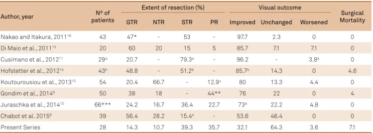
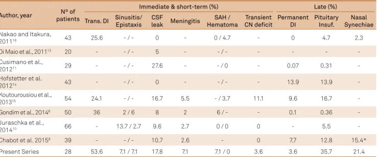
heir main surgical outcomes and complications rates are de
-tailed on Tables 3 and 4. Our group has recently analyzed the ini-tial experience (2000–2010) in the management of 35 giant non-functioning pituitary adenomas operated on by the open and the endoscopic routes, but the main objective was to correlate Ki-67 expression with tumor recurrence12.
Extent of resection of the endoscopic endonasal transsphenoidal approach
As aforementioned, GTR is not the goal for such large tumors because of neurovascular structures involvement
and the recent success of adjuvant therapies8. hus, the ex
-tent of resection is diicult to evaluate as an outcome for
large and giant pituitary adenomas8. Moreover, there is no
general consensus on the deinition for near total (NTR),
subtotal (STR), and partial resection (PR), as also noted by Chabot et al.8. Overall, the reported rate of GTR in the mod-ern purely endoscopic series published after the systematic review by Komotar et al.7 ranges from 14% in our series to 60% in the Naples series13 (average of 36.7%) (Table 3).
Such higher rates of GTR are quite encouraging in com-parison to the open and microscopic transsphenoidal groups (9.6% and 30.9%, respectively)7. It is worth discussing that cavernous sinus involvement is reported by most of the studies as the major limitation to GTR with the endoscop-ic approach6,8,14,16, whereas for the microscopic transsphe-noidal approach is the suprasellar extension25. Besides cav -ernous sinus involvement, tumor size greater than 10 cm3,14
Figure 3. Preoperative axial (A) and coronal (B) T1-weighted gadolinium-enhanced MRI of a rounded giant pituitary tumor. (C, D) The patient underwent near-total resection of the adenoma, but experienced a severe subarachnoid/tetraventricular haemorrhage after cavernous sinus violation and internal carotid artery injury, which ultimately lead to diffuse cerebral vasospasm and brain death.
A
B
C
D
Table 3. Outcomes of main series of endoscopic endonasal transsphenoidal approach for large and giant pituitary adenomas.
Author, year Nº of
patients
Extent of resection (%) Visual outcome
Surgical Mortality
GTR NTR STR PR Improved Unchanged Worsened
Nakao and Itakura, 201116 43 47* - 53 - 97.7 2.3 0 0
Di Maio et al., 201113 20 60 20 15 5 85.7 7.1 7.1 0
Cusimano et al., 201211 29a 20.7 - 79.3a - 96.2 - 3.8a 0
Hofstetter et al., 201214 43b 48.8 - 51.2b - 85.7b 14.3 0 4.6
Koutourousiou et al., 201315 54 20.4 66.7 - 12.9c 80 13.3 4.4 0
Gondim et al., 20146 50 38 18 - 44** 76 22 0 4
Juraschka et al., 201410 66*** 24.2 16.7 36.4 22.7 73d 22.2 4.8 0
Chabot et al. 20158 39 56.4 28.2 15.4e - 53.6 46.4 0 0
Present Series 28 14.3 10.7 39.3 35.7 32.1 64.3 3.6 7.1
GTR: gross total resection; NTR: near total resection; PR: partial resection; STR: subtotal resection. *The extent of resection considered STR for any residual tumor; **The extent of resection considered PR for any residual tumor greater than 10%; ***Preoperative and postoperative MRI were available in 66 of the 72 patients reported; aPurely endoscopic transsphenoidal approach was done in 29 of the 72 patients reported. Residual tumor was not defined in NTR, STR or PR. One patient
experienced no improvement on visual function, but it was not informed whether it was unchanged or worsened; bPurely endoscopic transsphenoidal approach for
large (> 3 cm) and giant (> 10 cm3) adenomas was done in 43 of the 71 patients reported. Residual tumor was not defined in NTR, STR or PR. Visual acuity was not
included in the analysis. The reported rates of visual outcomes reflect visual field defect of the patients suffering from giant tumors only. c The extent of resection
considered PR for any residual tumor greater than 10%. dPostoperative visual fields were available for 55 patients, of whom 61.8% experienced improvement. eThe
multilobular coniguration and extension to the middle fos -sa15 are also reported to have a negative impact on surgical resection rates by the endoscopic technique.
Taking together the results of GTR, NTR and STR as an arbitrary threshold to provide satisfactory neurovas-cular structures decompression, the endoscopic cohort was able to achieve decompression rates of 78.9% (range 63–95%)10,13,Present. Even though a fair comparison cannot be
performed, because of a diferent deinition for STR (residu
-al tumor > 10–20%)7, the endoscopic rates of decompression are at least similar to open and microscopic transsphenoidal groups (78.8% and 81.2%, respectively)7.
Finally, the surgical technique used by most of the endoscop-ic endonasal series is the standard approach, in whendoscop-ich a wide
exposure of the sellar loor is obtained from the medial walls
of the cavernous sinus laterally, the tuberculum sellae cranially and the dural indentation of the clivus/inferior transcavernous sinus caudally6,8,10,11,14,16. Some studies have used the extended transplanum/transtuberculum endonasal approach to address
some dumbbell-shaped or suprasellar adenomas or even ibrous
tumors6,8,10,13,14,15. he role of the extended approach is yet to be
deined, but the results are promising taken into consideration
the higher rates of GTR in comparison to the standard endona-sal approach (average of 41.3% vs. 27.3%, respectively; Table 3). Further studies are, however, necessary to clarify this issue.
Clinical outcomes of the endoscopic endonasal transsphenoidal approach
Nine patients in our series experienced visual acuity im-provement and only one patient had visual deterioration. Similar results were described by recent endoscopic endona-sal series, in which the rates of visual improvement achieved a mean of 75.5% (range 32.1%–97.7%; Table 3). Given that visual
improvement is observed in up to 40% in the open and micro-scopic transsphenoidal cohorts7, visual outcomes are by far the major advantage of the endoscopic endonasal approach over the remaining surgical techniques8. Chabot et al.8 hypoth-esized that the endoscope provides a better visualization and therefore protection of the optic apparatus and its blood sup-ply, which ultimately leads to such improved outcomes.
Conversely, the endocrine outcomes are generally not so encouraging as observed with visual outcomes. Our results indicate the occurrence of 35.7% of new postoperative
pitu-itary insuiciency, which is similar to other endoscopic en
-donasal series (mean 17.2%; range 4.7%–36%; Table 4). he
open and microscopic cohorts revealed better outcomes, however (9.1% and 9.5%, respectively)7. Even though Nakao and Itakura16 and Juraschka et al.10 reported lower rates of new postoperative hypopituitarism, the endocrine outcomes seems to correlate inversely with the greater extent of resec-tion and therefore with the greater surgical manipularesec-tion provided by the endoscope.
Complications of the endoscopic endonasal transsphenoidal approach
he complications related to the endoscopic transsphenoi
-dal surgery in our series show converging rates with published studies (Tables 3 and 4), suggesting the technical safety and
eicacy compared to the open transcranial and microscop -ic transsphenoidal approaches. We have didact-ically divided postoperative complications into two major groups, namely the immediate/short-term and late complications. From the
irst group the occurrence of CSF leak, transient DI and nasosi -nusal disorders (sinusitis/epistaxis) are the most frequent.
CSF leak has been considered one of the major disadvan-tages of the endoscopic endonasal approach, especially in the
Table 4. Immediate, short- and late postoperative complications of main series of endoscopic endonasal transsphenoidal approach for large and giant pituitary adenomas.
Author, year Nº of
patients
Immediate & short-term (%) Late (%)
Trans. DI Sinusitis/ Epistaxis
CSF
leak Meningitis
SAH / Hematoma
Transient CN deficit
Permanent DI
Pituitary Insuf.
Nasal Synechiae
Nakao and Itakura,
201116 43 25.6 - / - 0 - 0 / 4.7 - 0 4.7 2.3
Di Maio et al., 201113 20 - - / - 5 - - / - - - -
-Cusimano et al.,
201211 29 - - / - 27.6 - - / 0 - 0.07 0.31
-Hofstetter et al.
201214 43 - - / - 0 - - / - - 13.9 13.9
-Koutourousiou et al.,
201315 54 24.1 - / - 16.7 5.5 - / 3.7 11.1 9.6 16.7
-Gondim et al., 20146 50 36 2 / 6 8 2 6 / - - 0.1 0.36
-Juraschka et al.,
201410 66 - 13.7 / 2.7 9.6 2.7 0 / 0 0 - 5.5
-Chabot et al. 20158 39 - - / - 10.7 2.6 - 0 7.7 12.8 15.4*
Present Series 28 53.6 7.1 / 7.1 17.8 7.1 7.1 / 0 3.6 3.6 35.7 21.4
case of extended approaches8. As the matter in fact, it should
be noted that it is extremely diicult to resect such complex
tumors without violating the arachnoid layer8. he advent of
nasoseptal laps has contributed enormously to decrease its
incidence10,15. But, even after the routine use of nasoseptal
laps, 17.8% of our patients experienced a CSF leak postopera
-tively. he overall rates of postoperative CSF leaks among the
endoscopic surgical series are 10.6% (range 0%–27.6%; Table 4).
Several diferent reconstruction techniques were used by the
endoscopic reports in the way that a direct comparison cannot be done. However, we did not use routinely any inlay besides abdominal fat, which could be a potential source for our in-creased rates of CSF leak. Further studies are necessary to
ad-dress this issue deinitely. Open and microscopic cohorts ren -dered CSF leak rates of 7.1% and 5.1%, respectively7.
Transient DI is a common complication in the im-mediate postoperative period with a reported inci-dence of 24.1%–53.6% of the patients (Table 4). It is considered to occur as a result of direct surgical ma-nipulation, especially associated to hypothalamic in-jury6. Still in the immediate/short-term complication group, we have faced a cavernous sinus violation and consequently internal carotid artery injury, which lead to subarachnoid/tetraventricular haemorrhage followed by severe cerebral vasospasm and death. This is a rare
References
1. Guiot J, Rougerie J, Fourestier M, Fournier A, Comoy C, Vulmiere J et al. [Intracranial endoscopic explorations]. Presse Med. 1963;71:1225-8. French.
2. Xue-Fei S, Yong-Fei W, Shi-Qi L, Jing-Song W, Yao Z, Ying M et al. Microsurgical treatment for giant and irregular pituitary adenomas in a series of 54 consecutive patients. Br J Neurosurg. 2008;22(5):636-48. doi:10.1080/02688690802346083
3. Berker M, Hazer DB, Yücel T, Gürlek A, Cila A, Aldur M, et al. Complications of endoscopic surgery of the pituitary adenomas: analysis of 570 patients and review of the literature. Pituitary. 2012;15(3):288-300. doi:10.1007/s11102-011-0368-2
4. Santos RP, Zymberg ST, Abucham Filho JZ, Gregório LC, Weckx LLM. Endoscopic transnasal approach to sellar tumors. Rev Bras Otorrinolaringol. 2007;73(4):463-75. doi:10.1590/S0034-72992007000400005
5. Agrawal A, Cincu R, Goel A. Current concepts and controversies in the management of non-functioning giant pituitary macroadenomas. Clin Neurol Neurosurg. 2007;109(8):645-50. doi:10.1016/j.clineuro.2007.06.007
6. Gondim JA, Almeida JP, Albuquerque LA, Gomes EF, Schops M. Giant pituitary adenomas: surgical outcomes of 50 cases operated on by the endonasal endoscopic approach. World Neurosurg. 2014;82(1-2):e281-90. doi:10.1016/j.wneu.2013.08.028
7. Komotar RJ, Starke RM, Raper DM, Anand VK, Schwartz
TH. Endoscopic endonasal compared with microscopic transsphenoidal and open transcranial resection of giant pituitary adenomas. Pituitary. 2012;15(2):150-9. doi:10.1007/s11102-011-0359-3
8. Chabot JD, Chakraborty S, Imbarrato G, Dehdashti AR. Evaluation of outcomes after endoscopic endonasal surgery for large and
giant pituitary macroadenoma: a retrospective review of 39 consecutive patients. World Neurosurg. 2015;84(4):978-88. doi:10.1016/j.wneu.2015.06.007
9. Cappabianca P, Cavallo LM, de Divitiis O, de Angelis M, Chiaramonte C, Solari D. Endoscopic Endonasal Extended Approaches for the Management of Large Pituitary Adenomas. Neurosurg Clin N Am. 2015;26(3):323-31. doi:10.1016/j.nec.2015.03.007
10. Juraschka K, Khan OH, Godoy BL, Monsalves E, Kilian A, Krischek B, et al. Endoscopic endonasal transsphenoidal approach to large and giant pituitary adenomas: institutional experience and predictors of extent of resection. J Neurosurg. 2014;121(1):75-83. doi:10.3171/2014.3.JNS131679
11. Cusimano MD, Kan P, Nassiri F, Anderson J, Goguen J, Vanek I, et al. Outcomes of surgically treated giant pituitary tumours. Can J Neurol Sci. 2012;39(4):446-57. doi:10.1017/S0317167100013950
12. Landeiro JA, Fonseca EO, Monnerat AL, Taboada GF, Cabral GA, Antunes F. Nonfunctioning giant pituitary adenomas: invasiveness and recurrence. Surg Neurol Int. 2015;6:179. doi 10.4103/2152-7806.170536
13. Di Maio S, Cavallo LM, Esposito F, Stagno V, Corriero OV, Cappabianca P. Extended endoscopic endonasal approach for selected pituitary adenomas: early experience. J Neurosurg. 2011;114(2):345-53. doi:10.3171/2010.9.JNS10262
14. Hofstetter CP, Nanaszko MJ, Mubita LL, Tsiouris J, Anand VK, Schwartz TH. Volumetric classification of pituitary macroadenomas predicts outcome and morbidity following endoscopic endonasal transsphenoidal surgery. Pituitary. 2012;15(3):450-63.
doi:10.1007/s11102-011-0350-z
15. Koutourousiou M, Gardner PA, Fernandez-Miranda JC, Paluzzi A, Wang EW, Snyderman CH. Endoscopic endonasal surgery for giant
complication of the transsphenoidal surgery with an es-timated incidence of 1.58%–4.6%15,26.
From the late complication group, permanent DI and naso-sinusal disorders are a common occurrence. Of note, however, is the frequent underreported description of nasosinusal com-plications. Only two previous studies addressed this compli-cation8,16, which has a signiicant negative impact on patient’s quality of life27. Nakao and Itakura16 reported an incidence of 2.3% of nasal synechiae without the development of a
naso-septal lap, while Chabot et al.8 use the nasoseptal lap for skull base reconstruction rendering a rate of 15.4% of late nasosi-nusal symptoms, which is very similar to our results (Table 4).
Limitations
he retrospective data analysis and a reduced sample
were the main limiting factors of this study.
In conclusion, endoscopic endonasal transsphenoidal surgery is a valuable treatment option for large or giant pi-tuitary adenomas, which results in high rates of surgical decompression of cerebrovascular structures. Immediate, short-term and late complication rates are at least similar, if not less, to other surgical techniques, and well accepted in terms of disease complexity. Postoperative nasosinusal
pituitary adenomas: advantages and limitations. J Neurosurg. 2013;118(3):621-31. doi:10.3171/2012.11.JNS121190
16. Nakao N, Itakura T. Surgical outcome of the endoscopic endonasal approach for non-functioning giant pituitary adenoma. J Clin Neurosci. 2011;18(1):71-5. doi:10.1016/j.jocn.2010.04.049
17. Hardy J. Transphenoidal microsurgery of the normal and pathological pituitary. Clin Neurosurg. 1969;16:185-217.
18. Mohr G, Hardy J, Comtois R, Beauregard H. Surgical management of giant pituitary adenomas. Can J Neurol Sci. 1990;17(1):62-6.
19. Giustina A, Chanson P, Bronstein MD, Klibanski A, Lamberts S, Casanueva F et al. A consensus on criteria for cure of acromegaly. J Clin Endocrinol Metab. 2010;95(7):3141-8. doi:10.1210/jc.2009-2670
20. Colao A, Attanasio R, Pivonello R, Cappabianca P, Cavallo LM, Lasio G, et al. Partial surgical removal of growth hormone-secreting pituitary tumors enhances the response to somatostatin analogs in acromegaly. J Clin Endocrinol Metab. 2006;91(1):85-92. doi:10.1210/jc.2005-1208
21. Jho HD, Carrau RL. Endoscopic endonasal transsphenoidal surgery: experience with 50 patients. J Neurosurg. 1997;87(1):44-51. doi:10.3171/jns.1997.87.1.0044
22. Tabaee A, Anand VK, Barrón Y, Hiltzik DH, Brown SM, Kacker A, et al. Endoscopic pituitary surgery: a systematic review and meta-analysis. J Neurosurg. 2009;111(3):545-54. doi:10.3171/2007.12.17635
23. Cappabianca P, Cavallo LM, Esposito F, De Divitiis O, Messina A, De Divitiis E. Extended endoscopic endonasal approach to the midline skull base: the evolving role of transsphenoidal surgery. Adv Tech Stand Neurosurg. 2008;33:151-99. doi:10.1007/978-3-211-72283-1
24. Paiva Neto MA, Vandergrift A, Fatemi N, Gorgulho AA, Desalles AA, Cohan P et al. Endonasal transsphenoidal surgery and multimodality treatment for giant pituitary adenomas. Clin Endocrinol (Oxf). 2010;72(4):512-9. doi:10.1111/j.1365-2265.2009.03665.x
25. McLaughlin N, Eisenberg AA, Cohan P, Chaloner CB, Kelly DF. Value of endoscopy for maximizing tumor removal in endonasal transsphenoidal pituitary adenoma surgery. J Neurosurg. 2013;118(3):613-20. doi:10.3171/2012.11.JNS112020
26. Zhao B, Wei YK, Li GL, Li YN, Yao Y, Kang J, et al. Extended transsphenoidal approach for pituitary adenomas invading the anterior cranial base, cavernous sinus, and clivus: a single-center experience with 126 consecutive cases. J Neurosurg.
2010;112(1):108-17. doi:10.3171/2009.3.JNS0929


