Original article
Rett syndrome with and without detected MECP2 mutations: An
attempt to redefine phenotypes
Teresa Temudo
a,*, Mo´nica Santos
b,c, Elisabete Ramos
d, Karin Dias
e,
Jose´ Pedro Vieira
e, Ana Moreira
e, Eula´lia Calado
e, Ineˆs Carrilho
f, Guiomar Oliveira
g,
Anto´nio Levy
h, Clara Barbot
f, Maria Fonseca
i, Alexandra Cabral
g, Pedro Cabral
j,
Jose´ Monteiro
i, Luı´s Borges
g, Roseli Gomes
k, Gracßa Mira
l, Susana Aires Pereira
m,
Manuela Santos
f, Anabela Fernandes
b, Jorg T. Epplen
n, Jorge Sequeiros
c,o,
Patrı´cia Maciel
baUnidade de Neuropediatria, Servicßo de Pediatria, Hospital Geral de Santo Anto´nio, Porto, Portugal
bInstituto de Investigacßa˜o em Cieˆncias da Vida e da Sau´de (ICVS), Escola de Cieˆncias da Sau´de, Universidade Minho, Braga, Portugal c
ICBAS, Universidade do Porto, Portugal
d
Servicßo de Higiene e Epidemiologia, Faculdade de Medicina, Universidade do Porto, Portugal
e
Servicßo de Neuropediatria, Hospital Dª Estefaˆnia, Lisboa, Portugal
f
Servicßo de Neuropediatria, Hospital de Criancßas Maria Pia, Porto, Portugal
g
Centro de Neuropediatria, Hospital Pedia´trico, Coimbra, Portugal
h
Servicßo de Pediatria, Hospital Santa Maria, Lisboa, Portugal
i
Servicßo de Pediatria, Hospital Garcia da Horta, Almada, Portugal
j
Servicßo de Neurologia, Hospital Egas Moniz, Lisboa, Portugal
k
Servicßo de Pediatria, Hospital Pedro Hispano, Matosinhos, Portugal
lServicßo de Pediatria Hospital Espı´rito Santo, E´vora, Portugal mServicßo de Pediatria, Hospital Vila Nova de Gaia, Portugal nDepartment of Human Genetics, Ruhr-University, Bochum, Germany oUnIGENe, IBMC – Institute for Molecular and Cell Biology, Porto, Portugal
Received 4 August 2009; received in revised form 2 January 2010; accepted 7 January 2010
Abstract
Background: The diagnosis of Rett syndrome (RTT) is based on a set of clinical criteria, irrespective of mutation status. The aims of this study were (1) to define the clinical differences existing between patients with Rett syndrome with (Group I) and without a MECP2 mutation (Group II), and (2) to characterize the phenotypes associated with the more common MECP2 mutations. Patients and methods: We analyzed 87 patients fulfilling the clinical criteria for RTT. All were observed and videotaped by the same paediatric neurologist. Seven common mutations were considered separately, and associated clin-ical features analysed. Results: Comparing Group I and II, we found differences concerning psychomotor development prior to onset, acquisition of propositive manipulation and language, and evolving autistic traits. Based on age at observation, we found differences in eye pointing, microcephaly, growth, number of stereotypies, rigidity, ataxia and ataxic-rigid gait, and severity score. Patients with truncating differed from those with missense mutations regarding acquisition of propositive words and independent gait, before the beginning of the disease, and microcephaly, growth, foot length, dystonia, rigidity and sever-ity score, at the time of observation. Patients with the R168X mutation had a more severe phenotype, whereas those with
0387-7604/$ - see front matterÓ 2010 The Japanese Society of Child Neurology. Published by Elsevier B.V. All rights reserved. doi:10.1016/j.braindev.2010.01.004
* Corresponding author. Address: Unidade de Neuropediatria, Servicßo de Pediatria, Hospital de Santo Anto´nio, SA, Largo Abel Salazar, 4099/001
Porto, Portugal. Tel.: +351 22 207 75.00; fax: +351 22 332 03 18. E-mail address:[email protected](T. Temudo).
R133C showed a less severe one. Patients with R294X had a hyperactive behaviour, and those with T158M seemed to be particularly ataxic and rigid. Conclusion: A clear regressive period (with loss of prehension and language, deceleration of growth) and the presence of more than three different stereotypies, rigidity and ataxic-rigid gait seemed to be very helpful in differentiating Group I from Group II.
Ó 2010 The Japanese Society of Child Neurology. Published by Elsevier B.V. All rights reserved.
Keywords: Autism; Mental retardation; Movement disorder; Cerebellum; Clinical criteria; Clinical stage
1. Introduction
Rett syndrome (RTT) was first described by Andreas Rett, in 1966[1], and rediscovered in 1983, by Bent Hag-berg et al.[2]. Since 1999, after the description of muta-tions in the MECP2 gene as the cause of RTT [3], we know that more than 90% of the patients with classical, but only 30% with variant RTT have a MECP2 muta-tion. Thus, we can consider two groups of patients: Group I with clinical features of RTT and a mutation in the MECP2 gene and Group II with clinical features of RTT without a mutation in the MECP2 gene, and speculate that maybe, in addition to Group I patients, different diseases are currently under the large RTT umbrella. Widespread availability of the molecular diag-nosis creates a great opportunity for this to be clarified, based on clinical and genetic grounds.
Several series of patients with genetic studies have been published, and attempts of phenotype-genotype correlations made. The results, however, were not con-clusive, as different methodologies were applied in the various studies, concerning clinical and mutation classi-fications and severity score systems[4–6]. Nevertheless, the larger studies pointed to a broad correlation between type of mutation and phenotype, patients with truncat-ing mutations havtruncat-ing a more severe phenotype, than those with missense mutations[5–12].
Two different genetic factors are likely to influence the phenotype in RTT: the type and location of the muta-tions, and the X inactivation pattern [13]. Concerning the phenotype of specific mutations, several studies
[14–17] showed that patients with the R133C and
R306C mutations have better overall function. In general, the most severe outcomes were found with the R270X and R255X mutations[11,16]. Mutations toward the N termi-nus, including T158M and R168X, were also associated with a more severe phenotype[12,17]; however, no specific neurological or behavioural findings were identified in patients with these mutations, with the exception of an association of fear and anxiety with R133C and R306C
[18], and mood abnormalities in patients with R294X[16]. The aims of this study were (1) to define the clinical differences between patients with (Group I) and without (Group II) known mutations in the MECP2 gene, in order to delineate the clinical boundaries of the disease; (2) to analyze the genotype-phenotype correlation in
Group I patients and characterize phenotypes associated with the more common MECP2 mutations; and (3) to identify sub-groups of patients with RTT that might be candidates for mutational search in other genes. 2. Subjects and methods
All Portuguese paediatric neurologists were asked to indicate their patients with possible RTT. Patients included in the study (n = 87) fulfilled the revised clinical criteria for RTT [19]. They were observed and video-taped by the same paediatric neurologist (TT), and a clinical checklist for RTT was completed. Informed con-sent was obtained from all parents, for blood sampling and to take and use videos and photographs; 36 patients were observed and videotaped at least twice, at 6 months intervals.
Blood samples from patients and their parents were received at the laboratory, and genomic DNA was extracted using the Puregene DNA isolation kit (Gentra, Minneapolis, MN). The coding region and exon–intron boundaries of the MECP2 gene were amplified by PCR and sequenced. The RD-PCR method was used, as described[20], for the detection of large rearrangements in the MECP2 gene. Primers and PCR conditions are available upon request.
X-chromosome inactivation (XCI) assays were per-formed with genomic DNA isolated from leukocytes of peripheral blood. The assay was based on a previously described method [21], which allows the determination of the X-inactivation status, using a trinucleotide repeat polymorphism in the androgen receptor gene (AR, Ref-Seq ID: NM_000044.2), flanked by two methylation-sen-sitive restriction enzyme sites. The restriction enzyme (HhaI) will hydrolyze only the unmethylated alleles. Scoring of the XCI pattern was made by densitometry of the amplified DNA bands.
MECP2 mutations were categorized and grouped for analysis according to: (1) type of sequence change – mis-sense or truncating (including nonmis-sense and frameshift, but also large deletions) and (2) location of the mutation (affected domain) – TRD, MBD. The seven most com-mon mutations were considered individually, and their associated features analysed.
For statistical analysis, patients were divided into two groups: those with (Group I) and without a molecular
diagnosis (Group II). Within the mutation-positive group, comparisons were made between patients carry-ing missense and truncatcarry-ing mutations. Cases were scored using the Pineda severity scale [6]. The SPSS (v.14) statistical package was used to analyse the data. Proportions were compared using Chi-square and Fish-er’s exact test. Quantitative variables were compared using the Mann–Whitney test and were presented as median (25–75th percentile).
3. Results
In this study we analyzed the entire coding sequence of the MECP2 gene in 87 patients, who fulfilled the revised clinical criteria for RTT[19]. Cases were classi-fied as classical (58.6%) and variant (41.4%) forms (con-genital, preserved speech, forme fruste, early epileptic encephalopathy and male patients)[22].
3.1. Clinical differences between MECP2 positive and negative RTT patients
Mutations in MECP2 were found in 67.8% (n = 59) of all patients, corresponding to 96.0% of classical forms and of 27.8% variants (seven congenital forms; three preserved speech). In 28 patients MECP2 mutations were not found. Among these patients 60.7% were congenital, 10.7% males, 10.7% early epileptic encephal-opaties, 7.1% classical, 7.1% forme frustre and 3.6% pre-served speech forms.
We identified a total of 29 different mutations in MECP2 including two large deletions and a mutation in exon 1; most mutations were located in exons 3 and 4
(Fig. 1). Nine of these mutations were never described
before: A7fsX37, K39fsX43, Q110X, S113P, T184fsX
185, R253fsX275, I303fsX399, V380M and L386fsX 399. All were de novo mutations, except V380M which was present in the unaffected mother (who had a balanced XCI pattern) and in 4 X chromosomes in a control popu-lation of Portuguese origin including 230 X chromo-somes. We assumed the truncating mutations as causative of the disease; the new missense mutation S113P was excluded in the parents and in a control population of Portuguese origin including 230 X chromosomes.
XCI patterns were analysed for all patients carrying a MECP2 mutation, though data was not informative for 14; among the 46 patients remaining, only two showed a skewed XCI pattern. Of these, one patient with a R168X mutation had a less severe form (and a lower severity score) compared with the group of patients with the same mutation: she acquired and maintains independent gait, had neither epilepsy nor microcephaly. The other patient carried the T158M mutation, and had a pheno-type similar to those carrying the same mutation. Med-ian age at first observation and medMed-ian duration of the disease were not significantly different between Group I and II, neither between groups with missense and trun-cating mutations. A significant difference was found when comparing clinical stage: more patients with Group I, especially those with truncating mutations, were in stage IV of the disease, which may mean that their rate of evolution was faster.
Comparison between Group I and II showed some important clinical differences since birth (Table 1). Dystocic delivery (caesarean or forceps) seemed to be more common in Group I than Group II patients, though this did not reach significance. Additionally, none of the Group II patients presented microcephaly at birth, but 6 (10.2%) of Group I had an
frontal circumpherence (OFC) below the 5th percentile (3 truncating, 3 missense mutations). Also, 63.6% of Group II patients presented an OFC at birth above the 25th percentile, against 26.6% of Group I patients. Stagnation of OFC growth occurred in 71.7% of Group I and 53.6% of Group II, at a median age of 6 months in both groups. Later, microcephaly was
acquired in 45.8% of Group I (67.6% among truncat-ing, 19.2% for missense mutations) and 39.3% of Group II patients. Interestingly, 13.3% of Group I patients presented an OFC above the 25th percentile (3% above the 75th percentile); 26.9% of patients with missense mutations presented an OFC above the 25th percentile, against only 2.9% of those with truncating Table 1
Clinical data of Group I and Group II patients.
Group I (n = 59) Group II (n = 28) p Missense (n = 26) Truncanting (n = 33) p Median age (years)a 7.6 (4.1–14.3) 7.8 (5.1–12.7) 0.730 7.0 (3.7–10.6) 11.3 (4.5–14.8) 0.27 5 Median duration of disease (months)a 92 (55–140) 82 (35–139) 0.411 72 (26–111) 124 (44–157) 0.240 Stage (%) Stage II Stage III Stage IV A and IV B 8.5 61.0 30.5 3.6 82.1 14.3 0.153 7.7 80.8 11.5 9.0 45.5 45.5 0.008 Perinatal data (%) Abnormal delivery 28.8 21.4 0.466 23.1 33.3 0.566 Microcephaly at birth Yes Missing information 10.2 16.9 0.0 21.4 0.263 11.5 15.4 9.1 18.2 0.999
Reported psychomotor development (%)
Normal until 12 months of age 59.3 17.9 <0.001 69.2 51.5 0.268
Stagnation 93.2 92.9 0.999 92.3 93.9 0.999
Median age of stagnation (months)a 12.0 (10.0–18.0) 6.0 (4.0–11.3) <0.001 16.0 (10.0–18.0) 12.0 (9.0–18.0) 0.172
Regression 86.4 25.0 <0.001 84.6 87.9 0.999
Median age of regression (months)a 18.00 (14.0–24.0) 14.00 (11.0–24.0) 0.202 18.0 (18.0–24.5) 18.0 (13.0–23.0) 0.160
Acquisition of autistic traits 83.1 50.0 0.002 76.9 87.9 0.222
Median age of acquisition of autistic traits 18.0 (12.5–18.0) 7.5 (3.75–18.0) 0.005 18.0 (14.0–21.0) 15.0 (12.0–18.0) 0.175
Propositive manipulation 91.5 57.1 <0.001 92.3 93.8 0.999
Median age of acquisition of stereotypies 20.0 (14.5–25.0) 24.0 (14.5–36.0) 0.425 24.0 (18.0–28.5) 18.00 (13.3–24.0) 0.086
Propositive words 71.2 17.9 <0.001 88.5 57.6 0.021
Acquisition of independent gait 63.3 50.00 0.200 84.6 48.5 0.009
Data at observation
Eye pointing 96.6 57.1 <0.001 92.3 100.0 0.190
Mycrocephaly at time of observation 45.8 39.3 0.569 19.2 66.7 0.001
Low weight (<5th percentile) 43.3 18.5 0.031 23.1 57.6 0.009
Low height (<5th percentile) 55.2 30.8 0.038 38.5 68.8 0.033
Low foot length (<3th percentile) 67.9 45.8 0.112 47.8 83.3 0.008
Agitation 56.7 67.9 0.497 50.0 63.6 0.426
Laughing spells 69.5 53.6 0.227 61.5 75.8 0.372
Abnormal sleep pattern 55.9 53.6 0.836 46.2 63.6 0.199
Hyperpnea/Apnea 78.0 82.1 0.868 69.2 84.8 0.209
Epilepsy 57.6 75.0 0.183 46.2 66.7 0.184
Controlled 58.8 76.2 0.306 69.2 52.4 0.477
Vaso-motor disturbances 71.2 78.6 0.639 53.8 84.8 0.019
Neurogenic muscle atrophy 49.2 28.6 0.114 42.3 54.5 0.435
Chewing difficulties 78.0 71.4 0.691 65.4 87.9 0.080
Pyramidal signs 28.8 39.3 0.3465 23.1 33.3 0.566
Ataxia 35.6 10.7 0.030 46.2 27.3 0.219
Number of stereotypies per patienta 4.00 (3.0–6.0) 3.00 (2.2–4.0)
0.050 3.50 (2.8–7.0) 4.00 (3.0–6.0) 0.999 Dystonia 64.6 75.0 0.458 46.2 78.8 0.0209 Rigidity 49.2 7.1 <0.001 34.6 60.6 0.046 Tremor 49.2 35.7 0.344 50.0 48.5 0.999 Ataxic/rigid gait 43.6 0.0 0.001 36.8 50.0 0.523 Scoliosis 72.9 67.9 0.818 69.2 75.8 0.791 Gastro–intestinal (%) Gastro-esophageal reflux 11.9 14.3 0.999 11.5 12.1 0.999 Pineda’s scalea 13 (9–15) 14 (12–16) 0.034 11 (8–13) 14 (11–16) 0.001
mutations. Differences were also found for height and weight, comparing the two groups, and for head size, height, weight and foot length, comparing the group of patients with missense versus truncating mutations (Table 1).
Clinical classification in these groups was also differ-ent: 83.0% of Group I were classified as classical, against only 7.1% of Group II patients. Significant differences were found when comparing psychomotor development before the onset of the disease: in a higher percentage of Group II patients, this was not considered to be normal, by the parents, before the age of 12 months; a clear regression period was evident in 86.4% of Group I, but only 25.0% of Group II patients; acquisition of pro-positive words was more common in patients with Group I, specially those with missense mutations; evolv-ing of autistic traits (loss of social communication) was described in 83.1% of Group I and 50% of Group II, and it occurred earlier in life in Group II; in 42.9% of Group II, propositive manipulation was never acquired (against 8.5% in Group I); independent gait was acquired by 84.6% of patients with missense, but only in 48.5% of those with truncating mutations (Table 1).
Besides growth failure, we found other important dif-ferences concerning clinical data of the patients at obser-vation: eye pointing was present in 96.6% of Group I, but only in 57% of Group II patients; rigidity and ataxia were more frequent in Group I, and an ataxic-rigid gait was observed only in them. They also displayed a greater num-ber of stereotypies. Dystonia, rigidity, vaso-motor distur-bances and oral motor difficulties were more frequent in patients with truncating mutations (Table 1). We found no significant differences between the group I and II con-cerning pyramidal signs, dystonia, neurogenic muscle atrophy, vaso-motor disturbances, scoliosis, epilepsy or gastro-intestinal disturbances. Abnormal sleep pattern, agitation, laughing spells and hyperpnea/apnea were also not more frequent in patients with known mutations.
The Pineda’s severity score showed that the median severity was greater in the Group I group and, within this group, in those with truncating mutations (Table 1). 3.2. Clinical presentation among MECP2 positive RTT patients (group i)
In general, we identified three neurological presenta-tions in Group I patients:
(1) Some had a severe form (n = 27; 45,8% of all Group I patients), with major axial hypotonia, in which dystonia and rigidity were present after few years of evolution (median, 11 years). These patients either regressed earlier (median, 15.0; range, 6–36 months), or never had been normal (18.5%); 74.0% did not acquire independent gait; those who did, lost it at a median age of 66 months (range, 24–120). Epilepsy was present in 70.4% of the cases. Among patients with the severe form, 81,4% had
truncat-ing mutations, and all but one with the R168X mutation presented it; they showed a median severity score of 15 (range, 12–20).
(2) In an intermediate form, ataxia predominated (n = 17; 28.8% of all Group I patients). Most acquired an independent gait (94.1%), but this was both ataxic and rigid; they regressed at a median age of 18 months (13–26), and 52.9% had epilepsy. Their median severity score was 11 (range, 8–14); 68.8% of the patients with this presentation had missense mutations, particularly T158M.
(3) A milder form was present in 15 (25,4%) of Group I patients, with few neurological signs except for mental retardation and autistic features. All acquired indepen-dent gait and maintained it during the period of fol-low-up; 40% developed focal dystonia after some years of evolution, and gait became rigid in two patients (one with the R133C mutation, older than 20 years; one six years old with the R294X mutation). Epilepsy was present in 33.3% of these patients. They regressed at a median age of 25 months (range, 13–60), and had a median severity score of 9 (range, 4–11); 60% had mis-sense mutations. Most of our patients with the R133C mutation illustrate this presentation well. Three patients with R294X mutation also presented this phenotype.
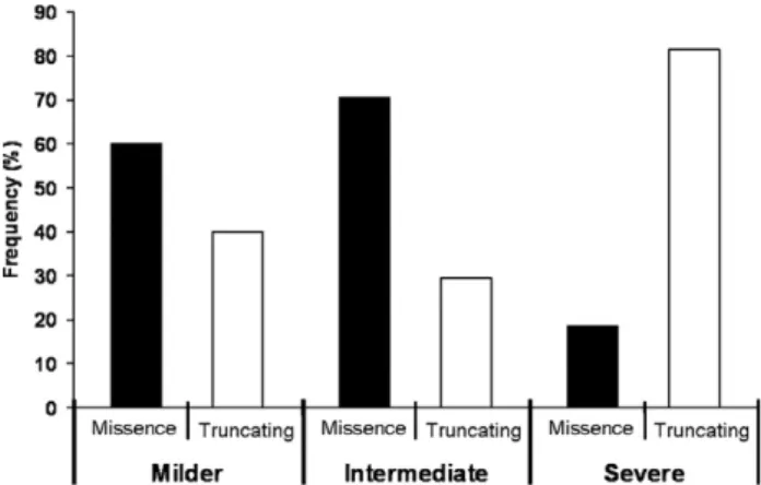
Interestingly, the distribution of mutation types was significantly different between these clinical groups. In the milder group, 60% of the patients had missense mutations. In the intermediate group, 70,6% of the patients had missense mutations; and in the more severe group 81.5% of the cases had a truncating mutation. The frequency of missense versus truncating mutations was significantly different between the three clinical subtypes (Fisher’s exact test, p = 0.001) (Fig. 2).
3.3. Clinical presentation associated with specific MECP2 mutations
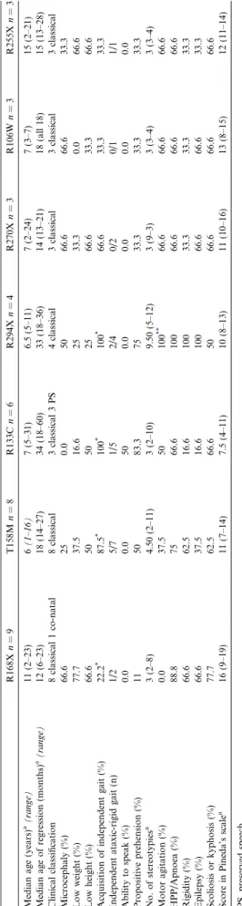
The clinical features associated with specific muta-tion groups found in Group I patients are presented in
Fig. 2. Frequency of missense versus truncating mutations according to the three clinical subtypes.
Table 2. Overall, we observed that those with the R168X mutation presented the most severe phenotype. Only two of nine patients with this mutation acquired independent gait; one lost it one year later, while the other had a very rigid and ataxic gait. Two presented microcephaly at birth and six developed it within the 2nd year of life. The majority had low height and weight, and epilepsy. They also had continuous hands-apart stereotypies.
Group I patients with the R294X mutation seemed to have a peculiar phenotype with an extremely unusual hyperactive motor behaviour. They had a lot of different and exuberant hand stereotypies and with other topog-raphies (including vocal), and all had epilepsy. All dis-played focal or segmental dystonia, and, in three, gait became rigid and broaded based, in the earlier stages of the disease; however, they maintained a good eye con-tact, and seemed less profoundly retarded than other patients with better motor function.
Patients with the R133C mutation had the mildest phenotype among all those within the Group I. None had microcephaly, and all but one acquired and main-tained independent gait. Three could make phrases, albeit with a peculiar voice tone, and answered to simple questions, after a considerable delay of time. Also, only one had epilepsy (the same patient who lost independent gait), and five maintained propositive prehension (three fed themselves with a spoon).
Some particular clinical features were also found in patients with the T158M mutation; the majority could walk without support, but their gait tended to be very rigid and ataxic, since the beginning of the disease; pos-tural and intention tremor was evident, and they had many different stereotypies.
4. Discussion
4.1. Clinical features: redefining MECP2 positive and negative RTT
The diagnosis of Rett syndrome remains clinical, and this may lead clinicians to decide whether or not, to request a laborious MECP2 mutation screening. Both from a scientific and economic point of view, it is very important to define more restrictive criteria, to improve the rate of finding a MECP2 mutation, and to identify groups of patients that may have a different disease.
Comparing patients with and without a MECP2 mutation (Group I and Group II), we concluded that there are significant clinical differences between them. Among Group II patients, only a minority had a normal development during the first year of life, and a regressive period was not evident to their parents; also, autistic behaviour, when noticed, occurred very early in life, which means that these children may never have been
normal. Table 2 Clinical characteristics of patients with the seven most frequent mutations. R168X n = 9 T158M n = 8 R133C n = 6 R294X n = 4 R270X n = 3 R106W n = 3 R255X n =3 Median age (years) a(range) 11 (2–23) 6 (1–16) 7 (5–31) 6.5 (5–11) 7 (2–24) 7 (3–7) 15 (2–21) Median age of regression (months) a(range) 12 (6–23) 18 (14–27) 34 (18–60) 33 (18–36) 14 (13–21) 18 (all 18) 15 (13–28) Clinical classification 8 classical 1 co-natal 8 classical 3 classical 3 P S 4 classical 3 classical 3 classical 3 classical Microcephaly (%) 66.6 25 0.0 50 66.6 66.6 33.3 Low weight (%) 77.7 37.5 16.6 25 33.3 0.0 66.6 Low height (%) 66.6 50 50 25 66.6 33.3 66.6 Acquisition of independent gait (%) 22.2 * 87.5 * 100 * 100 * 66.6 33.3 33.3 Independent ataxic-rigid gait (n) 1/2 5/7 1/5 2/4 0/2 0/1 1/1 Ability to speak (%) 0.0 0.0 50 0.0 0.0 0.0 0.0 Propositive prehension (%) 11 50 83.3 75 33.3 33.3 33.3 No. of stereotypies a 3 (2–8) 4.50 (2–11) 3 (2–10) 9.50 (5–12) 3 (9–3) 3 (3–4) 3 (3–4) Motor agitation (%) 0.0 37.5 50 100 ** 66.6 66.6 66.6 HPP/Apnoea (%) 88.8 75 66.6 100 66.6 66.6 66.6 Rigidity (%) 66.6 62.5 16.6 100 33.3 33.3 33.3 Epilepsy (%) 66.6 37.5 16.6 100 66.6 66.6 33.3 Scoliosis or kyphosis (%) 77.7 62.5 66.6 50 66.6 66.6 66.6 Score in Pineda’s scale a 16 (9–19) 11 (7–14) 7.5 (4–11) 10 (8–13) 11 (10–16) 13 (8–15) 12 (11–14) PS, preserved speech. * One patient lost independent gait, 1 year after acquisition. ** Very agitated. a Data presented as median (25–75th percentile) .
Propositive manipulation was acquired in the vast majority of Group I, but less frequently in Group II patients. On the whole, we observed that patients with no detected MECP2 mutation had worse social contact and less stereotypies. Our results suggest that eye point-ing seems to be an important sign to suspect for Group I. Rigidity, and especially the presence of an ataxic-rigid gait, seems to be very specific of Group I: none of the patients without a MECP2 mutation presented it. Growth failure was also more evident in the group with detected mutations. Surprisingly, hyperpnea/apnea were not more frequent in patients with known mutations, which suggests that this sign is not particularly helpful to differentiate group I from group II patients.
4.2. Genotype-phenotype correlations in MECP2 positive RTT
At a comparable age of observation, patients with truncating mutations had a higher probability of dis-playing microcephaly, as well as low weight, low height, small foot length and dystonia. Rigidity and oral motor difficulties, albeit not significant, were also more fre-quent in the group of patients with truncating muta-tions. Overall, the mean severity score of the patients with truncating mutations was higher, and these patients were more likely to display a severest than a milder or intermediate phenotype as was already described by other authors [11–16].
Regarding the most common MECP2 mutations in our series, patients with the R294X mutation seemed to have an extremely unusual hyperactive motor behav-iour. They had many different stereotypies, and all had epilepsy. Robertson et al. [18] described also that patients with this mutation were more likely to have mood difficulties, but they did not report hyperactive motor behaviour; they also reported that hand stereoty-pies were less frequent in these patients, which is in con-tradiction with our data.
Ambulant Group I patients may have a normal gait for decades, mainly those with less severe mutations, as is the case of R133C; however, the majority of ambu-lant patients have a particular wide-based and rigid gait, unique of this disease. Bradykinesia and rigidity appear at different times of evolution, some patients presenting these neurological signs very early in life. As we showed in previous work, stereotypies other than those with the hands tend to disappear, and even hand stereotypies slo-wed down with age, which may reflect the neurochemi-cal evolution of this complex disease[23].
In Group I patients, motor disability was not neces-sarily correlated with severity of the behavioural pheno-type or mental impairment. In our sample, some girls who never acquired independent gait had a good social contact and seemed to be calmer than others with very good motor function.
4.3. MECP2 positive RTT: explaining the symptom clusters
It is not clear why different MECP2 mutations can produce a particular phenotype, but one hypothesis is that some CNS areas are more susceptible than others to the degree of dysfunction of this protein.
The different timing of disease onset among patients with a MECP2 mutation and the different neurological pictures – previously recognized by others, but with a different classification[24] – may be partially explained by the preferential function of MECP2 in mature, rather than immature neurons[25]. One could speculate that a less functional protein may affect more dramatically the brainstem and cerebellar structures, provoking, for instance, such a severe hypotonia that patients can never acquire an independent gait. On the other hand, a more functional (or less disruptive) protein may not affect so severely the cerebellar structures, but absence of its full functionality may still be critical for the cortical function and, thus, provoke a milder phenotype where mental retardation predominates. In the human brain, although the number of MeCP2-expressing neurons increases dra-matically throughout gestation, the percentage of neu-rons expressing MeCP2 in the cortex continues to increase from birth to age 10 years [25]. This may also contribute to explain the progressive features of this disorder.
5. Final remarks
Some important questions remain, nevertheless: why are some classical forms of RTT not associated with MECP2 mutations? How do different gene mutations produce a similar phenotype? Detailed analysis of larger series of mutation-negative RTT patients may help define clinically homogeneous sub-groups that are good candidates for the search of mutations in other genes. Disclosure
The author report no conflict of interest. References
[1] Rett A. Uber ein eigarties hinartrophisches Syndrom bei Hyper-ammoniamie in Kindesalter. Wien Med Wochenschr 1966;116: 723.
[2] Hagberg B, Aicardi J, Dias K, Ramos O. A progressive syndrome of autism, dementia, ataxia and loss of purposeful hand use in girls: Rett’s syndrome: report of 35 cases. Ann Neurol 1983;14:471–9. [3] Amir RE, Van den Veyver IB, Wan M, Tran CQ, Francke U,
Zoghbi HY. Rett syndrome is caused by mutations in X-linked MECP2, encoding methylCpG-binding protein 2. Nat Genet 1999;23:185–8.
[4] Kerr AM, Nomura Y, Armstrong D, Anvret M, Belichenko PV, Budden S, et al. Guidelines for reporting clinical features in cases with MECP2 mutations. Brain Dev 2001;23:208–11.
[5] Monros E, Armstrong J, Aibar E, Poo P, Canos I, Pineda M. Rett syndrome in Spain: mutation analysis and clinical correlations. Brain Dev 2001;23(suppl 1):S251–3.
[6] Amir RE, Van den Veyver IB, Schultz R, Malicki DM, Tran CQ, Dahle EJ, et al. Influence of mutation type and X chromosome inactivation on Rett syndrome phenotypes. Ann Neurol 2000;47: 670–9.
[7] Cheadle JP, Gill H, Fleming N, Maynard J, Kerr A, Leonard H, et al. Long–read sequence analysis of the MECP2 gene in Rett syndrome patients: correlation of disease severity with mutation type and location. Hum Mol Genet 2000;9:1119–29.
[8] Weaving LS, Williasmson S, Bennetts B, Davis M, Ellaway C, Leonard H, et al. Effects of MECP2 mutation type, location and X-inactivation in modulating Rett syndrome phenotype. Am J Med Genet A 2003;118A:103–14.
[9] Hoffbuhr K, Devaney JM, LaFleur B, Sirianni N, Scacheri C, Giron J, et al. MeCP2 mutations in children with and without the phenotype of Rett syndrome. Neurology 2001;56:1486–95. [10] Huppke P, Laccone F, Kramer N, Engel W, Hanelfeld F. Rett
syndrome: analysis of MECP2 and clinical characterization of 31 patients. Hum Mol Genet 2000;9:1369–75.
[11] Bebbington A, Anderson A, Ravine D, Fyfe S, Pineda M, de Klerk N, et al. Investigating genotype-phenotype relationships in Rett syn-drome using an international data set. Neurology 2008;70:868–75. [12] Neul JL, Fang P, Barrish J, Lane J, Caeg EB, Smith EO, et al.
Specific mutations in methyl-CpG-binding protein 2 confer different severity in Rett syndrome. Neurology 2008;70:1313–21. [13] Huppke P, Held M, Hanelfeld F, Engel W, Laccone F. Influence
of mutation type and location on phenotype in 123 patients with Rett syndrome. Neuropediatrics 2002;33:63–8.
[14] Leonard H, Colvin L, Christodoulou J, Schiavello T, Williamson S, Davis M, et al. Patients with R133C mutation: is their phenotype different from patients with Rett syndrome with other mutations? J Med Genet 2003;40:e52.
[15] Colvin L, Leonard H, de Klerk N, Davis M, Weaving L, Williamson S, et al. Refining the phenotype of common mutations in Rett syndrome. J Med Genet 2004;41:25–30.
[16] Charman T, Neilson T, Mash V, Archer H, Gardiner M, Knudsen G. Dimensional phenotypic analysis and functional categorisation of mutations reveal novel genotype-phenotype associations in Rett syndrome. Eur J Hum Genet 2005;13:1121–30.
[17] Schanen C, Houwink E, Dorrani N, Lane J, Everett R, Feng A, et al. Phenotypic manifestations of MECP2 mutations in classical and atypical Rett syndrome. Am J Med Genet A 2004;126A: 129–40.
[18] Robertson L, Hall S, Jacoby P, Ellaway C, Klerk N, Leonard H. The association between behavior and genotype in Rett syndrome using the Australian Rett Syndrome Database. Am J Med Genet B Neuropsychiatric Genet 2006;141B:177–83.
[19] Hagberg B, Hanefeld F, Percy A, Skjeldal O. An update on clinically applicable diagnostic criteria in Rett Syndrome Com-ments to Rett Syndrome Clinical. Eur J Pediatr Neurol 2002;6:293–7.
[20] Shi J, Shibayama A, Liu Q, Nguyen VQ, Feng J, Santos M, et al. Detection of heterozygous deletions and duplications in the MECP2 gene in Rett syndrome by Robust Dosage PCR (RD-PCR). Hum Mutat 2005;25:505.
[21] Allen RC, Zoghbi HY, Moseley AB, Rosenblatt HM, Belmont JW. Methylation of HpaII and HhaI sites near the polymorphic CAG repeat in the human androgen-receptor gene correlates with X chromosome inactivation. Am J Hum Genet 1992;51:1229–39.
[22] Hagberg BA, Skjedal O. Rett variants: a suggested model for inclusion criteria. Pediatric Neurol 1994;11:5–11.
[23] Temudo T, Oliveira P, Santos M, Dias K, Vieira J, Moreira A, et al. Stereotypies in Rett syndrome: analysis of 83 patients with and without detected MECP2 mutations. Neurology 2007;68:1183–7. [24] Kerr AM, Stephenson JB. Rett’s syndrome in the west of
Scotland. Br Med J. 1985;291:579–82.
[25] Shahbazian MD, Antalffy B, Armstrong D, Zoghbi HY. Insight into Rett syndrome: MeCP2 levels display tissue- and cell-specific differences and correlate with neuronal maturation. Hum Mol Genet 2002;11:115–24.