Sonia Cristina de Magalhães Souza Fialho
Atividade de doença como principal fator de risco
para osteonecrose no lúpus eritematoso sistêmico de
diagnóstico recente
Tese apresentada à Faculdade de Medicina da Universidade de São Paulo para obtenção do título de Doutor em Ciências
Área de concentração: Reumatologia Orientadora: Rosa Maria Rodrigues Pereira
AGRADECIMENTOS
Ao meu querido marido Guilherme, “amor meu” e melhor amigo; companheiro e participante ativo nesta e em outras empreitadas; personagem sem o qual este projeto certamente nunca teria sido concluído.
Aos meus queridos pais: Sônia, Jairo, Olívia e Almir, exemplos de ser humano e de persistência, pessoas a quem devo grande parte do que sou hoje. Obrigada pelo incentivo, mas principalmente pelo apoio e amor incondicionais.
Ao meu querido irmão, Rafael, “orgulho meu”, por todo amor, apoio e carinho; pelo olhar atento, tímido, porém vigilante, apesar da distância.
À minha querida irmã Livinha, pelo apoio incondicional a todos os meus projetos.
À Profa Eloísa, pela oportunidade de aperfeiçoamento técnico e aprendizado.
Ao Dr Célio, pelo carinho e por tudo que me ensinou sobre a vida e a Reumatologia.
À Profa Rosa, exemplo de profissional e ser humano, pela amizade e pelo apoio nos momentos difíceis.
NORMALIZAÇÃO ADOTADA
Esta tese esta de acordo com:
Referências: adaptado de International Committee of Medical Journals Editors (Vancouver)
Universidade de São Paulo. Faculdade de Medicina. Serviço de Biblioteca e Documentação. Guia de apresentação de dissertações, teses e monografias. São Paulo: Serviço de Biblioteca e Documentação; 2004.
SUMÁRIO
Resumo Summary
1. INTRODUÇÃO ... 1
2. OBJETIVO... 4
3. METODOLOGIA... 6
Diagnóstico de ONA ... 8
Avaliação clínica... 9
Medicações ... 9
Avaliação da atividade da doença ... 10
Avaliação laboratorial geral ... 10
Avaliação laboratorial de trombofilia ... 11
Avaliação da massa óssea ... 11
Análise estatística... 12
4. RESULTADOS ... 13
5. DISCUSSÃO ... 22
6. CONCLUSÃO... 28
7. ANEXO ... 30
RESUMO
Fialho SCMS. Atividade de doença como principal fator de risco para osteonecrose no Lúpus Eritematoso Sistêmico de diagnóstico recente [tese]. São Paulo: Faculdade de Medicina, Universidade de São Paulo; 2006. 41 p. OBJETIVO. Identificar fatores preditivos para o desenvolvimento da osteonecrose (ONA) em pacientes com Lúpus Eritematoso Sistêmico (LES) de diagnóstico recente. METODOLOGIA. Quarenta e seis pacientes consecutivos, de uma coorte informatizada no ambulatório de LES do serviço de Reumatologia do Hospital das Clínicas de São Paulo, participaram deste protocolo que ocorreu entre julho de 2004 e julho de 2005. Os critérios de inclusão foram: pacientes do sexo feminino; menos de cinco anos de diagnóstico de LES; e idade maior que 18 anos. Todas as pacientes foram submetidas à ressonância nuclear magnética (RNM) dos quadris para o diagnóstico de ONA, independente da sintomatologia. Variáveis clínicas foram obtidas através de prontuários médicos, entrevista e exame clínico. Variáveis laboratoriais incluíram: lipoproteínas séricas, auto-anticorpos, fatores trombofílicos e de hipofibrinólise. Densidade mineral óssea foi medida através da densitometria de dupla emissão de raios-X. Fraturas vertebrais foram investigadas através da realização de radiografias da coluna. RESULTADOS. A ONA foi encontrada em 10 das 46 pacientes. Idade, duração de doença e raça não diferiram entre pacientes lúpicas com e sem ONA. Comparações envolvendo as várias manifestações clínicas do LES, perfil lipoprotéico e de auto-anticorpos, freqüência de trombofilia e hipofibrinólise também não foram estatisticamente diferentes entre os grupos. A freqüência de pacientes com SLEDAI ≥ 8 no ano anterior ao diagnóstico clínico de ONA foi significativamente maior (60%) do que no grupo sem ONA considerando-se o ano anterior à entrada no estudo (19,4%), p=0,011. Corroborando com esse achado, a dose cumulativa de glicocorticóide (GC) utilizada no anterior ao diagnóstico de ONA foi maior quando comparada ao ano anterior à entrada no estudo (p=0,045). Não foram observadas diferenças com relação aos dados densitométricos e radiográficos da coluna. Na análise multivariada somente o SLEDAI permaneceu como fator de risco independente para ONA (OR=6,6, IC=1,07-41,29, p=0,042). CONCLUSÃO. Este estudo revela que a atividade de doença no ano anterior ao diagnóstico clínico de ONA é fator de risco preponderante para o desenvolvimento desta complicação no LES recente.
SUMMARY
Fialho SCMS. Disease activity as a major risk factor for osteonecrosis in early systemic lupus erythematosus [thesis]. São Paulo: “Faculdade de Medicina, Universidade de São Paulo”; 2006. 41 p.
Introdução 2
A osteonecrose (ONA) é uma entidade clínica de patogênese pouco definida que se caracteriza por morte das células ósseas. Freqüentemente resulta em colapso da estrutura arquitetônica do osso levando à dor articular e perda da função (1).
Um possível prejuízo circulatório para o osso afetado tem sido postulado como o denominador comum para todos os casos de ONA (2). Inúmeras teorias foram propostas como prováveis causas desta interrupção circulatória na ONA não traumática.
Introdução 3
envolvimento do sistema nervoso central (SNC) (9), tromboflebite (8), anticorpos antifosfolípides ou síndrome antifosfolípide (7-9,26-29), trombofilia e hipofibrinólise (8,12,21,30).
Entre esses fatores, a atividade de doença é talvez o fator de mais difícil avaliação, visto que a maioria dos pesquisadores não utilizou instrumentos validados para este fim (5,24), ou realizou a análise em momento diferente ao momento do real diagnóstico da ONA (13,31).
Todavia, a interpretação desses trabalhos pode ter sido prejudicada pela não realização de ressonância nuclear magnética (RNM) em pacientes com ONA e/ou nos pacientes controles (3-16,20,21,24,26-28,30-32); e este fator, possivelmente, é o principal responsável pelos dados conflitantes obtidos nos diferentes estudos. De fato, o diagnóstico de ONA não pode ser baseado somente em sintomas clínicos, cintilografia óssea, ou imagens radiográficas convencionais (33-36).
Além disso, os estudos que utilizaram RNM para o diagnóstico de ONA silenciosa, analisaram um número limitado de possíveis fatores de risco (17-19,22,23), o que prejudica uma visão geral do problema e consequentemente dificulta uma conclusão definitiva.
Objetivo 5
Metodologia 7
Metodologia 8
positivo, proteinúria nefrótica ou insuficiência renal crônica, hemoglobinopatia, história de abuso de álcool, coagulopatia conhecida incluindo a síndrome do anticorpo antifosfolípide, uso de anticoagulante oral e radioterapia próxima à região do quadril.
Este estudo foi aprovado pela Comissão de Ética Médica da instituição e foi obtido consentimento informado de todas as participantes.
Diagnóstico de ONA
As pacientes foram submetidas à RNM dos quadris num aparelho de 1,5 T Medical Systems (General Electric) com as seguintes sequências: axial e coronal T2 com supressão de gordura, e T1, ambas com 4 mm de espessura e 1 mm de intersecção. Imagens contrastadas após a injeção de
Metodologia 9
Avaliação clínica
O banco de dados foi revisado e complementado através de entrevista e exame físico em todas as pacientes elegíveis. Com relação ao domínio social e demográfico, as variáveis incluíram: etnia; idade; sexo; tabagismo; e etilismo. Com relação às variáveis clínicas, foram observados: duração da doença; índice de massa corpórea (IMC); dor típica de ONA; diagnóstico prévio de ONA; Cushing prévio ou atual; hipertensão arterial sistêmica; diabetes mellitus; dislipidemia; história prévia de fraturas (fraturas clínicas); história ginecológica e obstétrica; trombose prévia; vasculite; Síndrome de Sjogren; fenômeno de Raynaud; livedo reticular; e envolvimento do SNC.
Medicações
Metodologia 10
Avaliação da atividade da doença
O SLEDAI foi registrado no momento de entrada no estudo (para pacientes controles), ou no momento do diagnóstico para o grupo com ONA; e nos dois anos anteriores às duas situações colocadas. Um escore ≥ 8 foi arbitrariamente definido como um indicador de alto nível de atividade de doença. Para a análise estatística, a presença de um SLEDAI ≥ 8, em pelo menos uma ocasião, no ano anterior ou no segundo ano anterior ao diagnóstico clínico de ONA ou à entrada no estudo, foi categorizada como positivo. O dano da doença foi acessado utilizando-se o SLICC que foi definido como ausente quando = 0 e presente quando >0 (o item ONA foi removido para a obtenção do escore).
Avaliação laboratorial geral
Metodologia 11
recomendações do Subcommittee on Lupus Anticoagulant/Antiphospholipid antibody of the Scientific and Standardization Committee of the International
Society of Thrombosis and Haemostasis. Pacientes foram considerados como positivos para os antifosfolípides, se tivessem pelo menos dois resultados positivos (títulos moderados ou altos de ACL, ou AL) separados em pelo menos seis semanas.
Avaliação laboratorial de trombofilia
Esta avaliação foi realizada em todas as pacientes na entrada ao estudo. Proteína C (PC) e anti-trombina III (ATIII) foram analisadas através de ensaio cromogênico (Spectrolyse®); tempo de protrombina (método de Quick), fator VIII e tromboplastina parcial ativada foram medidos por métodos de coagulação; proteína S (PS) foi medida por ELISA (Helena Laboratories®); Mutação dos fatores V e II foram acessados por reação de polimerase em cadeia (PCR); homocisteína foi medida por imunoensaio (IMX® system); e vitaminas B12 e ácido fólico foram determinadas através de quimiluminescência (Beckman Coulter®). Também foram analisados os níveis séricos de lipoproteína-a (Lp (a)), que têm sido associados à hipofibrinólise.
Avaliação da massa óssea
Metodologia 12
fontes de raios-X), usando um aparelho Hologic QDR 2000 Plus. Todos os exames foram realizados na entrada do estudo. A composição corpórea também foi avaliada. Fraturas vertebrais foram diagnosticadas utilizando um método semi-quantitativo através da utilização de radiografias da coluna torácica e lombar (T4-L4) em perfil (40).
Análise estatística
Resultados 14
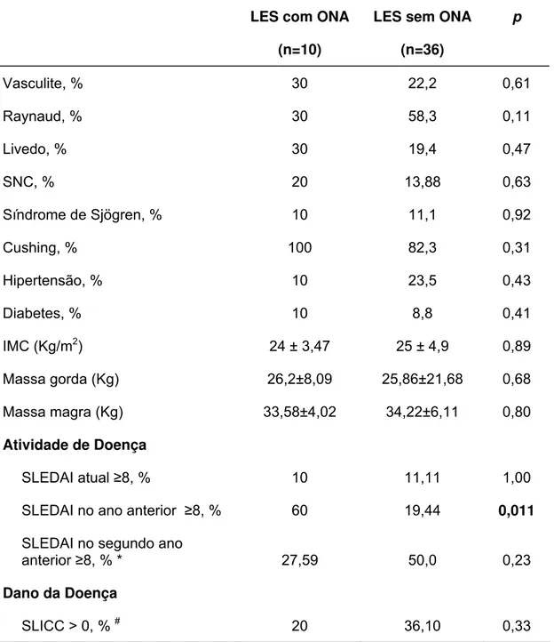
ONA foi encontrada em 10 de 46 pacientes, 80% nos primeiros 2 anos de diagnóstico de LES. Dessas, 8 pacientes já tinham diagnóstico de ONA (confirmado por RNM) anteriormente à entrada no estudo; e 2 manifestaram sintomas durante o período do estudo. Trinta e seis pacientes não apresentaram evidência clínica ou de imagem de ONA, e foram desta forma, denominadas de grupo controle.
Dados demográficos de pacientes com e sem ONA revelaram dados semelhantes com relação à média de idade (26,7 ± 11,8 vs. 32,11 ± 11,7 anos, p=0,08), média de duração de doença (17,6 ± 6,72 vs. 26,11 ± 18,67 meses, p=0,55) e à freqüência de caucasianos (50% vs. 44,4%, p= 0,75).
Resultados 15
Resultados 16
Tabela 1: Características clínicas de pacientes lúpicas com e sem osteonecrose (ONA)
LES com ONA
(n=10)
LES sem ONA
(n=36)
p
Vasculite, % 30 22,2 0,61
Raynaud, % 30 58,3 0,11
Livedo, % 30 19,4 0,47
SNC, % 20 13,88 0,63
Síndrome de Sjögren, % 10 11,1 0,92
Cushing, % 100 82,3 0,31
Hipertensão, % 10 23,5 0,43
Diabetes, % 10 8,8 0,41
IMC (Kg/m2) 24 ± 3,47 25 ± 4,9 0,89 Massa gorda (Kg) 26,2±8,09 25,86±21,68 0,68 Massa magra (Kg) 33,58±4,02 34,22±6,11 0,80
Atividade de Doença
SLEDAI atual ≥8, % 10 11,11 1,00 SLEDAI no ano anterior ≥8, % 60 19,44 0,011 SLEDAI no segundo ano
anterior ≥8, % * 27,59 50,0 0,23
Dano da Doença
SLICC > 0, % # 20 36,10 0,33
Valores estão expressos como médias ± DP ou percentuais; g: gramas.
*13°-24° meses anteriores ao diagnóstico de ONA ou à entrada ao estudo (grupo controle). Dez pacientes não foram incluídos nesta análise visto que apresentavam apenas 1 ano de duração de doença (2 no grupo com ONA e 8 no grupo controle).
#
Resultados 17
A extensa análise de tratamento do LES está ilustrada na tabela 2. Os 2 grupos foram similares no que diz respeito à dose cumulativa de GC no ano anterior, e nos 3 e 6 meses anteriores ao diagnóstico de ONA ou entrada no estudo. De forma semelhante, não foram encontradas diferenças com relação à dose cumulativa total (oral ou pulso), dose máxima diária oral ou de pulso e à dose média diária. Por outro lado, a dose cumulativa de GC no ano anterior ao diagnóstico de ONA foi significativamente maior quando comparado com a dose cumulativa no ano anterior à entrada no estudo (10.503 ± 5.100 vs. 7.050 ± 5.699 mg, p=0,045). No entanto, a análise da dose cumulativa do 13o ao 24o mês (anteriores ao diagnóstico de ONA ou à entrada no estudo) não revelou diferença estatística (4.470 ± 4.428 vs. 5.017 ± 5.005 mg, p= 0,75).
Resultados 18
Tabela 2: Tratamento nas pacientes lúpicas com e sem osteonecrose (ONA)
LES com ONA
(n=10)
LES sem ONA
(n=36)
p
Glicocorticosteróide
Mês anterior (mg) 618 ± 406 790 ± 1.164 0,32 3 meses anteriores (mg) 2.103 ± 1.179 1.816 ± 1.773 0,31 6 meses anteriores (mg) 4.713 ± 2.428 3.661 ± 3.102 0,16 Ano anterior (mg) 10.503 ± 5.100 7.050 ± 5.699 0,045 13o-24o mês anterior (mg) 4.470 ± 4.428 5.017 ± 5.005 0,75 Dose cumulativa total (mg) 17.823 ± 8.386 20.924 ±10.772 0,40 Dose cumulativa oral (mg) 15.823 ± 7.493 18.008 ± 9.825 0,52 Dose cumulativa em pulsos (mg) 2.000 ± 1.882 2.917 ± 3.123 0,55 Dose media oral (mg/dia) 26 ± 9,5 23,76 ± 11,51 0,54 Dose máxima oral (mg/dia) 59 ± 23 51 ± 18 0,65 Dose máxima em pulso (mg/dia) 688 ± 622 712 ± 619 0,90 Dose no diagnóstico de ONA ou
na entrada do estudo (mg/dia)
20 ± 14 19 ± 21 0,42
Imunossupressor, % 70 66,6 1,00
Cloroquina - uso atual, % 80 86 0,64
Estatina - uso atual, % 0 16,7 0,13
Contraceptivo oral - uso atual, % 0 5,55 1,00
Resultados 19
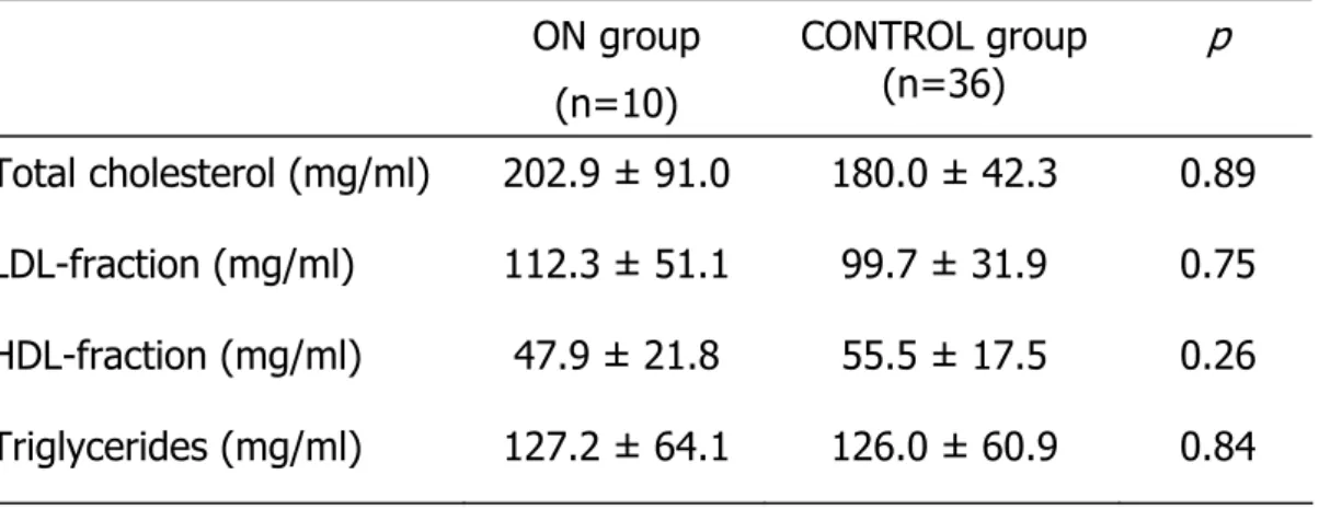
A tabela 3 ilustra que o perfil de lipoproteínas não diferiu entre os grupos com e sem ONA. A freqüência de auto-anticorpos (DNA, anti-Ro/SSA, anti-La/SSB, anti-Sm, anti-RNP e anti-P) também foi semelhante (Tabela 3).
Tabela 3: Dados laboratoriais (no momento do diagnóstico de ONA ou na entrada do estudo) para as lipoproteínas e perfil de auto-anticorpos nas pacientes lúpicas com e sem ONA
LES com ONA
(n=10)
LES sem ONA
(n=36)
p
Colesterol total (mg/ml) 202,9 ± 91 180 ± 42,3 0,89 LDL-colesterol (mg/ml) 112,3 ± 51,1 99,72 ± 31,9 0,75 HDL-colesterol (mg/ml) 47,9 ± 21,8 55,5 ± 17,5 0,26 Triglicerídeos (mg/ml) 127,2 ± 64,1 126 ± 60,9 0,84
Anti-DNA, % 30 42 0,50
Anti-Ro, % 60 42 0,30
Anti-P, % 10 28 0,24
Anti-La, % 30 17 0,35
Anti-Sm, % 20 33 0,42
Anti-RNP, % 40 53 0,47
Resultados 20
A avaliação de trombofilia revelou que a freqüência de positividade de ACL e AL e a elevação do fator VIII e homocisteína foram semelhantes nos grupos com e sem ONA (Tabela 4). Nenhum dos pacientes apresentava mutações dos fatores V de Leiden ou do gene da protrombina. Além disso, nenhuma diferença foi encontrada com relação à freqüência de deficiências de proteína C, S e anti-trombina III (Tabela 4).
Tabela 4: Dados laboratoriais (na entrada do estudo) para trombofilia e hipofibrinólise em pacientes lúpicas com e sem osteonecrose (ONA)
LES com ONA
(n=10)
LES sem ONA
(n=36)
p
AL, % 0 8,33 0,59
ACL IgG, % 10 2,77 0,39
ACL IgM, % 10 2,77 0,39
Fator VIII elevado, % 60 66 0,69 Homocisteína elevada, % 40 41,6 0,92 Lp (a) > 30mg/dl, % 50 44,11 0,74 AT III deficiente, % 10 8,33 1,00 PC deficiente, % 20 5,55 0,20 PS deficiente, % 20 2,8 0,11
Resultados 21
A avaliação densitométrica demonstrou que pacientes com e sem ONA apresentavam massa óssea semelhante em quadril, antebraço, coluna e corpo total. Apenas 4 pacientes apresentaram fraturas radiográficas, todas elas no grupo sem ONA (Tabela 5).
Tabela 5: Densidade mineral óssea (DMO); fraturas clínicas e radiográficas em pacientes lúpicas com e sem osteonecrose (ONA)
LES com ONA
(n=10)
LES sem ONA
(n=36)
p
DMO L1-L4, g/cm2 0,930 ± 0,122 0,968 ± 0,143 0,44 DMO colo femural, g/cm2 0,838 ± 0,106 0,801 ± 0,139 0,45 DMO fêmur total, g/cm2 0,834 ± 0,160 0,859 ± 0,141 0,65 DMO corpo total, g/cm2 0,976 ± 0,151 0,982 ± 0,087 0,87 DMO rádio proximal, g/cm2 0,677 ± 0,069 0,654 ± 0,064 0,45 DMO 1/3 distal rádio, g/cm2 0,395 ± 0,07 0,426 ± 0,07 0,25
Fraturas clínicas, % 0 0 NA
Fraturas radiográficas, % 0 11,43 1,00
Valores estão expressos como médias ± DP ou percentagens; NA: não é aplicável.
Discussão 23
Este é o primeiro estudo que avaliou simultaneamente vários possíveis fatores de risco para ONA num grupo homogêneo de pacientes com LES, especialmente no que diz respeito à duração da doença e ao sexo.
A ONA parece ocorrer precocemente no LES (19,23,31) e a escolha de inclusão de pacientes com menos de 5 anos de doença pode ter minimizado o efeito de variáveis de confusão envolvidas com o longo tempo de duração de doença. Além disso, uma vez que a predileção de ONA pelo sexo é controversa (31,41,42), o desenho de estudo incluiu somente pacientes do sexo feminino, o que excluiu essa possível influência do gênero.
Adicionalmente, foram excluídas todas as condições clínicas independentemente associadas com a ONA, tais como, gravidez, doenças infiltrativas da medula óssea, trauma recente em quadril, displasia congênita do quadril, gota, síndrome da imunodeficiência adquirida, hemoglobinopatias, história de abuso de álcool, radioterapia próxima à região de quadril, síndrome antifosfolípide e outras coagulopatias.
O diagnóstico de ONA foi definido por meio de RNM. ONA silenciosa pode ocorrer em 12%-35% de pacientes lúpicos (14,17,18,23,33) e um método de
Discussão 24
apresentando excelente correlação com a histologia (34-36). A grande variabilidade de resultados em estudos anteriores (no que diz respeito aos fatores de risco para ONA) poderia ser explicada pela utilização de outros procedimentos diagnósticos menos sensíveis tais como, sintomas clínicos, cintilografia óssea e/ou radiografias convencionais (3-14,16,20-24, 26,27,30,32).
O prejuízo circulatório, que pode levar à isquemia óssea, tem sido postulado como o denominador comum para todos os casos de ONA não traumática (2). Alterações na hemostasia causam trombose e embolia e podem ter um papel neste processo. Entretanto, poucos estudos analisaram fatores de coagulação nos pacientes com LES e ONA (8,12,30).
Discussão 25
Desde que Dubois e Cozen (3) descreveram o LES em associação com a ONA, pesquisadores têm tido dificuldades em diferenciar os possíveis efeitos deletérios causados pelo uso de GC, das manifestações inerentes à doença. De fato, um grande número de estudos encontrou que o uso de GC é um fator de risco importante para o desenvolvimento de ONA em paciente com LES (3-23). Em acordo com estes estudos, detectamos que pacientes com ONA utilizaram maior dose cumulativa total de GC no ano anterior ao diagnóstico clínico de ONA. No entanto, não obtivemos os mesmos resultados quando comparamos a dose cumulativa total, considerando toda a duração do tratamento. De fato, estudos prévios demonstraram que a ONA é precocemente detectada após o início do tratamento com altas doses de GC, através da realização de RNM (19,45). As comparações entre: a dose máxima diária de GC (oral ou em pulso), dose média diária e a dose no momento do diagnóstico foram semelhantes nos dois grupos. Os dados relacionados com os vários parâmetros de utilização de GC são conflitantes na literatura atual. A explicação mais plausível para essas discrepâncias é a definição imprecisa do tempo do desfecho, o que ocorre como conseqüência da utilização de metodologia diversa para o diagnóstico de ONA.
Discussão 26
mineral óssea e freqüência de fraturas, entre os dois grupos de pacientes. Estudos em animais e humanos mostraram que possivelmente o aumento do conteúdo de gordura na medula óssea, paralelamente associado à elevação dos níveis de colesterol total induzido por GC, poderia ter um papel para o desenvolvimento de ONA (23). O fato de não termos encontrado associação entre as frações de lipídeos no momento do diagnóstico de ONA não exclui a possibilidade desse efeito causal precedendo a complicação.
Discussão 27
elevações na atividade de doença no ano anterior ao diagnóstico clínico de ONA podem ter sido atenuadas pelos outros anos em que a doença pode ter permanecido relativamente inativa.
Conclusão 29
Anexo 31
Anexo I
HOSPITAL DAS CLÍNICAS DA
FACULDADE DE MEDICINA DA UNIVERSIDADE DE SÃO PAULO
TERMO DE CONSENTIMENTO LIVRE E ESCLARECIDO (Instruções para preenchimento no verso)
I - DADOS DE IDENTIFICAÇÃO DO SUJEITO DA PESQUISA OU RESPONSÁVEL LEGAL
1. NOME DO PACIENTE... ... DOCUMENTO DE IDENTIDADE :...SEXO: M F
DATA NASCIMENTO.: .../.../...
ENDEREÇO:... Nº ... APTO:... BAIRRO:...CIDADE: ... CEP:...TELEFONE:DDD(...)... 2.RESPONSÁVEL LEGAL ...
NATUREZA(grau de parentesco, tutor, curador etc.) ... DOCUMENTO DE IDENTIDADE :...SEXO: M F DATA NASCIMENTO.: .../.../...
ENDEREÇO:... Nº ... APTO:... BAIRRO:...CIDADE: ... CEP:...TELEFONE:DDD(...)... __________________________________________________________________________________
II - DADOS SOBRE A PESQUISA CIENTÍFICA 1.TÍTULO DO PROTOCOLO DE PESQUISA
AVALIAÇÃO DE OSTEONECROSE EM PACIENTES LÚPICOS PESQUISADOR: Sonia Cristina de Magalhães Souza
CARGO/FUNÇÃO: Pós-graduanda
INSCRIÇÃO CONSELHO REGIONAL Nº 99.245 UNIDADE DO HCFMUSP: Reumatologia
2. AVALIAÇÃO DO RISCO DA PESQUISA:
SEM RISCO RISCO MÍNIMO X RISCO MÉDIO
RISCO BAIXO RISCO MAIOR
(probabilidade de que o indivíduo sofra algum dano como consequência imediata ou tardia do estudo)
Anexo 32
III - REGISTRO DAS EXPLICAÇÕES DO PESQUISADOR AO PACIENTE OU SEU REPRESENTANTE LEGAL SOBRE A PESQUISA
CONSIGNANDO: 1. justificativa e os objetivos da pesquisa:
Pacientes com lúpus e que tomam corticóide (meticorten), tem um risco grande de ter problema no osso do quadril (a osteonecrose, que é a morte do osso), e isto causa dor e com o passar do tempo, dificuldade para andar. Esse problema pode ser visto por um exame chamado: Ressonância Nuclear Magnética, antes mesmo que o paciente sinta dor ou qualquer outro problema. Assim, você está convidada para participar deste estudo que irá fazer Ressonância Magnética em pacientes com lúpus tomando corticóide para ver se você tem ou não esse problema nos ossos do quadril, e tentar descobrir porque isto aconteceu.
2. procedimentos que serão utilizados e propósitos, incluindo a identificação dos procedimentos que são experimentais:
1.Ressonância nuclear magnética nos dois quadris, para ver se você tem ou não osteonecrose. Neste exame a pessoa se deita numa mesa que se movimenta dentro de uma espécie de túnel, não há dor e o exame dura de 10 a 30 minutos;
2.Raio-X simples dos quadris para comparar com o exame de Ressonância Magnética; 3.Densitometria óssea em coluna e fêmur para ver se você tem osteoporose ou não, porque as pessoas que tem osteonecrose podem também ter osteoporose. Este exame não tem nenhum incômodo e dura de 10 a 30 minutos;
4.Exames de sangue que você realiza normalmente para saber se você tem atividade de doença e para saber se você tem problemas de coagulação (sangue grosso);
3. desconfortos e riscos esperados:
Para realização destes exames, você será internada na enfermaria de Reumatologia, no 8º andar do Instituto Central do Hospital das Clínicas, por no máximo 3 dias. Será feita a coleta dos exames de sangue, a densitometria óssea, o raio-X dos quadris, e o exame de Ressonância Nuclear Magnética. Você ficará internada para facilitar a realização da Ressonância que é muito difícil de ser marcada pelo ambulatório.
4. benefícios que poderão ser obtidos:
A ressonância nuclear magnética irá ver se você tem osteonecrose do osso do quadril, mesmo antes de você sentir dor, e isto vai ajudar o médico a tratar você mais rapidamente, antes que você comece a sentir dor ou a ter dificuldade para andar. A densitometria óssea poderá ver se você tem osteoporose, doença também muito freqüente nas pessoas que tem lúpus e usam meticorten e com isso também poder começar o tratamento. Os exames de sangue também poderão mostrar se você tem tendência para alteração da coagulação (trombose).
5. procedimentos alternativos que possam ser vantajosos para o indivíduo:
Anexo 33
IV - ESCLARECIMENTOS DADOS PELO PESQUISADOR SOBRE GARANTIAS DO SUJEITO DA PESQUISA CONSIGNANDO:
1. acesso, a qualquer tempo, às informações sobre procedimentos, riscos e benefícios relacionados à pesquisa, inclusive para dirimir eventuais dúvidas.
2. liberdade de retirar seu consentimento a qualquer momento e de deixar de participar do estudo, sem que isto traga prejuízo à continuidade da assistência.
3. salvaguarda da confidencialidade, sigilo e privacidade.
4.disponibilidade de assistência no HCFMUSP, por eventuais danos à saúde, decorrentes da pesquisa.
5. viabilidade de indenização por eventuais danos à saúde decorrentes da pesquisa.
O paciente, ou responsável, terá acesso para questões sobre seus direitos como indivíduo do estudo, entrando em contato com a Comissão de Ética do Hospital das Clínicas, no telefone 3069-6442. Para qualquer pergunta sobre o estudo, entrar em contacto com a médica do estudo Dra Sonia Cristina de Magalhães Souza no telefone 3066-7213. A participação do paciente neste estudo é voluntária, podendo escolher não fazer parte deste estudo. Caso o paciente concorde em participar deste estudo, isto será mantido em sigilo. Esta pesquisa não envolve dano à saúde do paciente.
V. INFORMAÇÕES DE NOMES, ENDEREÇOS E TELEFONES DOS RESPONSÁVEIS PELO ACOMPANHAMENTO DA PESQUISA, PARA CONTATO EM CASO DE INTERCORRÊNCIAS CLÍNICAS E REAÇÕES
ADVERSAS.
Dra. Sonia Cristina de Magalhães Souza-Faculdade de Medicina da USP-3º andar Reumatologia sala 3107
Fone: 3066-7213, 3066-7490
Anexo 34
VII - CONSENTIMENTO PÓS-ESCLARECIDO
Declaro que, após convenientemente esclarecido pelo pesquisador e ter entendido o que me foi explicado, consinto em participar do presente Protocolo de Pesquisa
São Paulo, de de 2004.
Referências 36
1. Mont MA, Carbone JJ. Fairbank AC. Core decompression versus nonoperative management for osteonecrosis of the hip. Clin Orthop Relat Res. 1996;324: 169-78.
2. Ficat RP. Idiopathic Bone Necrosis of the femoral head. J Bone Joint Surg. 1985;67B: 3-9.
3. Dubois EL, Cozen L. Avascular (aseptic) bone necrosis associated with systemic lupus erythematosus. JAMA. 1960;174: 966-71.
4. Zizic TM, Marcoux C, Hungerford DS, Dansereau JV, Stevens MB. Corticosteroid therapy associated with ischemic necrosis of bone in systemic lupus erythematosus. Am J Med. 1985;79: 596-604.
5. Weiner ES, Abeles M. Aseptic necrosis and glucocorticosteroids in systemic lupus erythematosus: a reevaluation. J Rheumatol. 1989;16: 604-8.
6. Massardo L, Jacobelli S, Leissner M, Gonzalez M, Villarroel L, Rivero S. High-dose intravenous methylprednisolone therapy associated with osteonecrosis in patients with systemic lupus erythematosus. Lupus. 1992;1: 401-5.
Referências 37
9. Mok CC, Lau CS, Wong RW. Risk factors for avascular bone necrosis in systemic lupus erythematosus. Br J Rheumatol. 1998;37: 895-900.
10. Zizic TM, Hungerford DS, Stevens MB. Ischemic bone necrosis in systemic lupus erythematosus. The early diagnosis of ischemic necrosis of bone. Medicine. 1980;59: 134-42.
11. Abeles M, Urman JD, Rothfield NF. Aseptic necrosis of bone in systemic lupus erythematosus. Relationship to corticosteroid therapy. Arch Inter Med. 1978;138: 750-4.
12. Sheik JS, Retzinger GS, Hess EV. Association of osteonecrosis in systemic lupus erythematosus with abnormalities of fibrinolysis. Lupus. 1998;7: 42-8.
13. Rascu A, Manger K, Kraetsch HG, Kalden JR, Manger B. Osteonecrosis in systemic lupus erythematosus, steroid-induced or a lupus-dependent manifestation? Lupus. 1996;5: 323-7.
14. Klippel JH, Gerberg LH, Pollak L, Decker JL. Avascular necrosis of bone in systemic lupus erythematosus: Silent symmetric osteonecrosis. Am J Med. 1979;67: 83-7.
15. Zizic TM. Avascular necrosis of bone. Curr Opin Rheumatol. 1990;2: 26-37. 16. Bergstein JM, Wiens C, Fish AJ, Vernier RL, Michael A. Avascular
necrosis of bone in systemic lupus erythematosus. J Pediatr. 1974;85: 31-5.
Referências 38
18. Aranow C, Zelicof S, Leslie D, Solomon S, Barland P, Norman A, et al. Clinically occult avascular necrosis of the hip in systemic lupus erythematosus. J Rheumatol. 1997;24: 2318-22.
19. Oinuma K, Harada Y, Nawata Y, Takabayashi K, Abe I, Kamikawa K, et al. Osteonecrosis in patients with systemic lupus erythematosus develops very early after starting high dose corticosteroid treatment. Ann Rheum Dis. 2001;60: 1145-8.
20. Gladman DD, Urowitz MB, Chaudry-Ahluwalia V, Hallet DC, Cook RJ. Predictive factors for symptomatic osteonecrosis in patients with systemic lupus erythematosus. J Rheumatol. 2001;28: 761-5.
21. Nagasawa K, Ishii YI, Mayumi T, Tada Y, Ueda A, Yamauchi Y, et al. Avascular necrosis of bone in systemic lupus erythematosus: possible role of haemostatic abnormalities. Ann Rheum Dis. 1989;48: 672-6.
22. Houssiau FA, Toukap NA, Depresseux G, Maldague BE, Malghem J, Devogelaer JP, et al. Magnetic resonance-imaging-detected avascular osteonecrosis in systemic lupus erythematosus: lack of correlation with antiphospholipid antibodies. Br J Rheumatol. 1998;37: 448-53.
23. Nagasawa K, Tada Y, Koarada S, Horiuchi T, Tsukamoto H, Murai K, et al. Very early development of steroid-associated osteonecrosis of femoral head in systemic lupus erythematosus: prospective study by MRI. Lupus. 2005;14: 385-90.
24. Nilsen KH. Systemic lupus erythematosus and avascular bone necrosis.
N Z Med J. 1977;8: 472-5.
25. Hungerford DS, Zizic TM. The treatment of ischemic necrosis of bone in systemic lupus erythematosus. Medicine. 1980;59: 143-8.
Referências 39
27. Asherson RA, Liote F, Page B, Meyer O, Buchanan N, Khamashta MA, et al. Avascular necrosis of bone and antiphospholipid antibodies in systemic lupus erythematosus. J Rheumatol. 1993;20: 284-8.
28. Seleznic MJ, Silveira LH, Espinoza LR. Avascular necrosis associated with anticardiolipin antibodies. J Rheumatol. 1991;18: 1416-7.
29. Tektonidou MG, Malagari K, Vlachoyiannopoulos PG, Kelekis DA, Moutsopoulos HM. Asymptomatic avascular necrosis in patients with primary antiphospholipid syndrome in the absence of corticosteroid use: a prospective study by resonance imaging. Arthritis Rheum. 2003;48: 732-6.
30. Jones LC, Mont MA, Le TB, Petri M, Hungerford DS, Wang P, Glueck CJ. Progoagulants and Osteonecrosis. J Rheumatol. 2003;30: 783-91. 31. Ibanez D, Gladman DD, Urowitz MB. Adjusted mean Systemic Lupus
Erythematosus Disease Activity Index-2K is a predictor of outcome in SLE. J Rheumatol. 2005;32: 824-7.
32. Mok MY, Farewell VT, Isenberg DA. Risk factors for avascular necrosis of bone in patients with systemic lupus erythematosus: is there a role for antiphospholipid antibodies? Ann Rheum Dis. 2000;59: 462-7.
33. Halland AM, Klemp P, Botes D, Van Heerden BB, Loxton A, Scher AT. Avascular necrosis of the hip in systemic lupus erythematosus: the role of magnetic resonance imaging. Br J Rheumatol. 1993;32: 972-6.
34. Kalunian KC, Hahn BH, Bassett L. Magnetic resonance imaging identifies early femoral head ischemic necrosis in patients receiving systemic glucocorticoid therapy. J Rheumatol. 1989;16: 959-63.
35. Mitchell DG, Rao VM, Dalinka MK, Spritzer CE, Alavi A, Steinberg ME, et al. Femoral head avascular necrosis: correlation of MR imaging,
Referências 40
36. Markisz JA, Knowles RJ, Altchek DW, Schneider R, Whalen J, Cahill PT. Segmenal patterns of avascular necrosis of the femoral heads: early detection with MR imaging. Radiol. 1987;162: 717-20.
37. Hochberg MC. Updating the American College of Rheumatology revised criteria for the classification of systemic lupus erythematosus [letter].
Arthritis Rheum. 1997;40: 1725.
38. Bombardier C, Gladman DD, Urowitz MB, Caron D, Chang CH, and the Committee on Prognosis Studies in SLE. Derivation of the SLEDAI: a disease activity index for lupus patients. Arthritis Rheum. 1992;35: 630-40. 39. Gladman D, Ginzler E, Goldsmith C, Fortin P, Liang M, Urowitz M, et al.
The development and initial validation of the Systemic Lupus International Collaborating Clinics/American College of Rheumatology damage index for systemic lupus erythematosus. Arthritis Rheum. 1996;39: 363-9.
40. Genant HK. Radiographic assessment of the effects of intermittent cyclical treatment with etidronate. In: Christiasen C, Overgaard K (eds). Osteoporosis. Copenhagen: Osteopress ApS; 1990. p.2047-54.
41. Talamo G, Angtuaco E, Walker RC, Dong L, Miceli MH, Zangari M, et al. Avascular necrosis of femoral and/or humeral heads in multiple myeloma: results of a prospective study of patients treated with dexamethasone-based regimes and high-dose chemotherapy. J Clin Oncol. 2005;23(22): 5217-23.
42. Schulte CM, Beelen DW. Avascular osteonecrosis after allogenic hematopoietic stem-cell transplantation: diagnosis and gender matter.
Transplantation. 2004;78(7): 1055-63.
Referências 41
44. Bjorkman A, Svensson PJ, Hillarp A, Burtscher IM, Runow A, Benoni G. Factor V Leiden and prothrombin gene mutation: risk factors for osteonecrosis of femoral head in adults. Clin Orthop Relat Res. 2004;425: 168-72.
45. Sakamoto M, Shimizu K, Lida S, Akita T, Moriya H, Nawata Y. Osteonecrosis of the femoral head: a prospective study with MRI. J Bone Joint Surg Br. 1997;79: 213-9.
46. Weinstein RS, Jilka RL, Parfitt AM, Manolagas SC. Inhibition of Osteoblastogenesis and Promotion of Apoptosis of Osteoblasts and Osteocytes by Glucocorticoids: Potential Mechanisms of Their Deleterious Effects on Bone. J Clin Invest. 1998;102(2): 274-282.
Apêndice
ARTIGO ENVIADO PARA PUBLICAÇÃO
Disease Activity as a Major Risk Factor for Osteonecrosis in Early Systemic Lupus Erythematosus
Sonia CMS Fialho, MD1, Eloisa Bonfá, MD, PhD1, Luis F Vitule, MD2,
Elbio D’Amico, MD, PhD3, Valeria Caparbo1, Sandra Gualandro, MD, PhD3,
Rosa MR Pereira, MD, PhD1
Rheumatology1 and Hematology3 Division and Radiology Department2,
School of Medicine, University of São Paulo, São Paulo, Brazil
Grant support: CNPq # 304756/2003-2 (to EB)
Address correspondence and reprint request to Rosa M.R. Pereira, MD, PhD,
Faculdade de Medicina USP, Reumatologia, Avenida Dr Arnaldo, 455 – 3°
andar- Reumatologia, sala 3107, São Paulo SP, 01246-000 Brazil.
E-mail: rosamariarp@yahoo.com, reumato@usp.br
Apêndice
SUMMARY
Objective. Identify possible risk factors for osteonecrosis in a homogenous
group of early SLE.
Methods. 46 consecutive SLE patients (<5 years duration) followed at the
Lupus Clinic, were enrolled between 2004-2005. An extensive clinical and
laboratory evaluation using a standard electronic protocol established since
1999, including osteonecrosis symptoms and appropriate magnetic
resonance imaging (MRI), were carried out at 1-6 months intervals. All
other asymptomatic for osteonecrosis patients at study entry underwent
MRI.
Results. Osteonecrosis confirmed by MRI was found in 10 of 46 patients
(22%). Age, disease duration, clinical vascular features, the frequency of
thrombophilia and hypofibrinolysis factors and the lipoprotein profile were
comparable in patients with and without osteonecrosis (p>0.05).
Remarkably, the frequency of patients with SLEDAI ≥8 in the previous year
of osteonecrosis clinical diagnosis was significantly higher when compared
to patients without this manifestation (60.0% vs. 19.4%, p=0.011),
supported by the higher glucocorticoid cumulative dose in the same period
(p=0.045). In contrast, these two parameters evaluated in 13th-24th months
preceding osteonecrosis diagnosis was similar in patients with and without
osteonecrosis (p>0.05). In the multivariate analysis only SLEDAI remained
as an independent risk factor for osteonecrosis (OR=6.6, CI=1.07-41.29, p=
0.042).
Conclusion. Disease activity in the previous year of osteonecrosis clinical
diagnosis is the main predictor factor for the development of this
complication in early SLE.
Keywords: systemic lupus erythematosus; osteonecrosis; SLEDAI; disease
Apêndice
INTRODUCTION
Osteonecrosis is a clinical entity of unclear pathogenesis characterized
by death of bone marrow and trabecular bone. It often results in the
collapse of the architectural bone structure, leading to joint pain, bone
destruction, and loss of function (1). Circulatory impairment to the affected
bone has been postulated to be the common denominator for all cases of
osteonecrosis (2).
Among the rheumatic diseases, osteonecrosis is strongly associated
with systemic lupus erythematosus (SLE). Most investigators have found
glucocorticoid (GC) use to be a major risk factor for osteonecrosis in SLE
(3-23), however this complication has also been reported in lupus patients who
have never received therapy with this drug (3,9,13,15). Moreover, a
significant lower frequency of osteonecrosis has been reported in other
clinical conditions requiring chronic corticosteroid therapy (15).
Alternatively, disease associated factors have been proposed to
enhance the risk for osteonecrosis development, such as: disease activity
(5,24), vasculitis (8,10,16,25), Raynaud’s phenomenon (4,10,18,25),
thrombophlebitis (8), antiphospholipid antibodies or syndrome (7-9,26-29),
thrombophilia and hypofibrinolysis (8,12,21,30).
Among those, disease activity is perhaps the most difficult to elucidate
Apêndice
(5,24) and more importantly they have not evaluated flares at the time or
close to the osteonecrosis clinical onset (13,31).
In addition, the interpretation of all these risk factors has been
hampered by the lack of magnetic resonance imaging (MRI) in patients
and/or controls groups (3-16,20,21,24,26-28,30-32). In fact, clinical
symptoms, bone scintigraphy, and conventional radiographic images
(33-36) are not as sensitive to diagnose early osteonecrosis. Moreover, the
simultaneous analysis of a limited number of potential risk factors
associated with osteonecrosis (17-19,22,23) in previous reports, may have
hampered an overall view of the problem and consequently precludes a
definitive conclusion.
We therefore have evaluated simultaneously several risk factors for
osteonecrosis in early SLE patients in whom diagnosis was confirmed by
Apêndice
PATIENTS AND METHODS
Since 1999, 870 patients with SLE who fulfilled the American College
of Rheumatology 1997 revised criteria for SLE (37) had been registered and
followed at the Lupus Outpatient Clinic, University of São Paulo. The
standard electronic protocol was carried out at 1-6 months intervals and
consisted of an extensive clinical and laboratory evaluation, including those
relevant for this study such as, osteonecrosis symptoms and appropriate
magnetic resonance imaging (MRI), Systemic Lupus Erythematosus Disease
Activity Index (SLEDAI) (38) and glucocorticoid therapy. All information
collected on these patients is entered into a database.
Between July 2004 and July 2005, forty six consecutive patients
followed in this cohort were enrolled for the present study according to the
following criteria. The inclusion criteria were female gender, age > 18
years-old and less than 5 years of disease duration. The exclusion criteria were
previous history of endovenous contrast allergy or claustrophobia, acute
thrombosis, pregnancy, current nephrotic proteinuria or renal chronic
insufficiency, HIV positive or acquired immunodeficiency syndrome,
hemoglobinopathy, known coagulopathy (including the antiphospholipid
syndrome), recent trauma or radiotherapy next to the hip region, congenital
hip dysplasia, infiltrative bone marrow disease, previous or current history of
alcohol abuse, and oral anticoagulant use. This study received institutional
Apêndice
All 46 patients underwent MRI of both hips on a 1,5 T Medical
Systems (General Electric) with the following sequences: axial and coronal
T2-weighted fat suppressed, and T1-weighted, both with 4mm section
thickness, and 1mm intersection gap. Contrast-enhanced images were
obtained after administration of gadolinium-DTPA and images were acquired
with coronal T2-gradient, and sagital, coronal and axial T1 with fat
suppression. Patients with known osteonecrosis diagnosis confirmed by
previous MRI at study entry were not exposed to a new examination.
Lupus patients with clinical osteonecrosis confirmed by MRI were
subsequently compared to the control group (patients without clinical and
MRI evidence of osteonecrosis) regarding clinical and laboratorial
parameters as well as therapies formerly reported to be associated with
osteonecrosis. Disease activity, Raynaud’s phenomenon, vasculitis, livedo
reticularis, thrombophilia and fibrinolysis factors, lipoprotein profile (12 hour
fasting serum total cholesterol, high-density lipoprotein-HDL, low density
lipoprotein-LDL, triglycerides), drugs (corticosteroid, chloroquine and
cytotoxic) were therefore evaluated as possible associated factors. All
clinical, laboratorial and therapy parameters were evaluated at
osteonecrosis clinical onset or at study entry for the control group, except
when otherwise stated.
Disease activity was assessed by SLEDAI (38) and a score ≥ 8 was
Apêndice
using SLICC damage index (39) which was categorized as absent when =0
and present when > 0 (osteonecrosis was not included for the SLICC score).
Laboratory variables were determined according to the routine
examination and included: 12 hour fasting serum lipoproteins (total
cholesterol, high-density lipoprotein-HDL, low density lipoprotein-LDL,
triglycerides); autoantibodies [anti-nuclear antibodies, anti-dsDNA, anti-Sm
anticardiolipin antibodies (ACL) IgG and IgM and lupus anticoagulant (LA)].
The later was detected following the recommendations of the Subcommittee
on Lupus Anticoagulant/Antiphospholipid antibody of the Scientific and
Standardization Committee of the International Society of Thrombosis and
Haemostasis.
Laboratory thrombophilic factors were performed after enrollment for
all patients. Protein C (PC) and anti-thrombin III (ATIII) were measured by
chromogenic assay (Spectrolyse®); prothrombin time (Quick method),
factor VIII and activated partial thromboplastin time were measured by
coagulation methods; free protein S (PS) was measured by an enzyme
linked immunosorbent assay (Helena Laboratories®); factors V and II
mutations were assessed by the polymerase chain reaction; homocysteine
was measured by polarized fluorescence immunoassay (IMX® system);
vitamins B12 and folic acid were determined by chemiluminescence
(Beckman Coulter®). We also analyzed serum lipoprotein-a (Lp(a)), which
Apêndice
Statistical analysis. The distribution of each continuous variable was
examined graphically and statistically for normality. Numerical data are
summarized as the mean and standard deviation (SD). Variables not
normally distributed were compared using the Wilcoxon nonparametric test
for differences. Variables normally distributed were compared using
Student’s test. Categorical data among groups were compared by the
chi-square statistic. Some results were evaluated according to the established
normal values and were subsequently ranked as elevated or depressed.
Multivariate logistic regression was used to analyze association between
osteonecrosis (dependent variable) and variables found to be statistically
significant on the univariate analysis. Statistical significance was set at p<
Apêndice
RESULTS
Osteonecrosis was found in 10 out of 46 patients (22%), 80% in the
first two years of lupus diagnosis (ACR criteria). These patients comprised
the osteonecrosis group (ON group), eight of them had osteonecrosis
diagnosis confirmed by MRI before study enrollment and two had the
clinical manifestation confirmed by MRI during the study period. The other
36 patients without clinical symptoms/MRI osteonecrosis were denominated
the CONTROL group.
Demographic data of osteonecrosis patients and SLE controls revealed
a similar mean age (26.7 ± 11.8 vs. 32.11 ± 11.7 years, p=0.08), mean
disease duration (17.6 ± 6.72 vs. 26.11 ± 18.67 months, p=0.55) and
frequency of Caucasians (50 vs. 44.4%, p= 0.75).
A similar frequency of clinical features, such as, vasculitis, Raynaud’s
phenomenon, livedo reticularis was observed between the two groups as
illustrated in Table 1. Moreover, clinical signs that are associated with
steroid therapy, including a Cushing appearance and BMI were comparable
in both groups. In addition, none of the individuals had a history of alcohol
use and the use of tobacco (pack-years) was similar in patients with (4.5 ±
12.5) and without osteonecrosis (4.9 ± 10.2).
Regarding disease activity, the frequency of patients with SLEDAI ≥8
in the previous year of osteonecrosis clinical diagnosis was significantly
higher when compared to patients without osteonecrosis in the previous
Apêndice
patients with SLEDAI >8 at the osteonecrosis clinical diagnosis was alike the
control group at study entry (10 vs. 11%. p=1.00). Moreover, the frequency
of patients with SLEDAI >8 at the remote (13th-24th preceding months)
osteonecrosis clinical diagnosis was comparable to the control group in the
same period (28 vs. 50%, p=0.23). The frequency of SLICC damage index
greater than zero did not differ between groups as well (20 vs. 36%,
p=0.33).
The extensive analysis regarding GC revealed a significant higher
cumulative dose during the previous year in ON group compared CONTROL
group (10,503 + 5,100 vs. 7,050 + 5,699 mg, p=0.040). In contrast, the
prior 13th-24th months GC cumulative doses (4,470 ± 4,428 vs. 5,017±
5,005 mg, p=0.75), the total GC cumulative doses (17,823 + 8,386 vs.
20,924 + 10,772 mg, p=0.40) and the GC doses at osteonecrosis clinical
onset (ON group)/entry (CONTROL group) (20 + 14 vs. 19 + 21, p=0.42)
were alike in both groups (Table 2).
The frequency of cytotoxic drugs (ever used) and chloroquine use at
osteonecrosis onset (ON group)/entry (CONTROL group) was comparable
(p>0.05) (Table 2). Likewise, the frequency of oral contraceptive at
osteonecrosis onset (ON group)/entry (CONTROL group) (0 vs. 5.5%,
p=1.0) was similar in both groups (Table 2).
Table 3 illustrates that lipoprotein profile at ON clinical diagnosis (ON
group) and study entry (CONTROL group) did not distinguish patients with
Apêndice
± 42.3 mg/ml, p=0.89), LDL-fraction (112.3 ± 51.1 vs. 99.7 ± 31.9 mg/dl,
p=0.75), HDL-fraction (47.9 ± 21.8 vs. 55.5 ± 17.5 mg/dl, p=0.26),
triglycerides (127.2 ± 64.1 vs. 126.0 ± 60.9 mg/dl, p=0.84) were
comparable. The frequency of autoantibodies (anti-dsDNA, anti-Ro/SSA,
anti-La/SSB, anti-Sm, anti-RNP and anti-P) was also alike in both groups
(data not shown).
Likewise evaluation of thrombophilia at study entry for both groups
revealed that the frequency of lupus anticoagulant, anticardiolipin
antibodies, high factor VIII, high homocysteine and high LPL were
comparable in ON and CONTROL groups (Table 4). None of the SLE patients
presented factor V or II mutations. Also no differences in the frequency of
low protein C, low protein S and anti-thrombin III deficiency was observed
in both groups (Table 4).
Multivariate analysis was performed (including SLEDAI and cumulative
GC dose in the previous year of osteonecrosis clinical diagnosis or study
entry) and only SLEDAI remained as an independent risk factor for
Apêndice
DISCUSSION
Our study demonstrated that disease activity is a major risk factor for
osteonecrosis development in female SLE patients.
The advantage of this study is to evaluate simultaneously several
possible risk factors for osteonecrosis in homogenous SLE patients
regarding short disease duration. In fact, osteonecrosis seems to occur early
in SLE (19,23,31) and the inclusion criteria of less than five years of disease
duration might have minimized the effect of other confounding variables. In
addition, since osteonecrosis predilection for sex is still controversial
(31,40,41), the study design including only female patients, excluded this
possible gender influence.
Moreover, osteonecrosis diagnosis was confirmed and excluded herein
by MRI, which is the gold standard method for early evaluation of bone
marrow changes indicative of osteonecrosis (34-36) and has a good
correlation with histology (34-36). The great variability in the results of
previous studies regarding osteonecrosis risk factors could be explained by
the use of other less sensitive screening diagnostic procedures such as
clinical symptoms, bone scintigraphy and/or conventional radiographic
images (3-14,16,20-24,26,27,30,32).
Disruption of vascular supply has been proposed as the major
underlying triggering event of non-traumatic osteonecrosis (2). Altered
hemostasis leading to thrombosis and embolism is thought to play a role in
Apêndice
patients with osteonecrosis (8,12,30). Interestingly, this complication has
been reported in 20% of primary antiphospholipid syndrome (29), although
the association with antiphospholipid antibodies is still controversial
(8,22,32). We therefore excluded patients with established antiphospholipid
syndrome in order to avoid this known risk factor. Interestingly, the
particular association with the hallmark antibodies of this syndrome was not
confirmed in the present study. Similarly, there were no differences
concerning thrombophilia and hypofibrinolysis abnormalities between the
two groups. Moreover, hereditary procoagulant abnormalities, such as factor
V Leiden and prothrombin gene mutation, which are possible risk factors for
osteonecrosis were not observed in this study (42,43). However, the small
sample size may preclude a definitive conclusion since these are uncommon
abnormalities in the general population.
On the other hand, several investigators have suggested that steroid
use is the major risk factor for osteonecrosis in SLE patients since the first
report of Dubois and Cozen (3-23). Accordingly, we have observed a
significant higher cumulative dose of GC in the previous year of
osteonecrosis clinical diagnosis, but not with the total cumulative dose over
the years. Supporting this finding, previous studies have demonstrated that
osteonecrosis is detected by MRI very early after steroid therapy (19,44).
The lack of association of total cumulative dose over the years observed in
Apêndice
patients with osteonecrosis compared to patients without this complication,
although this difference did not reach statistical significance.
It is also important to emphasize that the incidence of osteonecrosis
is low when GC is the only risk factor, reinforcing the possible relevance of
other contributing factors in lupus. Indeed, Colwell et al did not find a direct
relationship between GC dosage and the development of femoral head
osteonecrosis in 1420 hip-years at 10-year follow up of asthma or
inflammatory arthritis patients (45).
In this regard, the most relevant aspect raised by the multifactorial
analysis performed herein is the role of disease activity as a significant
factor for the development of osteonecrosis. Supporting this finding, Mok et
al., observed that patients who require an initial high-dose steroid for
disease control are at risk of osteonecrosis (9). The lack of association with
SLEDAI reported by these authors is probably due to the fact that this
measurement was performed at a single point in time.
A relevant issue concerning SLEDAI measurement is raised by the
present work. The lack of association with remote SLEDAI reinforces the
importance of evaluating flares close to osteonecrosis onset. On the other
hand, our data also suggest that this complication is a process overtime
which does not occur solely at the moment clinical ON onset.
In this regard, no association of osteonecrosis and the adjusted mean
SLEDAI score of an average length of follow up of 6.9 years was reported
Apêndice
of higher disease activity scores in the previous year of osteonecrosis clinical
diagnosis by the other years with lower SLEDAIs.
This multifactorial study provides therefore evidence that disease
activity in the previous year of osteonecrosis clinical diagnosis is the main
predictor factor for the development of this complication in early SLE. The
consequent use of high dose of GC in the same period has certainly a
synergistic role to the event.
Apêndice
REFERENCES
1. Mont MA, Carbone JJ, Fairbank AC. Core decompression versus
nonoperative management for osteonecrosis of the hip. Clin Orthop
Relat Res 1996; 324: 169-178.
2. Ficat RP. Idiopathic bone necrosis of the femoral head. J Bone Joint
Surg 1985; 67B: 3-9.
3. Dubois EL, Cozen L. Avascular (aseptic) bone necrosis associated with
systemic lupus erythematosus. JAMA 1960; 174: 966-971.
4. Zizic TM, Marcoux C, Hungerford DS, Dansereau JV, Stevens MB.
Corticosteroid therapy associated with ischemic necrosis of bone in
systemic lupus erythematosus. Am J Med 1985; 79: 596-604.
5. Weiner ES, Abeles M. Aseptic necrosis and glucocorticosteroids in
systemic lupus erythematosus: a reevaluation. J Rheumatol 1989; 16: 604-608.
6. Massardo L, Jacobelli S, Leissner M, Gonzalez M, Villarroel L, Rivero S.
High-dose intravenous methylprednisolone therapy associated with
osteonecrosis in patients with systemic lupus erythematosus. Lupus
1992; 1: 401-405.
7. Migliaresi S, Picillo U, Ambrosone L, et al. Avascular osteonecrosis in
patients with SLE: relation to corticosteroid therapy and anticardiolipin
Apêndice
8. Mont MA, Glueck CJ, Pacheco IH, Wang P, Hungerford DS, Petri M.
Risk factors for osteonecrosis in systemic lupus erythematosus. J
Rheumatol 1997; 24: 654-662.
9. Mok CC, Lau CS, Wong RW. Risk factors for avascular bone necrosis
in systemic lupus erythematosus. Br J Rheumatol 1998; 37: 895-900.
10. Zizic TM, Hungerford DS, Stevens MB. Ischemic bone necrosis in
systemic lupus erythematosus. The early diagnosis of ischemic
necrosis of bone. Medicine 1980; 59: 134-142.
11. Abeles M, Urman JD, Rothfield NF. Aseptic necrosis of bone in
systemic lupus erythematosus. Relationship to corticosteroid therapy.
Arch Inter Med 1978; 138: 750-754.
12. Sheik JS, Retzinger GS, Hess EV. Association of osteonecrosis in
systemic lupus erythematosus with abnormalities of fibrinolysis. Lupus
1998; 7: 42-48.
13. Rascu A, Manger K, Kraetsch HG, Kalden JR, Manger B. Osteonecrosis
in systemic lupus erythematosus, steroid-induced or a
lupus-dependent manifestation? Lupus 1996; 5: 323-327.
14. Klippel JH, Gerberg LH, Pollak L, Decker JL. Avascular necrosis of
bone in systemic lupus erythematosus: Silent symmetric
osteonecrosis. Am J Med 1979; 67: 83-87.
Apêndice
16. Bergstein JM, Wiens C, Fish AJ, Vernier RL, Michael A. Avascular
necrosis of bone in systemic lupus erythematosus. J Pediatr 1974;
85: 31-35.
17. Nagasawa K, Tsukamoto H, Tada Y, et al. Imaging study on the mode
of development and changes in avascular necrosis of the femoral
head in systemic lupus erythematosus: long term observations. Br J
Rheumatol 1994; 33: 343-347.
18. Aranow C, Zelicof S, Leslie D, et al. Clinically occult avascular necrosis
of the hip in systemic lupus erythematosus. J Rheumatol 1997; 24: 2318-2322.
19. Oinuma K, Harada Y, Nawata Y, et al. Osteonecrosis in patients with
systemic lupus erythematosus develops very early after starting high
dose corticosteroid treatment. Ann Rheum Dis 2001; 60: 1145-1148. 20. Gladman DD, Urowitz MB, Chaudry-Ahluwalia V, Hallet DC, Cook RJ.
Predictive factors for symptomatic osteonecrosis in patients with
systemic lupus erythematosus. J Rheumatol 2001; 28: 761-765. 21. Nagasawa K, Ishii YI, Mayumi T, et al. Avascular necrosis of bone in
systemic lupus erythematosus: possible role of haemostatic
abnormalities. Ann Rheum Dis 1989; 48: 672-676.
22. Houssiau FA, Toukap NA, Depresseux G, et al. Magnetic
resonance-imaging-detected avascular osteonecrosis in systemic lupus
erythematosus: lack of correlation with antiphospholipid antibodies.
Apêndice
23. Nagasawa K, Tada Y, Koarada S, et al. Very early development of
steroid-associated osteonecrosis of femoral head in systemic lupus
erythematosus: prospective study by MRI. Lupus 2005; 14: 385-390.
24. Nilsen KH. Systemic lupus erythematosus and avascular bone
necrosis. N Z Med J 1977; 8: 472-475.
25. Hungerford DS, Zizic TM. The treatment of ischemic necrosis of bone
in systemic lupus erythematosus. Medicine 1980; 59: 143-148.
26. Abeles M, Weiner ES, Parke A, Wilson D. The association of
osteonecrosis in SLE with anticardiolipin antibodies. Arthritis Rheum
1991; 34(suppl): R39.
27. Asherson RA, Liote F, Page B, et al. Avascular necrosis of bone and
antiphospholipid antibodies in systemic lupus erythematosus. J
Rheumatol 1993; 20: 284-288.
28. Seleznic MJ, Silveira LH, Espinoza LR. Avascular necrosis associated
with anticardiolipin antibodies. J Rheumatol 1991;18: 1416-1417.
29. Tektonidou MG, Malagari K, Vlachoyiannopoulos PG, Kelekis DA,
Moutsopoulos HM. Asymptomatic avascular necrosis in patients with
primary antiphospholipid syndrome in the absence of corticosteroid
use: a prospective study by resonance imaging. Arthritis Rheum
2003; 48: 732-736.
30. Jones LC, Mont MA, Le TB, Petri M, Hungerford DS, Wang P, Glueck CJ.
Apêndice
31. Ibanez D, Gladman DD, Urowitz MB. Adjusted mean Systemic Lupus
Erythematosus Disease Activity Index-2K is a predictor of outcome in
SLE. J Rheumatol 2005; 32: 824-827.
32. Mok MY, Farewell VT, Isenberg DA. Risk factors for avascular necrosis
of bone in patients with systemic lupus erythematosus: is there a role
for antiphospholipid antibodies? Ann Rheum Dis 2000; 59: 462-467.
33. Halland AM, Klemp P, Botes D, Van Heerden BB, Loxton A, Scher AT.
Avascular necrosis of the hip in systemic lupus erythematosus: the role
of magnetic resonance imaging. Br J Rheumatol 1993; 32: 972-976.
34. Kalunian KC, Hahn BH, Bassett L. Magnetic resonance imaging
identifies early femoral head ischemic necrosis in patients receiving
systemic glucocorticoid therapy. J Rheumatol 1989; 16: 959-963.
35. Mitchell DG, Rao VM, Dalinka MK, et al. Femoral head avascular
necrosis: correlation of MR imaging, radiographic staging, radionuclide
imaging, and clinical findings. Radiol 1987; 162: 709-715.
36. Markisz JA, Knowles RJ, Altchek DW, Schneider R, Whalen J, Cahill
PT. Segmenal patterns of avascular necrosis of the femoral heads:
early detection with MR imaging. Radiol 1987; 162: 717-720.
37. Hochberg MC. Updating the American College of Rheumatology
revised criteria for the classification of systemic lupus erythematosus.
Apêndice
38. Bombardier C, Gladman DD, Urowitz MB, Caron D, Chang CH, and the
Committee on Prognosis Studies in SLE. Derivation of the SLEDAI: a
disease activity index for lupus patients. Arthritis Rheum 1992; 35: 630-640.
39. Gladman D, Ginzler E, Goldsmith C, et al. The development and initial
validation of the Systemic Lupus International Collaborating
Clinics/American College of Rheumatology damage index for systemic
lupus erythematosus. Arthritis Rheum 1996; 39: 363-369.
40. Talamo G, Angtuaco E, Walker RC, et al. Avascular necrosis of femoral
and/or humeral heads in multiple myeloma: results of a prospective
study of patients treated with dexamethasone-based regimes and
high-dose chemotherapy. J Clin Oncol 2005; 23: 5217-5223.
41. Schulte CM, Beelen DW. Avascular osteonecrosis after allogenic
hematopoietic stem-cell transplantation: diagnosis and gender matter.
Transplantation. 2004; 78: 1055-1063.
42. Bjorkman A, Burtscher IM, Svensson PJ, Hillarp A, Besjakov J, Benoni G.
Factor V Leiden and prothrombin 20210A gene mutation and
osteonecrosis of the knee. Arch Orthop Trauma Surg 2005; 125: 51-55.
43. Bjorkman A, Svensson PJ, Hillarp A, Burtscher IM, Runow A, Benoni
G. Factor V Leiden and prothrombin gene mutation: risk factors for
osteonecrosis of femoral head in adults. Clin Orthop Relat Res 2004;
Apêndice
44. Sakamoto M, Shimizu K, Lida S, Akita T, Moriya H, Nawata Y.
Osteonecrosis of the femoral head: a prospective study with MRI. J
bone Joint Surg Br 1997; 79: 213-219.
45. Colwell CW Jr, Robinson CA, Stevenson DD, Vint VC, Morris BA.
Osteonecrosis of the femoral head in patients with inflammatory
arthritis or asthma receiving corticosteroid therapy. Orthopedics 1996;
Apêndice
Table1: Clinical features in SLE patients at osteonecrosis clinical diagnosis
(ON group) and SLE patients without osteonecrosis at study entry
(CONTROL group).
ON group
(n=10)
CONTROL group (n=36)
p
Vasculitis, % 30 22 0.61
Raynaud, % 30 58 0.11
Livedo reticularis, % 30 19 0.47
Cushing, % 100 82 0.31
BMI (Kg/m2) 24.0 ± 3.5 25.0 ± 4.9 0.89
Disease activity ( SLEDAI > 8)
ON clinical diagnosis ,% 10 11 1.00
Previous year, % 60 19 0.01
Remote(13-24th mo. prior ON)*, % 28 50 0.23
Disease damage
SLICC index > 0, % # 20 36 0.33
Values are expressed as mean ± SD or percentages; g: grams; NA: not
applicable. *
Ten patients were not included in this analysis since they had only one year
of disease duration (2 in the ON group and 8 in the CONTROL group).
Apêndice
Table 2: Treatment in SLE patients at osteonecrosis clinical diagnosis (ON
group) and SLE patients without osteonecrosis at study entry (CONTROL
group).
ON group
(n=10)
CONTROL group (n=36)
p
Glucocorticoid treatment
Previous year (mg) 10,503 ± 5,100 7,050 ± 5,699 0.04
Previous 13th-24th mo. (mg) 4,470 ± 4,428 5,017± 5,005 O.75
Total cumulative (mg) 17,823 ± 8,386 20,924 ±10,772 0.40
ON clinical onset (mg/day) 20 ± 14 19 ± 21 0.42
I mmunosuppressive*, % 70 67 1.00
Chloroquine, % 80 86 0.64
Apêndice
Table 3: Lipoprotein profile in SLE patients at osteonecrosis clinical
diagnosis (ON group) and SLE patients without osteonecrosis at study entry
(CONTROL group).
ON group
(n=10)
CONTROL group (n=36)
p
Total cholesterol (mg/ml) 202.9 ± 91.0 180.0 ± 42.3 0.89
LDL-fraction (mg/ml) 112.3 ± 51.1 99.7 ± 31.9 0.75
HDL-fraction (mg/ml) 47.9 ± 21.8 55.5 ± 17.5 0.26
Triglycerides (mg/ml) 127.2 ± 64.1 126.0 ± 60.9 0.84
Apêndice
Table 4: Laboratory data at study entry for thrombophilia and
hypofibrinolysis in SLE patients with osteonecrosis (ON group) and SLE
patients without osteonecrosis (CONTROL group)
ON group
(n=10)
CONTROL group
(n=36)
p
LA, % 0 8 0.59
ACL IgG, % 10 3 0.39
ACL IgM, % 10 3 0.39
High Factor VIII, % 60 66 0.69
High Homocysteine, % 40 42 0.92
Lp (a) > 30mg/dl, % 50 44 0.74
Low AT III, % 10 8 1.00
Low PC, % 20 6 0.20
Low PS, % 20 3 0.11
Values are expressed as percentages. LA: lupus anticoagulant. ACL:
anticardiolipin antibodies, Lp(a): Lipoprotein a, AT: antithrombin, PC: