Clínica Universitária de Cardiologia
Prognostic relevance of exercise testing in
Hypertrophic Cardiomyopathy
Ana Sofia Carôla Cavaco
Clínica Universitária de Cardiologia
Prognostic relevance of exercise testing in
Hypertrophic Cardiomyopathy
Ana Sofia Carôla Cavaco
Orientado por:
Prof. Doutor Luís Miguel da Rocha Lopes
1
Abstract
Background: Resting echocardiography is fundamental in the diagnosis and
monitoring of Hypertrophic Cardiomyopathy (HCM), as well as in estimating its severity. Cardiopulmonary exercise testing (CPET) is indicated to complete the evaluation of HCM patients and stress echocardiography is often used to assess symptoms. However, current guidelines still do not incorporate any type of exercise testing for risk stratification in HCM, inclusively of sudden cardiac death.
Objective: The aim of this work is to review the evidence on the relevance of exercise
testing in determining the prognosis in HCM.
Methods: A systematic review was conducted for eligible publications, between 2010
and 2016, that included evaluation of outcomes and prognosis. In these studies, patients underwent exercise echocardiography and/or cardiopulmonary exercise testing, performed according to predefined protocols. Diverse parameters were assessed in order to determine which were relevant for the prognosis. Analysed outcomes included death from any cause (including SCD and other causes of cardiovascular death), equivalents of sudden death, heart failure requiring hospitalization or progression to New York Heart Association classes III or IV, cardiac transplantation, sustained ventricular tachycardia, stroke, myocardial infarction and need of therapy to relieve left ventricular outflow tract obstruction.
Results: 12 publications were included, corresponding to a total of 4655 patients. The
mean follow-up period varied between about 1 and 8 years. “Classical” risk factors for sudden death were generally present in less than half of the patients and these were not always considered predictors of long-term outcomes. The main findings of this set of studies revealed that the major predictors of outcomes were abnormal heart rate recovery, atrial fibrillation during exercise, exercise wall motion abnormalities, lower peak VO2 and higher VE/VCO2.
Conclusions: Although most studies concluded that exercise testing is a safe and useful
2 the question of whether it adds independent value to the current risk stratification strategies, is needed.
Keywords: Hypertrophic Cardiomyopathy; exercise echocardiography; cardiopulmonary exercise testing; prognosis; risk stratification.
3
Resumo
Introdução: A Miocardiopatia Hipertrófica (MCH) é uma doença miocárdica, na
grande maioria das vezes de causa genética, por mutações em genes que codificam proteínas sarcoméricas, com um padrão de hereditariedade autossómico dominante. É uma causa comum de morte súbita cardíaca (MSC), insuficiência cardíaca (IC) e arritmias, sobretudo fibrilhação auricular (FA). Embora os doentes possam permanecer assintomáticos ou com sintomas pouco significativos durante vários anos, muitas vezes também se apresentam com capacidade reduzida para o exercício, que pode dever-se a vários mecanismos, nomeadamente a obstrução do tracto de saída do ventrículo esquerdo (OTSVE), a isquémia do miocárdio e a disfunção sistólica e/ou diastólica do ventrículo esquerdo (VE). O diagnóstico é baseado em critérios estabelecidos, tais como os da Sociedade Europeia de Cardiologia (SEC), tendo em conta sobretudo a espessura da parede ventricular esquerda (≥15 milímetros num ou mais segmentos, medidos por qualquer técnica de imagem, que não seja explicado por aumento da pós-carga). A ecocardiografia em repouso é fundamental no diagnóstico e monitorização da MCH, bem como na avaliação da severidade da doença e no prognóstico. A prova de esforço cardiorrespiratória está, neste momento, indicada para completar a avaliação dos doentes com MCH e a ecocardiografia de esforço é frequentemente utilizada com o objectivo de identificar e avaliar os sintomas. Contudo, as guidelines actuais não incorporam nenhum tipo de prova de esforço na estratificação de risco na MCH, nomeadamente de MSC.
Objectivo: O objectivo deste trabalho é rever sistematicamente a evidência científica
publicada sobre a relevância dos testes de esforço (tanto ecocardiográficos como da prova de esforço cardiorrespiratória), na determinação do prognóstico na MCH.
Métodos: Foi realizada uma revisão sistemática com inclusão de publicações elegíveis,
cujos critérios de inclusão foram: estudos prospectivos ou retrospectivos, realizados em humanos, com idade superior a 18 anos, publicados nos últimos 5 anos (entre 2010 e 2016), em língua inglesa, no qual fossem satisfeitos os critérios de diagnóstico da MCH e em que fossem realizados testes de esforço, com electrocardiograma (ECG) e/ou ecocardiograma e/ou prova de esforço cardiorrespiratória e que incluíssem outcomes e prognóstico. Nestes estudos, os doentes foram submetidos a uma avaliação clínica inicial, tendo em conta antecedentes pessoais, sintomas cardiovasculares, história
4 familiar de MCH e/ou MSC em idade precoce, medicação habitual no momento do ensaio clínico, história de cirurgia cardíaca e/ou terapêutica médica para aliviar os sintomas de OTSVE, existência de uma janela acústica adequada para realizar ecocardiograma e a capacidade de realizar testes de esforço. A avaliação incluiu ainda, geralmente, a realização de ECG e ecocardiograma transtorácico em repouso (em modo M, bidimensional e estudo Doppler). Diversos parâmetros foram avaliados, de forma a determinar quais eram relevantes para o prognóstico: no caso do ecocardiograma, tanto em repouso como em esforço, a espessura máxima da parede ventricular esquerda, a fracção de ejecção do VE (FEVE), o gradiente de pressão máximo do tracto de saída do VE (TSVE), o movimento sistólico anterior da válvula mitral (SAM), a existência de regurgitação mitral, a sua gravidade e o seu aparecimento ou agravamento com o esforço, o diâmetro transversal da aurícula esquerda (AE), o volume indexado da AE e as alterações da contractilidade segmentar; no caso da prova de esforço cardiorrespiratória, o consumo de oxigénio no pico do esforço (VO2), a relação entre a
ventilação por minuto e a produção de dióxido de carbono (VE/VCO2), a relação de
trocas respiratórias no pico do esforço (rácio de trocas respiratórias), definido pelo rácio entre VCO2 e VO2 no pico do esforço e o limiar anaeróbio. Foram também avaliados, a
intervalos regulares durante os testes de esforço, a frequência cardíaca e a pressão arterial.
Os outcomes avaliados incluíram morte por qualquer causa (incluindo morte súbita, morte decorrente de IC progressiva ou outra), equivalentes de morte súbita (descarga apropriada de cardioversor desfibrilhador implantável – CDI – ou reanimação bem sucedida de paragem cardíaca), IC que requeresse internamento hospitalar ou progressão de classes I e II da New York Heart Association (NYHA) de IC para classes III e IV, transplante cardíaco, taquicardia ventricular mantida, acidente vascular cerebral (AVC), especialmente se no contexto de FA, enfarte do miocárdio (EM) e necessidade de terapêutica para aliviar a OTSVE.
Resultados: Foram incluídas 12 publicações, correspondendo a um total de 4655
doentes. A ecocardiografia de esforço foi utilizada na maioria dos artigos seleccionados, sendo que em alguns foi associada prova cardiorrespiratória, e em dois foi apenas realizada esta última. O tempo médio de follow-up variou entre 1 e 8 anos. Os factores de risco “clássicos” para morte súbita (síncope, história familiar de MSC, taquicardia ventricular não mantida, pressão arterial (PA) com resposta anormal ao exercício e
5 espessura da parede ventricular esquerda> 30mm) estavam presentes em menos de metade dos doentes e em dois estudos não foi comprovada associação a piores
outcomes(1,2). Os principais resultados deste conjunto de estudos revelaram que os
melhores preditores de outcomes foram a recuperação anormal da frequência cardíaca, FA durante o exercício, anomalias da contractilidade segmentar durante o esforço, decorrentes da ocorrência de isquémia miocárdica, o consumo máximo de oxigénio durante o exercício (VO2 no pico) (bem como a percentagem prevista de VO2 no pico)
reduzido e VE/VCO2 elevado, sendo os dois últimos parâmetros relacionados com a
intolerância ao esforço que, em dois dos artigos, só estavam associados a alguns dos
outcomes, nomeadamente IC e transplante cardíaco, e não a MSC, provavelmente
porque os mecanismos promotores de arritmia ventricular e disfunção da contractilidade são diferentes. O limiar anaeróbico reduzido não demonstrou ser tão bom preditor de
outcomes como os outros dois parâmetros anteriormente referidos. A FEVE reduzida,
que é um factor de risco de prognóstico cardiovascular global previamente identificado, é também relevante neste contexto. Relativamente a alguns dos parâmetros avaliados na ecocardiografia, tais como a OTSVE, os índices indirectos de disfunção diastólica (nomeadamente o diâmetro da AE) e a regurgitação mitral induzida pelo exercício, foram obtidos resultados contraditórios nos vários artigos, não permitindo concluir se estariam ou não associados a piores outcomes.
Conclusões: Apesar de a maioria dos estudos ter concluído que as provas de esforço
são uma ferramenta segura e útil na determinação do prognóstico da MCH, é necessária uma maior investigação relativamente ao valor adicional e eventual inclusão nas actuais estratégias de estratificação do risco.
Palavras-chave: Miocardiopatia Hipertrófica; ecocardiografia de esforço; prova de
esforço cardiorrespiratória; prognóstico; estratificação do risco.
O trabalho final exprime a opinião do autor e não da Faculdade de Medicina de Lisboa.
6
Contents
Abstract ... 1 Resumo ... 3 Introduction ... 7 Methods ... 9Search Strategy and Study Selection ... 9
Data extraction ... 9
Patients and baseline assessment ... 9
Resting and exercise echocardiography ... 10
Cardiopulmonary exercise testing ... 10
Follow-up and outcomes ... 11
Results ... 12
Study selection ... 12
Design of the study and baseline characteristics of the patients ... 13
Resting and exercise echocardiography ... 18
Cardiopulmonary exercise testing ... 21
Other clinical resting and exercise parameters ... 23
Discussion ... 27
Conclusion ... 30
Acknowledgments ... 31
7
Introduction
Hypertrophic Cardiomyopathy (HCM) is, in most cases, a genetic heart muscle disease, caused by mutations in genes coding for sarcomere proteins, with an autosomal dominant pattern of inheritance. Although not so frequent, there are other etiologies to consider, including other genetic (e.g. inherited metabolic and neuromuscular disorders) and non-genetic diseases(1). It is a common cause of sudden cardiac death (SCD), heart failure (HF) and arrhythmias (especially atrial fibrillation), even though patients may remain asymptomatic or minimally symptomatic for many years(2). However, it is common for patients with HCM to present with reduced exercise capacity(3), which can be explained by diverse mechanisms, including left ventricular outflow tract obstruction (LVOTO), myocardial ischemia and left ventricular systolic and diastolic dysfunction (1). This condition affects approximately 0.2% of the adult population worldwide(1). The diagnosis should be based on established criteria, such as those present in the latest guidelines published by the European Society of Cardiology (ESC)(3), namely: ≥15 mm in one or more LV myocardial segments, measured by any imaging technique, that is not explained solely by loading conditions, in the absence of any other condition that could have been responsible for the hypertrophy. A diagnosis is also considered in patients with maximal wall thickness 13-14 mm in the presence of a positive family history for HCM and/or ECG changes compatible with HCM. Other pathophysiologic features of the disease include myocardial fibrosis, mitral valve disease and coronary microcirculation dysfunction(1).
Resting echocardiography is fundamental both in the diagnosis and in the monitoring of HCM. It enables the measurement of many parameters that confirm the existence of the disease and also contribute to assess its severity and estimate prognosis, such as left ventricular wall thickness, systolic anterior motion of the mitral valve (that can cause LVOTO and mitral regurgitation), left ventricular outflow tract (LVOT) pressure gradient, diastolic and systolic function(1). Exercise echocardiography allows the detection and evaluation of symptoms.
Cardiopulmonary exercise testing (CPET) is currently indicated to complete the initial evaluation of patients with a diagnosis of HCM, when these report change in symptoms or when considering therapy to reduce LVOTO(3). It is useful to measure parameters
8 such as peak oxygen consumption (VO2) and the anaerobic threshold. When this is not
available, simple ergometry with electrocardiography (ECG) may be useful(1).
Some publications have reported on the use of ECG exercise testing, cardiopulmonary exercise testing and exercise echocardiography in predicting multiple aspects of the prognosis in HCM patients, although this indication is still not fully incorporated in the guidelines.
The aim of this work is to systematically review the current evidence concerning the relevance of electrocardiographic, echocardiographic and cardiopulmonary exercise testing in the prognosis of hypertrophic cardiomyopathy.
9
Methods
Search Strategy and Study Selection
In order to find suitable articles for this systematic review a search was made, using the Medline (PubMed) database, of the years 2010-2016 as well as searching for the publications included in the bibliography of the selected articles.
The keywords used were: "prognosis AND (exercise OR stress OR cardiopulmonary) AND hypertrophic cardiomyopathy".
The following inclusion criteria were applied: prospective or retrospective studies; performed in humans; participants older than 18 years; published in English; fulfilling the diagnostic criteria for HCM; making use of cardiac (ECG or echocardiography) or cardiopulmonary exercise tests; inclusion of outcomes and prognosis.
Data extraction
Patients and baseline assessment
Inclusion of patients required a previous diagnosis of HCM, based on specific criteria, from the ESC or other cardiology societies (such as American College of Cardiology Foundation/American Heart Association).
Baseline patient characteristics that were analyzed were: age(3–14); the presence of cardiovascular symptoms (syncope, angina, palpitations)(4–6,8,11–14) or disease (coronary disease, hypertension, arrhythmia, valve disease)(5,7–9,12,13), as well as other systemic diseases (genetic, metabolic) that could cause myocardial hypertrophy(3,5,7–10,12,13); family history of premature sudden death or HCM(3– 6,8,11–14); medication at the time of the study(3–7,10–14); history of cardiac surgery and/or medical therapy to relieve LVOTO(3,5,7,13,14); an adequate acoustic window(8,13) for echocardiographic studies and the ability and will to undergo exercise testing(8,13). Patients were always submitted to a written informed consent.
10 Important initial baseline evaluations included clinical assessment(4,5,7,8,10,12,14), New York Heart Association (NYHA) class of HF(3–8,10–14), 12-lead ECG at rest(4– 6,11,14) and 24h-ECG(4).
Resting and exercise echocardiography
In eleven studies (1,2,4–12), patients underwent resting transthoracic echocardiography, including M-Mode, bi-dimensional (2D) and, in some cases, Doppler evaluation, followed by symptom-limited exercise echocardiography in a treadmill.
Parameters of relevance were left ventricular (LV) maximal wall thickness (mm)(3– 6,8,12,14), LV ejection fraction (LVEF) (%)(3,4,7–14), LVOT gradient (mmHg)(3– 5,7,8,10–14), systolic anterior motion (SAM) of the mitral valve(7,8,12,13), mitral regurgitation (MR)(3,5,7,10–12,14), left atrial (LA) diameter (mm)(4,6,7,13,14) and LA indexed volume (mm/m²)(5,7,8), regional wall motion abnormalities (WMA)(11), among others.
Regarding exercise echocardiography, performed in all but two studies(1,13), generally medications were not withdrawn before the test. Heart rate (HR) and blood pressure (BP) were measured during the test and afterwards, as well as ECG monitoring. Some of the parameters measured in resting echocardiography, such as LVOT gradient, WMA and MR were measured during exercise, including at peak, and after the test.
Cardiopulmonary exercise testing
In those studies in which patients were submitted to cardiopulmonary exercise testing(5,7,9,10,12,14), the tests were generally performed in a treadmill, with regular HR and BP measurements and ECG monitoring, according to standard protocols (e.g. Bruce), with gas exchange measurement.
Parameters of interest were peak oxygen consumption (VO2) (mL/Kg/min); the
relationship between minute ventilation and carbon dioxide production (VE/VCO2
slope); peak respiratory exchange ratio (RER), defined as the ratio between VCO2 and
VO2 during peak exercise; and anaerobic threshold, calculated using the V-slope
11 Heart rate recovery (HRR), defined as the drop in heart rate (HR) from peak to 1 minute post-exercise(14) was measured in some of the studies, so as to ascertain the fraction of patients with abnormal HRR (<12 beat drop over 1 minute in recovery(14)). Abnormal BP response (usually defined as a failure of the systolic BP to increase >20 mmHg during exercise(15)) was also recorded in seven studies (1,2,5–7,10,11).
Follow-up and outcomes
Patients were followed up with a defined regularity, so as to determine the occurrence of any events. Death certificates were also analyzed in some of the studies(2,13,10,11). Primary outcomes of relevance were death from any cause(3–6,8,11–14), which could be included in one of three groups, namely: sudden death, death due to progressive CHF or “other”(3,4,6,8,11–13), sudden death equivalents (successful resuscitation from cardiac arrest and appropriate ICD discharge), heart failure (HF) requiring hospitalization(3,6,11–13) or progression from NYHA class I or II to class III or IV of HF and cardiac transplantation (10,11,14). Sudden death was defined as unexpected sudden collapse occurring <1h from the onset of symptoms in patients who had previously experienced a relatively stable course(15).
Secondary outcomes of relevance were sustained ventricular tachycardia(4,6,8,11); stroke in the context of atrial fibrillation (AF)(6,11,12); myocardial infarction(6,11) and clinical deterioration leading to need of therapy to relieve LVOTO (inclusively septal reduction)(10).
12
1364 unique
hits
• Inclusion criteria:
• Prospective or retrospective study • Humans
• Age > 18 years • English language
• Published in the last 5 years • HCM (satisfying diagnostic criteria) • Using ECG or echocardiography or
cardiopulmonary exercise tests • Inclusion of outcomes and prognosis
127 analyzed
by reading
the abstract
• 1237 excluded for not satisfying the age, language and date of publication
12 selected
for systematic
review
• 113 excluded for not satisfying the reimaining criteria
Results
Study selection
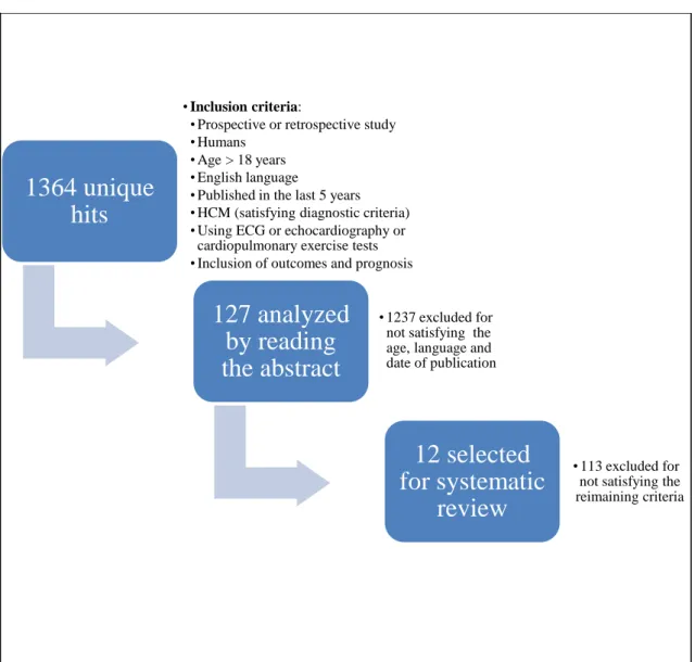
1364 unique publications were found and analyzed. 1237 of these publications were excluded for not satisfying the age, date of publication and language criteria. The remaining 127 were analyzed by reading the abstract, and 113 of them excluded for not satisfying the rest of the criteria. Therefore, 12 studies were selected for this systematic review(3–14) (Figure 1), corresponding to a total of 4655 patients.
13
Design of the study and baseline characteristics of the patients
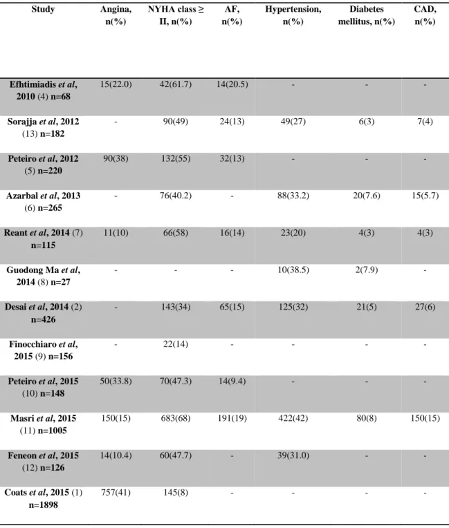
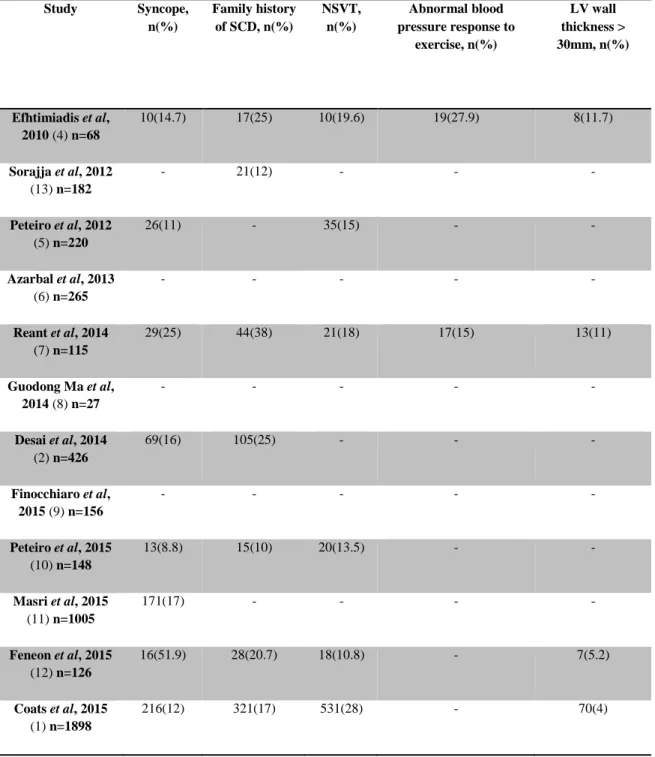
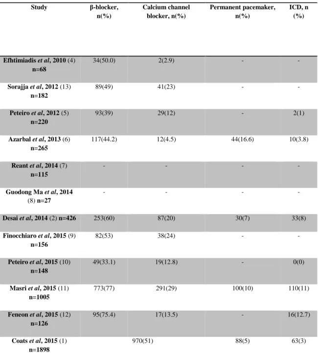
Most studies were single-center and observational. Only two studies included over 1000 patients(1,11). The mean follow-up was between 1.6±0.95 years and 8.7±3 years. Mean age of the patient populations varied between 44±14 and 54.3±12.4 years. A percentage between 57.7 and 78.1 were men, with a positive familial history of HCM present in at least 20% up to 51% of the study population. A percentage from 8% to 68% of the patients was in class NYHA≥II at baseline. Between 9.4% and 20.5% of patients had a diagnosis of AF and 20% to 42% had hypertension. Risk factors for sudden death, such as syncope, family history of SCD, non-sustained ventricular tachycardia (NSVT), abnormal BP response to exercise, and LV wall thickness>30mm varied, but were generally present in less than 30% of the study population in almost all studies. The percentage of patients under β-blockade was between 33.1% and 77%. A minority of patients had, at baseline, a permanent pacemaker or an ICD. Baseline characteristics of the patients included in the studies are summarized in Tables 1-4.
14
Table 1 – Demographic characteristics of the patients
Study Region Period Overall population, n Mean follow-up, years Mean age, years Men, n(%) Family history of HCM, n(%) Efhtimiadis et al, 2010 (4) n=68 Thessaloniki, Greece 2007-2009 68 2 44.8±14.6 45(67.1) 32(47) Sorajja et al, 2012 (13) n=182 Rochester (Minnesota), USA 1991-2008 182 4±3.2 53±15 119(65) 43(24) Peteiro et al, 2012 (5) n=220 A Coruña, Spain - 239 4.1±2.6 52±15 145(61) 76(32) Azarbal et al, 2013 (6) n=265 Stanford (California), USA 2006-2012 265 - 52±15 162(61.1) - Reant et al, 2014 (7) n=115 Bordeaux-Pessac, France 2009-2012 115 1.6±0.95 51.9±15.2 76(66) 59(51) Guodong Ma et al, 2014 (8) n=27 Beijing, China 2010-2011 27 - 54.3±12.4 16(57.7) - Desai et al, 2014 (2) n=426 Cleveland (Ohio), USA 1997-2007 426 8.7±3.0 44±14 310(73) 105(25) Finocchiaro et al, 2015 (9) n=156 Stanford (California), USA 2007-2012 156 2.25±0.92 51±14 96(62) - Peteiro et al, 2015 (10) n=148 A Coruña, Spain - 148 7.1±2.7 51±15 97(65.5) 44(29.7) Masri et al, 2015 (11) n=1005 Cleveland (Ohio), USA 1997-2012 1005 5.5±4 50±14 643(64) 201(20) Feneon et al, 2015 (12) n=126 Rennes and Tours, France 2009-2013 126 2.4±2.0 47.41±15.48 99(78.1) 42(34.1) Coats et al, 2015 (1) n=1898 London, United Kingdom 1998-2010 1898 5.6 46±15 1278(67) 778(42) HCM: hypertrophic cardiomyopathy
15
Table 2 - Symptoms and personal medical history disease of the patients Study Angina, n(%) NYHA class ≥ II, n(%) AF, n(%) Hypertension, n(%) Diabetes mellitus, n(%) CAD, n(%) Efhtimiadis et al, 2010 (4) n=68 15(22.0) 42(61.7) 14(20.5) - - - Sorajja et al, 2012 (13) n=182 - 90(49) 24(13) 49(27) 6(3) 7(4) Peteiro et al, 2012 (5) n=220 90(38) 132(55) 32(13) - - - Azarbal et al, 2013 (6) n=265 - 76(40.2) - 88(33.2) 20(7.6) 15(5.7) Reant et al, 2014 (7) n=115 11(10) 66(58) 16(14) 23(20) 4(3) 4(3) Guodong Ma et al, 2014 (8) n=27 - - - 10(38.5) 2(7.9) - Desai et al, 2014 (2) n=426 - 143(34) 65(15) 125(32) 21(5) 27(6) Finocchiaro et al, 2015 (9) n=156 - 22(14) - - - - Peteiro et al, 2015 (10) n=148 50(33.8) 70(47.3) 14(9.4) - - - Masri et al, 2015 (11) n=1005 150(15) 683(68) 191(19) 422(42) 80(8) 150(15) Feneon et al, 2015 (12) n=126 14(10.4) 60(47.7) - 39(31.0) - - Coats et al, 2015 (1) n=1898 757(41) 145(8) - - - -
16
Table 3 – “Classical” risk factors for sudden cardiac death Study Syncope, n(%) Family history of SCD, n(%) NSVT, n(%) Abnormal blood pressure response to exercise, n(%) LV wall thickness > 30mm, n(%) Efhtimiadis et al, 2010 (4) n=68 10(14.7) 17(25) 10(19.6) 19(27.9) 8(11.7) Sorajja et al, 2012 (13) n=182 - 21(12) - - - Peteiro et al, 2012 (5) n=220 26(11) - 35(15) - - Azarbal et al, 2013 (6) n=265 - - - - - Reant et al, 2014 (7) n=115 29(25) 44(38) 21(18) 17(15) 13(11) Guodong Ma et al, 2014 (8) n=27 - - - - - Desai et al, 2014 (2) n=426 69(16) 105(25) - - - Finocchiaro et al, 2015 (9) n=156 - - - - - Peteiro et al, 2015 (10) n=148 13(8.8) 15(10) 20(13.5) - - Masri et al, 2015 (11) n=1005 171(17) - - - - Feneon et al, 2015 (12) n=126 16(51.9) 28(20.7) 18(10.8) - 7(5.2) Coats et al, 2015 (1) n=1898 216(12) 321(17) 531(28) - 70(4)
17
Table 4 – Left ventricular outflow tract obstruction medications and implanted devices Study β-blocker, n(%) Calcium channel blocker, n(%) Permanent pacemaker, n(%) ICD, n (%) Efhtimiadis et al, 2010 (4) n=68 34(50.0) 2(2.9) - - Sorajja et al, 2012 (13) n=182 89(49) 41(23) - - Peteiro et al, 2012 (5) n=220 93(39) 29(12) - 2(1) Azarbal et al, 2013 (6) n=265 117(44.2) 12(4.5) 44(16.6) 10(3.8) Reant et al, 2014 (7) n=115 - - - - Guodong Ma et al, 2014 (8) n=27 - - - - Desai et al, 2014 (2) n=426 253(60) 87(20) 30(7) 33(8) Finocchiaro et al, 2015 (9) n=156 82(53) 38(24) - - Peteiro et al, 2015 (10) n=148 49(33.1) 19(12.8) - 0(0) Masri et al, 2015 (11) n=1005 773(77) 291(29) 100(10) 110(11) Feneon et al, 2015 (12) n=126 95(75.4) 17(13.5) - 16(12.7) Coats et al, 2015 (1) n=1898 970(51) 88(5) 63(3)
18
Resting and exercise echocardiography
Resting and exercise echocardiography main measurements are summarized in table 5. Exercise echocardiography was the only exercise test performed in five studies(2,10,5,7,12), and it was executed along with CPET in five other studies(11,4,6,8,9).
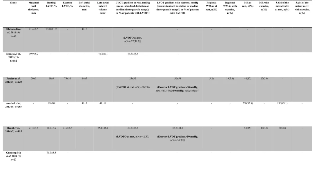
For resting echocardiography, measurements revealed that patients generally had a significant LV hypertrophy, with a maximal wall thickness between 17±5 and 21.4±6.5. LVEF was generally preserved (>50%), according to the current reference values(16), and in general was minimally or not changed with exercise. Mean LVOT pressure gradient at rest was >30mmHg in three of the studies(13,11,7), which is diagnostic of LVOTO(17), and during exercise it was >50mmHg in three studies(2,11,5). When evaluated both at rest and during exercise it worsened by, at least, 5mmHg. Regional WMAs appeared de novo or worsened with exercise in the two studies evaluating this parameter, in around four times more patients, when compared with the same parameter evaluated on resting echocardiography(10,5). Not all studies that evaluated MR at rest repeated this evaluation in exercise echocardiography and one study(12) only reported it with exercise. MR at rest was present in between 10% and 95.4% of patients in all studies evaluating this parameter and between 24% and 93% of patients had MR during exercise, since it appeared de novo in some cases. In others, MR progressed to higher degrees of severity. Between 25% and 49.1% of patients had SAM, defined as any contact of the leaflet with the septum during systole(6), at rest (in the studies where it was evaluated). In the studies that also evaluated this parameter during exercise it was shown that it generally appeared in patients who did not have it at rest and worsened in the ones who had(2,11).
19
Table 5 - Resting and exercise echocardiographic findings
Study Maximal wall thickness, mm Resting LVEF, % Exercise LVEF, % Left atrial diameter, mm Left atrial indexed volume, ml/m²
LVOT gradient at rest, mmHg (mean±standard deviation or median (interquartile range)) or % of patients with LVOTO
LVOT gradient with exercise, mmHg (mean±standard deviation or median (interquartile range)) or % of patients
with LVOTO Regional WMAs at rest, n(%) Regional WMAs with exercise, n(%) MR at rest, n(%) MR with exercise, n(%) SAM of the mitral valve at rest, n(%) SAM of the mitral valve with exercise, n(%) Efhtimiadis et al, 2010 (4) n=68 21.4±6.5 75.0±11.2 - 42±8 - - (LVOTO at rest, n(%)=27(39.7)) - - - - Sorajja et al, 2012 (13) n=182 19.9±5.2 - - - 46.6±8.1 46.3±38.5 - - - - Peteiro et al, 2012 (5) n=220 20±5 69±9 73±10 44±7 - 25±32 (LVOTO at rest, n(%)=60(25)) 50±54
(Exercise LVOT gradient>30mmHg, n(%)=103(43);>50mmHg, n(%)=83(35)) 5(2) 19(7.9) 40(17) 67(28) - - Azarbal et al, 2013 (6) n=265 - 69±10 - 41±7 41±18 - - - - 238(92.9) - 130(49.1) - Reant et al, 2014 (7) n=115 21.3±4.8 71.0±6.9 71.2±6.8 - 35.1±18.1 30.7±33.5 (LVOTO at rest, n(%)=42(37) 43.5±44.5
(Exercise LVOT gradient>50mmHg, n(%)=34(30)) - - 51(45) 49(43) 30(26) - Guodong Ma et al, 2014 (8) n=27 - 71.3±8.8 - - - -
20 Desai et al, 2014 (2) n=426 20±5 61±5 - 42±8 - 28±32 62±47 - - 381(89) 381(89) 105(25) 233(55) Finocchiaro et al, 2015 (9) n=156 17±5 67±11 - - 44±19 - (LVOTO at rest, n(%)=40(27)) -
(Exercise LVOT gradient>50mmHg, n(%)=54(35)) - - 15(10) - - - Peteiro et al, 2015 (10) n=148 20±5 71±9 73±10 44±6 - 10(9-25) (LVOTO at rest, n(%)=35(24)) 26(10-100)
(Exercise LVOT gradient>30mmHg, n(%)=66(45)) 3(2) 13(9) 23(15.5) 36(24) - - Masri et al, 2015 (11) n=1005 21±5 62±6 - 44±24 - 41±39 92±51 - - 958(95.4) 934(93) 391(39) 763(76) Feneon et al, 2015 (12) n=126 - 66±8 72±15 52±8 25[14] 7[8] (LVOT gradient at rest>50mmHg, n(%)=11(8.7)) 12[12]
(Exercise LVOT gradient>50mmHg, n(%)=16(12.7)) - - - 22(20.9) - - Coats et al, 2015 (1) n=1898 19±5 65±11 - 44±8 - - - 221(12) - - -
LVEF: left ventricular ejection fraction; LVOT: left ventricular outflow-tract; LVOTO: LVOT obstruction; WMAs: wall motion abnormalities; MR: mitral regurgitation; SAM: systolic anterior motion
21
Cardiopulmonary exercise testing
Cardiopulmonary exercise testing was performed exclusively in two studies(1,13) and together with exercise echocardiography in five(11,4,6,8,9). Main measurements regarding CPET are summarized in table 6.
Among the evaluated parameters in CPET, Peak VO2 and VE/VCO2 were the ones that
were consistently analyzed in all the studies that performed CPET, and also those that demonstrated a higher correlation with outcomes, as discussed below. Mean peak VO2
varied between 21±6 and 28.3±8.7 and mean VE/VCO2 varied between 20±17 and
32.6±7.3. In a study that compared an HCM group of patients to a control population(8), it was shown that in the HCM population peak VO2 was lower and
VE/VCO2 slope was higher than in controls. Percentage of predicted Peak VO2
22
Table 6 - Cardiopulmonary exercise testing findings Study Peak VO2,
ml/kg/min
% of Predicted Peak VO2
or % of patients with reduced peak VO2
VE/VCO2 Anaerobic threshold, ml/kg/min RER Efhtimiadis et al, 2010 (4) n=68 28.3±8.7 79.1±27.5 27.3±4.6 21.8±6.9 1.16±0.11 Sorajja et al, 2012 (13) n=182 22.7±7.6 75±21 31.9±4.7 - 1.13±0.12 Azarbal et al, 2013 (6) n=265 25.8±11.0 - 29±6 - 1.12±0.10 Guodong Ma et al, 2014 (8) n=27 27.7±3.9 - 26.9±2.7 - - Finocchiaro et al, 2015 (9) n=156 26±10 - 29.3±6.7 - - Masri et al, 2015 (11) n=1005 21±6 (Peak VO2<50%, n(%)=150(15)) 20±17 - 1.09±0.17 Coats et al, 2015 (1) n=1898 22.0±9.1 67±21 32.6±7.3 11.7±4.2 1.10±0.11
23
Other clinical resting and exercise parameters
Other resting and exercise parameters are summarized in table 7.
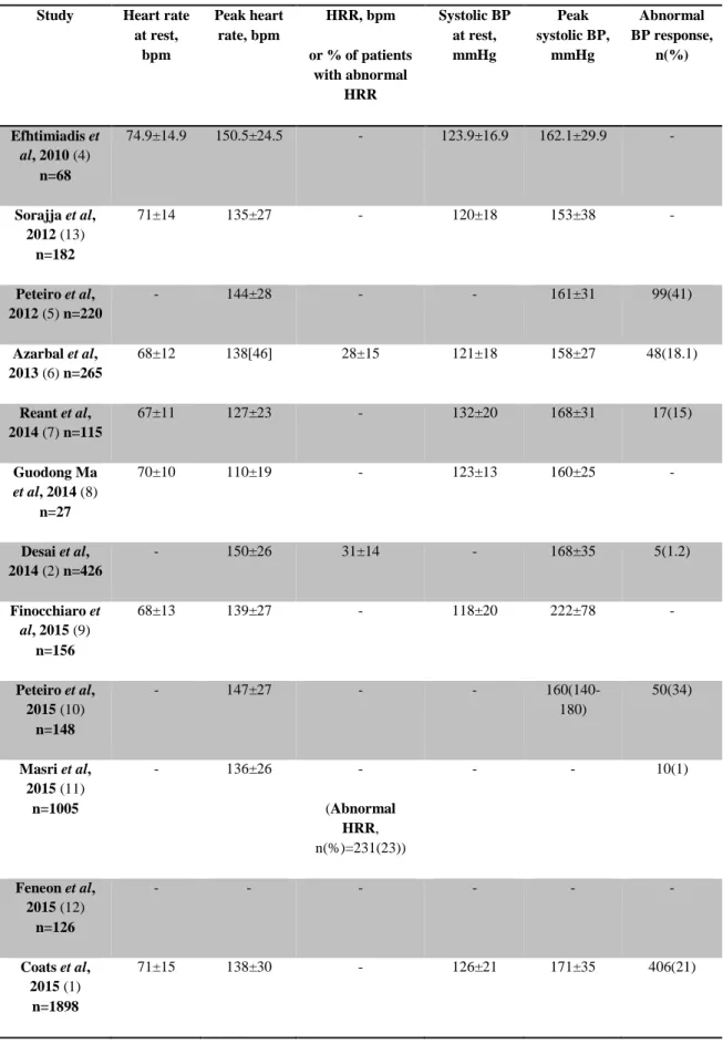
Mean HRR was, in the two studies that presented this parameter on the results, over 12 beats per minute (bpm) (the threshold below which it is considered abnormal), although this parameter demonstrated to be relevant in some of the studies, as discussed below. Abnormal BP response, which is considered a “classical” risk factor for SCD, occurred in between 1% and 41% of the patients.
24
Table 7 - Other resting and exercise parameters Study Heart rate
at rest, bpm Peak heart rate, bpm HRR, bpm or % of patients with abnormal HRR Systolic BP at rest, mmHg Peak systolic BP, mmHg Abnormal BP response, n(%) Efhtimiadis et al, 2010 (4) n=68 74.9±14.9 150.5±24.5 - 123.9±16.9 162.1±29.9 - Sorajja et al, 2012 (13) n=182 71±14 135±27 - 120±18 153±38 - Peteiro et al, 2012 (5) n=220 - 144±28 - - 161±31 99(41) Azarbal et al, 2013 (6) n=265 68±12 138[46] 28±15 121±18 158±27 48(18.1) Reant et al, 2014 (7) n=115 67±11 127±23 - 132±20 168±31 17(15) Guodong Ma et al, 2014 (8) n=27 70±10 110±19 - 123±13 160±25 - Desai et al, 2014 (2) n=426 - 150±26 31±14 - 168±35 5(1.2) Finocchiaro et al, 2015 (9) n=156 68±13 139±27 - 118±20 222±78 - Peteiro et al, 2015 (10) n=148 - 147±27 - - 160(140-180) 50(34) Masri et al, 2015 (11) n=1005 - 136±26 - (Abnormal HRR, n(%)=231(23)) - - 10(1) Feneon et al, 2015 (12) n=126 - - - - Coats et al, 2015 (1) n=1898 71±15 138±30 - 126±21 171±35 406(21)
25
Follow-up and outcomes
Adverse events during follow-up included both primary (death from any cause, HF requiring hospitalization or progression from NYHA class I or II to class III or IV and cardiac transplantation) and secondary (sustained ventricular tachycardia, stroke in the context of AF, myocardial infarction and need for therapy to reduce LVOTO) outcomes. The main predictors of worse outcomes were abnormal HRR(2,11), AF(2,11,6), exercise WMAs(10), higher VE/VCO2 slope and lower peak VO2(1,8,9).
While one study considered peak VO2 (and percentage of predicted Peak VO2) to be a
better predictor than VE/VCO2 (13), another one concluded the opposite(8). However,
in one study(11), ratio of VE/VCO2 was not associated with outcomes, which was most
likely due to the less symptomatic state of patients in that population.
On the other hand, one study verified that surgical relief of LVOTO was associated with less hard events, and, therefore, better outcomes(11).
Two studies(2,11) stated that previously reported variables of the patients, including traditional risk factors for SCD, were not predictive of long-term outcomes.
Limitations
Concerning the selected studies, the limitations were common to the majority of them and included studies being performed by only one centre(12), mostly in tertiary care centres for HCM, which means that the sample might not be representative of the overall HCM population(2,4,9). Selection biases were present, such as only including patients who were able to undergo exercise echocardiography(2) and exclusion of patients on NYHA class IV and with a prior LVEF<50%(11); it was also verified either a tendency to include more symptomatic patients, because of clinical indication, in some studies(1,4,9) or, on the other hand, not including more symptomatic patients, since they were referred to surgery before undergoing stress testing, in other studies(10,11), with only less sick patients included in these publications. Another limitation of some of the included articles was a small sample size(13,7,8,12). In some of the studies, the patients did not withdraw the medications (because it was normally considered unethical), especially drugs as beta-blockers and calcium channel blockers, which obviously influenced the hemodynamic response to exercise, diminishing the accuracy of defining an abnormal BP response(13), an abnormal chronotropic response and blunting the exercise-induced LVOT gradient(7,12). Many studies were also limited
26 due to having a short follow-up period(7,9) and/or a reduced number of events(10,7,9,12). Finally, some of the parameters were not uniformly assessed and hence not reported(2,11), which related to the fact that exercise echocardiography protocols are not standardized in HCM (some are performed in treadmill and others in semi-supine position, and the latter tends to require lower workload). Therefore, it might be challenging to compare different works(12).
27
Discussion
Exercise testing, either echocardiographic or cardiopulmonary, allows the evaluation of diverse relevant parameters in the assessment of HCM. The role of exercise testing is well defined in the symptom evaluation and management of HCM. However, its utility in the prediction of outcomes, in order to obtain a more accurate risk stratification and improve the prognosis assessment, is less well established.
The relevance of performing exercise echocardiography to study LVOTO during exercise(18) was firstly reported in 2006(18) but only few groups have made investigation in this area regarding prognosis. The data correlating peak VO2 and other
parameters obtained from cardiopulmonary exercise testing and prognosis(19) has also been scarce.
As expected, some of the parameters evaluated both at rest and with exercise echocardiography in the included studies, including SAM, LVOTO and MR appeared
de novo or worsened with exercise, due to the normal and physiologic cardiac response
to stress. However, correlation with hard events was the main aim of this systematic review. Therefore, we report a group of parameters, obtained from either exercise echocardiography or cardiopulmonary exercise testing, that consistently revealed to be predictors of worse outcomes. One of these parameters was an abnormal HRR(2,11,4), that can be possibly due to a blunted vagal reactivation in HCM patients(4), and might identify patients with a higher risk of death, malignant arrhythmias and HF progression. Abnormal BP response to exercise(7), defined as a failure to increase systolic blood pressure by at least 20mmHg from rest to peak exercise, has been previously considered a risk factor for SCD, although not included in the new risk score evaluation from the latest ESC guidelines(3). It was, however, correlated with worse outcomes in a few of the included studies.
AF induced by exercise also interestingly seems to be a relevant prognostic marker, correlating with higher rates of mortality, stroke and functional disability.
Regional exercise WMAs(5), probably explained by myocardial ischemia, were also associated with a worse prognosis for HCM patients(10,20,21), having incremental prognostic value over clinical and resting echocardiographic variables.
28 Lower peak VO2 (as well as lower percentage of predicted peak VO2) and higher
VE/VCO2 slope, that reflects ventilatory inefficiency, are parameters of exercise
tolerance and associate with a worse prognosis (1,8,9). However, two of the studies(1,9) concluded that these were only related with some of the outcomes, namely heart failure and heart transplantation, and not with sudden cardiac death, probably because mechanisms for ventricular arrhythmia and loss of contractile function are different, suggesting the importance of defining prediction parameters for specific outcomes. The largest study(1) concluded that CPET was useful for the risk stratification of patients with both obstructive and non-obstructive forms of the disease. Lower anaerobic threshold was not as much predictive of worse outcomes as peak VO2 and VE/VCO2.
Lower LVEF(11), which is a universal prognostic marker in cardiovascular disease, was confirmed to be also a relevant prognostic marker in the context of HCM.
Some of the assessed parameters were not consistently considered predictors of outcomes by all the studies, for various reasons. Indirect indices of diastolic dysfunction, such as LA diameter, were considered a predictor in two studies(9,12), but not in another one(5).
Severity and worsening of LVOTO (i.e. higher LVOT gradient at peak exercise) was predictive of outcomes in two studies(7,12), as well as in a previous work by Maron et
al(22), and not associated with events in two other(2,10). One of the studies pointed out
that peak LVOT gradient≥50mmHg was more predictive of outcomes than rest LVOT gradient≥30mmHg and that peak LVOT gradient was a better predictor than post-exercise measurement(7). There is still conflicting evidence regarding the prognostic impact of exercise-induced LVOTO in HCM.
MR has been described in patients with HCM since the first report of the disease and it is commonly associated with LVOTO and SAM. Since this is a dynamic phenomenon, it is important to evaluate this parameter during exercise. Exercise-induced MR was associated with adverse cardiovascular events in two studies(7,12), although in one of them(12) the result was not considered significant. As such, the increase in the degree of MR and its appearance de novo with exercise also seems to be of prognostic relevance.
29 Chronotropic incompetence (i.e. a blunted increase in heart rate during exercise, defined as a maximal HR during exercise that is less than 80% of the predicted value) is a predictor of clinical outcome in coronary artery disease, congenital heart disease and healthy populations(23–25). In HCM, it is possibly explained by autonomic dysfunction and was considered an independent predictor of exercise intolerance in one study(4), but whether it is a predictor of worse outcome is uncertain and requires further investigation.
30
Conclusion
Data derived from physiologic exercise, which can be assessed with exercise echocardiography and cardiopulmonary exercise testing, is able to evaluate functional status and help to ascertain limitations in exercise capacity that are not apparent on clinical examination, even before the development of significant symptoms(13,5). Thus, it has been suggested that these tests might have a role in the risk stratification of HCM patients, refining prognostic assessment.
Further investigation in this area is warranted, namely there is a need for larger, multi-center studies with longer follow-up periods. Additionally, methodological studies to standardize exercise protocols are also needed. Most importantly, the question of whether exercise testing adds value to the current risk assessment strategies is still open in HCM.
31
Acknowledgments
I would like to thank Professor Luís Rocha Lopes for everything he taught me and for helping me all through the process of writing this work.
I would also like to thank my parents and sisters, as well as the rest of my family, for all the opportunities they have given me, for accompanying me during all my academic years and in the challenges I have had throughout my life.
Lastly, I thank all my friends, both the ones from university and the ones from outside of it, and Davide for all the affection and support they have always given me.
32
Bibliography
1. Coats CJ, Rantell K, Bartnik A, Patel A, Mist B, Mckenna WJ, et al. (2015).
Cardiopulmonary Exercise Testing and Prognosis in Hypertrophic Cardiomyopathy. Circulation.
2. Task A, Elliott PM, Uk C, Anastasakis A, et al. (2014). 2014 ESC Guidelines on
diagnosis and management of hypertrophic cardiomyopathy. European Society
of Cardiology. 2733–79.
3. Sorajja P, Allison T, Hayes C, Nishimura RA, Lam CSP, Ommen SR. (2012)
Prognostic Utility of Metabolic Exercise Testing in Minimally Symptomatic Patients With Obstructive Hypertrophic Cardiomyopathy. American Journal
Cardiology. 1494–8.
4. Cole CR, Blackstone EH, Pashkow FJ, Snader CE, Lauer MS. (1999).
Heart-Rate Recovery Immediately after Exercise as a Predictor of Mortality. The New
England Journal of Medicine. 1351-1357.
5. Olivotto I, Maron BJ, Montereggi A, Mazzuoli F, Dolara A, Cecchi F. (1999).
Prognostic value of systemic blood pressure response during exercise in a community-based patient population with hypertrophic cardiomyopathy. Journal
of the American College of Cardiology. 2044–51.
6. Smedira NG, Thamilarasan M, Lytle BW, Lever HM. (2016). Exercise
Echocardiography in Asymptomatic HCM. Journal of the American College of
Cardiology - Cardiovascular Imaging. 26–36.
7. Rodriguez-garcia E, Soler R, Couto D, Castro-beiras A. (2015). Exercise
echocardiography and cardiac magnetic resonance imaging to predict outcome in patients with hypertrophic cardiomyopathy. European Heart Journal. 423–32.
8. Masri A, Pierson LM, Smedira NG, Agarwal S, Lytle BW. (2015). Predictors of
long-term outcomes in patients with hypertrophic cardiomyopathy undergoing cardiopulmonary stress testing and echocardiography. American Heart Journal.
33 9. Efthimiadis GK, Giannakoulas G, Parcharidou DG, Pagourelias ED, Kouidi EJ, Spanos G, et al. (2011). Chronotropic incompetence and its relation to exercise
intolerance in hypertrophic cardiomyopathy. International Journal of Cardiology.
179–84.
10. Bouzas-mosquera A, Fernandez X, Monserrat L. (2012). Prognostic Value of
Exercise Echocardiography in Patients with Hypertrophic Cardiomyopathy.
American Society of Echocardiography. 1–8.
11. Azarbal F, Singh M, Finocchiaro G, Le V, Schnittger I, Wang P, et al. (2014).
Exercise capacity and paroxysmal atrial fibrillation in patients with hypertrophic cardiomyopathy. Heart. 624–30.
12. Reant P, Reynaud A, Pillois X, Dijos M, Arsac F, Touche C, et al. (2014).
Comparison of Resting and Exercise Echocardiographic Parameters as Indicators of Outcomes in Hypertrophic Cardiomyopathy. Journal of the
American Society of Echocardiography. 1–10.
13. Ma G, Xu M, Gao W, Li Z, Li W, Chen B, et al. (2014). Left ventricular filling
pressure assessed by exercise TDI was correlated with early HFNEF in patients with non-obstructive hypertrophic cardiomyopathy. BMC Cardiovascular
Disorders. 1–7.
14. Pavlovic A, Homburger J, Shmargad Y, Sinagra G, Page SEE. (2015).
Cardiopulmonary Responses and Prognosis in Hypertrophic Cardiomyopathy.
Journal of the American College of Cardiology: Heart Failure. 1351-1357.
15. Galli E, Bernard A, Feneon D, Mabo P, Daubert J, Leclercq C. (2015). Impact of
exercise-induced mitral regurgitation on hypertrophic cardiomyopathy outcomes. European Heart Journal - Cardiovascular Imaging. (iii).
16. Lang RM, Badano LP, Mor-Avi V, Afilalo J, Armstrong A, Ernande L, et al. (2015). Recommendations for Cardiac Chamber Quantification by Echocardiography in Adults: An Update from the American Society of Echocardiography and the European Association of Cardiovascular Imaging.
34 17. Gersh BJ, Maron BJ, Bonow RO, Dearani JA, Fifer MA, Link MS, et al. (2011)
2011 ACCF/AHA guideline for the diagnosis and treatment of hypertrophic cardiomyopathy. Journal of Thoracic and Cardiovascular Surgery. 153–203.
18. Maron MS, Olivotto I, Zenovich AG, Link MS, Pandian NG, Kuvin JT, et al. (2006). Hypertrophic cardiomyopathy is predominantly a disease of left
ventricular outflow tract obstruction. Circulation. 2232–9.
19. Cohen-Solal A. (2002). A non-invasively determined surrogate of cardiac power
(’circulatory power’) at peak exercise is a powerful prognostic factor in chronic heart failure. European Heart Journal. 806–14.
20. O’Gara PT, Bonow RO, Maron BJ, Damske BA, Van Lingen A, Bacharach SL,
et al. (1987). Myocardial perfusion abnormalities in patients with hypertrophic cardiomyopathy: assessment with thallium-201 emission computed tomography.
Circulation. 1214–23.
21. Okeie K, Shimizu M, Yoshio H, Ino H, Yamaguchi M, Matsuyama T, et al. (2000). Left ventricular systolic dysfunction during exercise and dobutamine
stress in patients with hypertrophic cardiomyopathy. Journal of the American
College of Cardiology. 856–63.
22. Maron MS, Olivotto I, Betocchi S, Casey SA, Lesser JR, Losi MA, et al. (2003).
Effect of Left Ventricular Outflow Tract Obstruction on Clinical Outcome in Hypertrophic Cardiomyopathy. The New England Journal of Medicine. 295–303.
23. Diller GP, Dimopoulos K, Okonko D, et al. (2006). Heart rate response during
exercise predicts survival in adults with congenital heart disease. Journal of the
American College of Cardiology. 1250–6.
24. Jouven X, Empana JP, Schwartz PJ, Desnos M, Courbon D, Ducimetiere P. (2005). Heart-rate profile during exercise as a predictor of sudden death. The New England Journal of Medicine. 1951–8.
25. Lauer MS, Francis GS, Okin PM, Pashkow FJ, Snader CE, Marwick TH. (1999).
Impaired chronotropic response to exercise stress testing as a predictor of mortality. Journal of the American Medical Association. 524–9.