Increase in Systolic Blood Pressure during Exercise Testing after Heart
Transplantation: Correlation with the Clinical Condition and Ventricular
Function Assessed by Dobutamine Stress Echocardiography
Ana Fátima Salles, Cristiano Vieira Machado, Adriana Cordovil, Wagner Aparecido Leite, Valdir Ambrósio Moisés,
Dirceu Rodrigues de Almeida, Antonio Carlos Camargo Carvalho, Japy Angelini Oliveira Filho
Universidade Federal de São Paulo - UNIFESP-EPM, São Paulo, SP, Brazil
objective: Patients who underwent heart transplantation (HTX) experience a reduction in the elevation that is usual in systolic blood pressure during exercise testing. Of unknown origin, this phenomenon varies in frequency and intensity. The aim of this study was to analyze the relationship between systolic blood pressure increase (delta SBP) and clinical aspects, as well as variables measured during exercise testing (ET) and dobutamine stress echocardiography (DSE) in patients in the late post-transplantation course.
Methods: Forty-five men, mean age 49.04 ± 10.19, underwent clinical assessment, ET and DSE 40.91 ± 27.46 months after heart transplantation. Left ventricular wall motion score index and ejection fraction were assessed. Delta SBP < 35mmHg during ET was considered abnormal (SBC,1995).
Results: No significant correlation was found between delta SBP and post-transplantation time, graft ischemic time, history of rejection, diltiazem dosage, oxygen uptake, ejection fraction, and wall motion score index (WMSI). Delta SBP was normal in 17 patients (Group I) and abnormal in 28 (Group II). Patients of both groups did not differ significantly in regard to clinical features and ET and DSE results.
Conclusion: Unlike other populations, no correlation was found between delta SBP during exercise testing and clinical condition or left ventricular function in heart transplant patients. Pathophysiological factors associated with delta SBP reduction during exercise testing remain unknown.
key words: Heart transplantation, exercise testing, echocardiogram.
Mailing Address: Ana fátima Salles •
Rua Cantagalo, 229 - 03319-000 - São Paulo, SP, Brazil
Systolic blood pressure elevation (delta SBP) during exercise testing (ET) has been related to left ventricular (LV) performance1. In LV contractile dysfunction cases caused by
ischemia or LV outflow tract obstruction, lower delta SBP values have been described1. In the early and late
post-transplantation (HTX) course, a depressed response in SBP2-6
and mean BP7-9 during ET has been observed. In an analysis of
consecutive ET performed after heart transplantation showed that 69% of the heart recipients showed a depressed response of SBP during exercise testing10; delta SBP < 35mmHg
was considered abnormal (SBC,1995)11. The cause of SBP
depressed response during ET after heart transplantation is still unclear7. It may be associated with graft vascular disease,
one of the frequent causes of mortality among heart recipient survivors12.
The aim of this study was to determine indicators of attenuated behavior of SBP during ET in male patients who underwent heart transplantation using clinical variables and parameters derived from ET and DSE.
Methods
Forty-five men, mean age 49.04 ± 10.19, were studied 40.91 ± 27.46 months after heart transplantation. Causes for transplantation were: idiopathic dilated cardiomyopathy (40%), chronic Chagas cardiomyopathy (33%), chronic ischemic cardiomyopathy (25%), and hypertensive cardiomyopathy (2%). Study patients were in NYHA functional class I (n = 43) and II (n = 2) and using cyclosporine A, azathioprine, prednisone, anti-hypertensive, lipid-lowering, and hypoglycemic drugs regularly. No patient showed rejection episodes exceeding grade 3A (International Society Heart Lung Transplantation) for at least two months. Mean graft ischemic time was 114.24 ± 29.73 minutes.
predicted HRmax, VO2 max, FAI, endurance time (p = NS). Exercise testing was considered ischemic in two patients from group I and one patient from group II. SBP values at exercise peak and delta SBP were significantly lower in group II (p < 0.001).
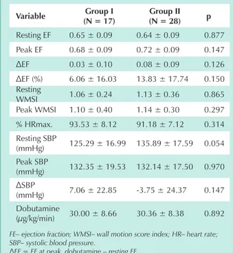
DSE results are presented in Tables 5 and 6. Both groups underwent stress with the same dose of dobutamine and reached equivalent percentages of predicted HRmax. One patient from group I and four patients from group II required atropine co-administration.
There were no significant differences between groups with respect to: [1] WMSI and EF values at rest and at peak infusion; [2] abnormal WMSI at rest and at peak infusion; [3] presence of myocardial ischemia.
Discussion
Blood pressure is governed by a complex mechanism involving hemodynamic, neural, and hormonal factors. Its determinants are cardiac output and peripheral resistance. SBP is primarily related to factors influencing ventricular performance, namely contractility, the degree to which myocardial fibers are stretched (Frank-Starling principle), blood volume, resistance to blood ejection (afterload), and heart rate.
The SBP rises during exercise, showing a 50% increase over its baseline value at maximal exercise16. In this study, 62% of
the patients experienced a depressed response in SBP during ET performed at late post-transplantation course. It has been suggested that abnormal delta SPB during ET is associated with reduced inotropic reserve secondary to changes in contractility caused by coronary disease, Chagas cardiomyopathy, hypertensive cardiomyopathy, dilated cardiomyopathy, and other heart diseases1,17-19.
Our results showed no correlation between abnormal delta SBP and post-transplantation time. On average, patients had undergone heart transplantation more than three years earlier was estimated by regression equation13. Functional Aerobic
Impairment (FAI) was defined by the following formula: FAI = (predicted VO2 max – measured VO2max) / predicted VO2 max x 102. FAI values between – 27% to + 26% were considered
normal1. Exercise test results were analyzed according to
criteria established by the Brazilian Society of Cardiology11.
During dobutamine stress echocardiography (Ultramark 9-HDI, ATL), intravenous dobutamine was infused with or without atropine14. Regional LV contractility was evaluated
by using the 16-segment model15, and the mean score was
considered as the wall motion score index (WMSI)15. Left
ventricular ejection fraction (EF) was determined by Simpson’s method, both at rest and at peak infusion (Image VueTM
DCRTM, Nova Microsonics). Resting EF values above 0.55
were regarded as normal15. No reference values for EF at peak
dobutamine infusion are reported in the literature
All procedures (clinical evaluation, ET, and DSE) were performed by independent observers.
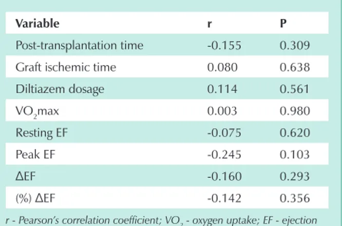
The correlation between delta SBP and the following variables were analyzed: post-transplantation time, graft ischemic time, diltiazem dosage, VO2max, resting EF, peak EF, delta EF, and % delta EF. Subsequently, patients were distributed into two groups: Group I - normal delta SBP (n = 17) and Group II - abnormal delta SBP (n = 28), so that possible markers of attenuated SBP responses between clinical variables and parameters measured by ET and DSE could be studied.
Pearson’s correlation coefficient and multiple linear regressions were used to evaluate the linear relationship between delta SBP and selected variables. The Student’s t-test, Fisher’s exact test, and Pearson’s chi-square test with Yates’ correction were used to compare groups I and II. The paired t test and McNemar’s test were applied to compare resting and peak values. P values < 0.05 were considered statistically significant.
The protocol was approved by the Institutional Research Ethics Committee, and all patients signed an informed consent before entering the study.
Results
No significant linear correlation was found between delta SBP and post-transplantation time, graft ischemic time, diltiazem dosage, VO2max, resting EF, peak EF, and EF variation
in absolute and relative values (Tab. 1).
The combined analysis of 10 variables, for which multiple linear regression (resting EF, peak EF, and % EF, WMSI at rest, WMSI at peak infusion, post-transplantation time, graft
ischemic time, history of rejection episodes ≥ 3 A, diltiazem
dosage, VO2max) were used, showed that no variable, at the 10% significance level, influenced delta SBP values (Tab. 2).
No significant differences were found between groups I and II regarding age, body weight, height, post-transplantation time, graft ischemic time, arterial hypertension, dyslipidemia
and obesity, history of rejection ≥ 3 A, and use of prednisone,
statins or diltiazem (Tab. 3).
Exercise test results are described in Table 4.Groups I and II showed similar resting heart rate (HR), resting BP, % of
variable r p
Post-transplantation time -0.155 0.309
Graft ischemic time 0.080 0.638
Diltiazem dosage 0.114 0.561
VO2max 0.003 0.980
Resting EF -0.075 0.620
Peak EF -0.245 0.103
ΔEF -0.160 0.293
(%) ΔEF -0.142 0.356
r - Pearson’s correlation coefficient; VO2 - oxygen uptake; EF - ejection
fraction.
ΔFE = EF at peak dobutamine – resting EF.
ΔEF (%) = Peak EF – Resting EF x 100 Resting EF
and, thereby, were subject to graft vascular disease.
Graft vascular disease is the major cause of death after the first year of heart transplantation20. Dobutamine stress
echocardiography has emerged as a promising non-invasive examination for detecting this condition21, with sensitivity of
67% to 100%, specificity of 55% to 89.5%, negative predictive value of 90% to 100% and positive predictive value of 33% to 76%22-25. Graft vascular disease may cause changes in LV
contractility, affecting delta SBP. The association between delta SBP and ischemia secondary to graft vascular disease is yet to be established. Myocardial ischemia incidence in groups I and II by DSE was 6% and 21%, respectively; however, no statistical significance was found between these values (p= 0.227), even though ischemia was three times higher in group II. This lack of significance may be related to the number of patients studied. Nor was significant difference found between groups I and II regarding, respectively, EF decrease at peak DSE (35.3% vs 17.8%) and mean EF increase during DSE (6.06 ± 16.03 vs 13.83 ± 17.74).
Rejection episodes are common after heart transplantation and, if repeated, may cause fibrosis and a decrease in ventricular cavity size9.Under dobutamine stress, Bellotti et
al. reported normal contractility in heart transplant recipients in whom there was no rejection. In the presence of rejection, contractility was reduced26. Our series did not corroborate
these findings, since history of rejection was similar in groups I and II (23% vs 18%, p = NS). Among the nine patients with
variable Coefficient Standard error p
Post-transplantation
time -0.008 0.641 0.990
Graft ischemic time 0.023 0.296 0.941
History of rejection
> 3 A (ISHLT) 4.349 15.037 0.779 Diltiazem dosage 0.030 0.129 0.821
VO2 max 0.686 1.521 0.663
Resting EF -3.671 4.100 0.394
Peak EF 1.912 3.418 0.590
ΔEF (%) -1,365 1,886 0,488 Resting WMSI 11.557 35.385 0.751
Peak WMSI -17.573 28.585 0.554
VO2 – oxygen uptake; FE – ejection fraction; WMSI - wall motion score
index
ΔEF (%) = Peak EF – Resting EF x 100 Resting EF
table 2 - Multiple linear regression results for Sbp increase during exercise test in heart transplant recipients (n = 45)
variable group i (n = 17)
group ii (n = 28) p
Age (years) 48.65 ± 11.58 49.29 ± 9.46 0.84 Body weight
(kg) 74.71 ± 11.25 70.86 ± 9.50 0.23
Height (cm) 168.88 ± 4.47 167.29 ± 5.42 0.31
Post-transplantation time (months)
32.88 ± 15.52 45.78 ± 31.95 0.13
Graft time
(min) 115.26 ± 22.90 113.55 ± 34.12 0.87 Arterial
hypertension 15 (88%) 24 (86%) > 0.99 Dyslipidemia 7 (41%) 14 (50%) 0.56
Obesity 5 (29%) 2 (7%) 0.09
History of rejection > 3 A (ISHLT)
4 (23%) 5 (18%) 0.94
Use of
prednisone 6 (35%) 8 (29%) 0.64
Use of statins 4 (23%) 11 (39%) 0.28
Diltiazem
dosage (mg) 165 ± 58.74 160 ± 46.02 0.80
Post-transplantation time - time elapsed since transplantation; Graft time - graft ischemic time.
table 3 - Clinical data of heart transplant recipients (n = 45)
variable group i (n = 17)
group ii
(n = 28) p
VO2max
(mL/kg/min) 29.70 ± 6.20 28.94 ± 4.71 0.642 FAI (%) 18.27 ± 24.91 19.82 ± 13.24 0.787
Endurance
time (min) 8.20 ± 2.46 8.21 ± 1.96 0.988
Resting HR
(bpm) 101.06 ± 9.32 94.68 ± 10.97 0.052
Peak HR
(bpm) 163.29 ± 21.29 154.96 ± 14.91 0.130 % HRmax. 95.29 ± 11.46 90.54 ± 7.42 0.097 Resting SBP
(mmHg) 128.82 ± 17.72 134.64 ± 19.19 0.316
Peak SBP
(mmHg) 176.47 ± 17.30 154.11 ± 19.72 < 0.001
ΔSBP
(mmHg) 47.64 ± 8.12 19.46 ± 8.53 < 0.001
Resting SBP
(mmHg) 89.71 ± 12.05 91.96 ± 9.75 0.495 Peak SBP
(mmHg) 90.88 ± 13.26 85.36 ± 12.17 0.160
VO2 – oxygen uptake; FAI - functional aerobic impairment; HR – heart rate; SBP – systolic blood pressure; DBP – diastolic blood pressure; ΔSBP
= peak SBP – resting SBP.
history of rejection, only three showed changes in contractility, two from Group I (normal delta SBP) and one from Group II (abnormal delta SBP). No case of ventricular fibrosis or reduction in ventricular cavity was identified.
Some authors have attributed the enhanced pressure response to exercise to a late sympathetic reinnervation. Wilson et al described a trend to increased delta SBP during late follow-up of patients with evidence of marked reinnervation after heart transplantation27.
Abnormal delta SBP values might be influenced by LV stiffness and dysfunction secondary to ventricular ischemia caused during cold preservation of the graft28,29. In our series,
mean graft ischemic time was 114 minutes and was not correlated with abnormal delta SBP. According to Kao et al, it is unlikely that two hours of cold ischemia would cause changes in the graft capable of persisting up to 16 months post-transplantation8.
Diltiazem hydrochloride has been frequently used for BP control after heart transplantation. In our series, 28 (62.2%) patients took diltiazem regularly at doses ranging from 60 to 240 mg/day. Drug dosage did not correlate with delta SBP during exercise testing. Both the percentage of patients on diltiazem and the dose used were similar in both groups.
No correlation was found between abnormal delta SBP and VO2max. According to the Fick principle, VO2 varies with HR, stroke volume, and arteriovenous oxygen difference. SBP
is a function of HR, stroke volume, contractility, preload and afterload. Therefore, it would be possible to detect abnormal delta SBP in the presence of the decreased VO2max values.
Douard et al30 found a significant correlation between SBP
peak values and VO2max. In our study, not only was this
relationship not observed, but groups I and II reached equal VO2max values. These results may have been affected by the estimated values used, calculated from formulas that were perhaps inadequate for transplant patients. Actually, the use of direct measurements of VO2maxin cardiopulmonary tests
would have been more appropriate.
Overall, LV systolic performance after heart transplantation has been shown to be satisfactory at rest and during exercise. Most studies have reported normal LV values at rest and at exercise peak, during both early and late follow-up8,9,31-37. In
our study, LV systolic function, assessed by the WMSI and EF, showed no correlation with abnormal delta SBP. WMSI and EF values were similar in patients of both groups. Our results were corroborated by other authors. Pflugfelder at al found no correlation between peak EF and peak BP during exercise in patients after thirteen months of transplantation32.
Other clinical, ergometric, and echocardiographic measurements also failed to characterize the abnormal delta SBP group. Groups I and II shared the same clinical features, and their results were similar on ET and DSE.
Limitations - Our study has some limitations, [1] namely, the small number of patients in groups I and II; [2] and population heterogeneity regarding different etiologies.
variable group i (n = 17)
group ii (n = 28) p
Resting EF 0.65 ± 0.09 0.64 ± 0.09 0.877
Peak EF 0.68 ± 0.09 0.72 ± 0.09 0.147
ΔEF 0.03 ± 0.10 0.08 ± 0.09 0.126
ΔEF (%) 6.06 ± 16.03 13.83 ± 17.74 0.150 Resting
WMSI 1.06 ± 0.24 1.13 ± 0.36 0.865 Peak WMSI 1.10 ± 0.40 1.14 ± 0.30 0.297
% HRmax. 93.53 ± 8.12 91.18 ± 7.12 0.314 Resting SBP
(mmHg) 125.29 ± 16.99 135.89 ± 17.59 0.054 Peak SBP
(mmHg) 132.35 ± 19.53 132.14 ± 17.50 0.970
ΔSBP
(mmHg) 7.06 ± 22.85 -3.75 ± 24.37 0.147 Dobutamine
(µg/kg/min) 30.00 ± 8.66 30.36 ± 8.38 0.892
FE– ejection fraction; WMSI– wall motion score index; HR– heart rate; SBP– systolic blood pressure.
ΔEF = EF at peak dobutamine – resting EF
ΔFE (%) = FE pico – FE repouso x 100
FE repouso
ΔSBP = peak SBP – resting SBP
table 5 - dobutamine stress echocardiogram variables in heart transplant recipients (n = 45)
variable group i (n = 17)
group ii (n = 28) p
WMSI (resting) 1 (6.0%) 5 (17.8%) 0.385
WMSI (peak) 1 (6.0%) 7 (25.0%) 0.132 Myocardial
ischemia 1 (6.0%) 6 (21.0%) 0.227 EF reduction
(peak) 6 (35.3%) 5 (17.8%) 0.284
WMSI- wall motion score index; EF- ejection fraction.
table 6 - Changes in dobutamine stress echocardiogram in heart transplant recipients (n = 45)
Conclusions
1. Bruce RA. Exercise testing of evaluation of ventricular function. N Engl J Med 1977; 296:671-5.
2. Hidalgo R, Alegria E, Castello R, et al. Stress testing in patients one year after orthotopic cardiac transplantation. Angiology 1989; 40(7): 650-5.
3. Braith RW, Wood CE, Limacher MC, et al. Abnormal neuroendocrine responses during exercise in heart transplant recipients. Circulation 1992; 86: 1453-63.
4. Rudas L, Pflugfelder PW, Kostuk WJ. Hemodynamic observations following orthotopic cardiac transplantation: hemodynamic responses to upright exercise at 1 year. Acta Physiol Hung 1992; 79(1):49-56.
5. Martin TW, Gaucher J, Pupa LE, Seaworth JF. Response to upright exercise after cardiac transplantation. Clin Cardiol 1994; 17:292-300.
6. Notarius CF, Levy RD, Tully A, Fitchett D, Magder S. Cardiac versus noncardiac limits to exercise after heart transplantation. Am Heart J 1998; 135:339-48.
7. Marzo KP, Wilson JR, Mancini DM. Effects of cardiac transplantation on ventilatory response to exercise. Am J Cardiol 1992; 69:547-53.
8. Kao AC, Trigt PV, Shaeffer-McCall GS, et al. Central and peripheral limitations to upright exercise in untrained cardiac transplant recipients. Circulation 1994; 89:2605-15.
9. Kao AC, Trigt PV, Shaeffer-McCall GS, et al. Allograft diastolic dysfunction and chronotropic incompetence limit cardiac output response to exercise two to six years after heart transplantation. J. Heart Lung Transplant 1995; 14:11-22.
10. Salles AF, Machado CV, Leite WA, et al. Teste ergométrico em transplantados: resposta inotrópica deprimida. Arq Bras Cardiol 1999; 73(supl.VI):58.
11. Mastrocolla LE, Brito AX, Brito FS, et al. Consenso Nacional de Ergometria. Arq Bras Cardiol 1995; 65(2): 189-211.
12. Chomette G, Auriol M, Cabrol C. Chronic rejection in human heart transplantation. J. Heart Transplant 1988; 7:292.
13. Bruce RA, Kusumi F, Hosmer D. Maximal oxygen intake and nomographic assessment of functional aerobic impairment in cardiovascular disease. Am Heart J 1973; 85:546-62.
14. Mcneill A, Fioretti PM, El Said SM, et al. Enhanced sensitivity for detection of coronary artery disease by addition of atropine to dobutamine stress echocardiography. Am J Cardiol. 1992; 70:41-6.
15. American Society of Echocardiography – Recommendations for quantitation of the left ventricle by two-dimensional echocardiography. J Am Soc Echo 1989; 2(5):358-67.
16. Aloan L. Hemodinâmica e angiocardiografia. Obtenção de dados, interpretação e aplicações clínicas. São Paulo: Ed. Atheneu, 1982: 58p.
17. Araujo WB. Ergometria e Cardiologia Desportiva. Rio de Janeiro: MEDSI, 1986: 149p.
18. Froelicher VF. Exercise and the Heart. Clinical Concepts. Chicago: Year Book, 1987.
19. Myers J, Froelicher VF. Teste de esforço e reabilitação cardíaca. Clin Cardiol 1993; 2:203-18.
20. Chomette G, Auriol M, Cabrol C. Chronic rejection in human heart transplantation. J Heart Transplant 1998; 7: 292.
21. Akosah KO, Mohanty PK. Role of dobutamine stress echocardiography in heart transplant patients. Chest 1998; 113:809-15.
22. Akosah KO, Mohanty PK, Funai JT, et al. Noninvasive detection of transplant coronary artery disease by dobutamine stress echocardiography. J Heart Lung Transplant 1994; 13:1024-38.
23. Derumeaux G, Redonnet M, Mouton-Schleifer D, et al. Dobutamine stress echocardiography in orthotopic heart transplant recipients. J Am Coll Cardiol 1995; 25:1665-72.
24. Spes CH, Klauss V, Rieber J, et al. Functional and morphological findings in heart transplant recipients with a normal coronary angiogram: an analysis by dobutamine stress echocardiography, intracoronary doppler and intravascular ultrasound. J Heart Lung Transplant 1999;18:391-8.
25. Machado CV. Valor da ecocardiografia sob estresse com dobutamina e da cintilografia de perfusão miocárdica com tetrofosmin no diagnóstico da doença vascular do enxerto pós- transplante cardíaco. Tese – Doutorado. São Paulo, 2000. Escola Paulista Medicina/Unifesp.
26. Bellotti G, Moraes AV, Bocchi EA, et al. Efeitos da rejeição na reserva de contratilidade do enxerto após o transplante cardíaco. Arq Bras Cardiol 1996; 67: 5-9.
27. Wilson RF, Johnson TH, Haidet GC, Kubo SH, Mianuelli M. Sympathetic reinnervation of the sinus node exercise hemodynamics after cardiac transplantation. Circulation 2000; 101:2727-33.
28. Marti V, Ballester M, Auge JM, Obrador D, Moya C, Caralps-Riera J M. Donor and recipient determinants of fatal and nonfatal cardiac dysfunction during the first week after orthotopic heart transplantation. Transplant Proc 1992; 24: 16-9.
29. Begona JA, Gundry SR, Razzouk AJ, Boucek MM, Bailey LL. Prolonged ischemic times in pediatric heart transplantation: early and late results. Transplant Proc 1993; 25: 1645-8.
30. Douard H, Parrens E, Billes MA, Labbe L, Baudet E, Broustet JP. Predictive factors of maximal aerobic capacity after cardiac transplantation. Eur Heart J 1997; 18: 1823-8.
31. Pflugfelder PW, Purves PD, Mckenzie FN, Kostuk WJ. Cardiac dynamics during supine exercise in cyclosporine-treated orthotopic heart transplant recipients: assessment by radionuclide angiography. J Am Coll Cardiol 1987; 10: 336-41.
32. Pflugfelder PW, Purves PD, Menkis AH, Mckenzie FN, Kostuk WJ. Rest and exercise left ventricular ejection and filling characteristics following orthotopic cardiac transplantation. Can J Cardiol 1989; 5: 161-67.
33. Stevenson LW, Sietsema K, Tillisch JH, et al. Exercise capacity for survivors of cardiac transplantation or sustained medical therapy for stable failure. Circulation 1990; 81: 78-85.
34. Younis LT, Melin JA, Schoevaerdts JC, et al. Left ventricular systolic function and diastolic filling at rest and during upright exercise after orthotopic heart transplantation: comparison with young and aged subjects. J Heart Transplant 1990; 9: 683-92.
35. Murali S, Carell ES, Uretsky BF, Estrada-Quintero T, Tokarczyk TR, Cannon Y M. Determinants of exercise performance early and late after cardiac transplantation. Chest 1992; 102: S77.
36. Tischler MD, Lee RT, Plappert T, Mudge GH, Sutton MJ, Parker JD. Serial assessment of left ventricular function and mass after orthotopic heart transplantation: A 4 year longitudinal study. J Am Coll Cardiol 1992; 19: 60-6.
37. Ohar J, Osterloh J, Ahmed N, Miller L. Diffusing capacity decreases after heart transplantation. Chest 1993; 103: 857-61.