University of Lisbon
Faculty of Science
Department of chemistry and biochemistry
Recruitment of Pyk2 during complement-mediated
phagocytosis and expression of kinase domains in
insect cells
by Carina Dinis SantosBiomedical Inorganic Chemistry – Applications in Diagnostic and Therapy
University of Lisbon
Faculty of Science
Department of chemistry and biochemistry
Recruitment of Pyk2 during complement-mediated
phagocytosis and expression of kinase domains in
insect cells
by Carina Dinis Santos1streferee: Prof. Dr. rer. nat. Christof R. Hauck 2nd referee: Prof. Dr. rer. nat. Helena Garcia
Biomedical Inorganic Chemistry – Applications in Diagnostic and Therapy
„All men by nature desire to know“ Aristotle
I Acknowledgments
In the first place I want to thank Alex, my supervisor, for all the patience and watch, and all the detailed explanations about the work. You were a God saver. Without you, of course I could never make it!
Prof. Dr. Cristof R. Hauck for the availability to receive me in the lab and the care he always had. Thank You for the opportunity to join your group and for all the support and new ideas for the project.
Prof. Dr. Helena Garcia, for all the support before the internship and for being my coordinator in this project.
I want also thank to all people working in the group that helped me when I was lost and always treat me with care.
My family that was always here for me, always make possible for me do different things and always support in everything. The family that always makes me take it all in front.
And of course the best person in the world, that always was here in the worst moments and always makes me smile. Always helped with everything and without him my life would be much more difficult. I´m glad you exist and are with me. Thanks Daniel.
II List of Figures
Figure 1 Innate and adaptive immunity ... 2
Figure 2 Innate and adaptive immunity ... 4
Figure 3 Types of adaptive immunity ... 4
Figure 4 Phagocytosis of bacteria by a neutrophil ... 6
Figure 5 A model for complement-receptor mediated phagocytosis ... 10
Figure 6 Non-receptor tyrosine kinase Pyk2 ... 11
Figure 7 Generation of recombinant proteins with Baculovirus Expression Vector System ... 15
Figure 8 Transfection of 293T cells with Pyk2 constructs ... 17
Figure 9 Analysis of Pyk2 expression by transfected cells using immunofluorescence ... 18
Figure 10 Analysis of fixation and permeabilization parameters for Pyk2 immunostaining ... 19
Figure 11 Actin recruitment during phagocytosis ... 21
Figure 12 Internalization of coated latex beads by mouse macrophages ... 22
Figure 13 Internalization of coated latex beads by mouse macrophages ... 22
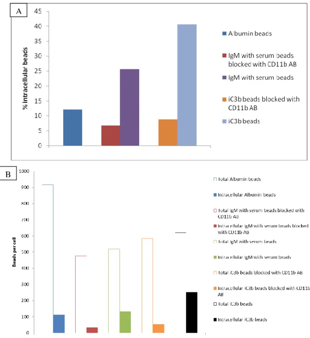
Figure 14 Intra- and extracellular assay with serum free medium ... 24
Figure 15 Intra- and extracellular assay with CD11b blocking ... 27
Figure 16 Recruitment of Pyk2 during CD11b or IgG-mediated phagocytosis ... 28
Figure 17 Recruitment of Pyk2 after infection with iC3b beads ... 30
Figure 18 Intra- and extracellular assay with iC3b beads... 31
Figure 19 Test the phosphorylation of Pyk2 ... 32
Figure 20 Amplification of GST cDNA by PCR ... 33
Figure 21 Digestion of pVL 1392 transfer vector and GST cDNA with BglII and NotI ... 34
Figure 22 Verification of the pVL1392-GST clones by restriction digest ... 34
Figure 23 Amplification of mFRNK-TAT cDNA and Hck-KD cDNA by PCR ... 35
Figure 24 Digestion of mFRNK-TAT, Hck-KD and pVL1392 N-term GST with NotI and EcoRI ... 35
Figure 25 PCR products... 36
Figure 26 Verification of the clones by restriction digest ... 36
Figure 27 Verification of the clones by restriction digest ... 36
Figure 28 Indirect Baculovirus titer determination by antibody staining ... 37
Figure 29 Analysis of target protein expression for GST-Hck-KD with NEF and GST antibodies and GST-mFRNK-TAT with GST antibody... 38
III Figure 30 Profiles from the purification by FPLC of Pyk2-KD, mFRNK-TAT and
GST-Hck-KD ... 40
Figure 31 Fractions analysis for FAK-KD, GST-mFRNK-TAT and GST-Hck-KD ... 42
Figure 32 Quantification of Pyk2-KD, FAK-KD, GST-mFRNK-TAT and GST-Hck-KD ... 43
Figure 33 In vitro Kinase Assay ... 45
IV List of Tables
Table 1 Amounts of reagents for the in vitro kinase assay using GST-p130Cas substrate ... 44
Table 2 Amounts of reagents for the in vitro kinase assay using GST-FAK-substrate ... 44
Table 3 Cells ... 50
Table 4 Medium for agar plates ... 50
Table 5 Medium for cell culture ... 50
Table 6 Primary antibodies ... 50
Table 7 Secondary antibodies ... 51
Table 8 Antibodies independent staining reagents ... 52
Table 9 Enzymes ... 52
Table 10 Proteins and complement components ... 52
Table 11 Inhibitors ... 53
Table 12 Primers ... 53
Table 13 Plasmids ... 53
Table 14 Media and Buffer ... 54
Table 15 Chemicals ... 58
Table 16 Kits ... 60
Table 17 Equipment ... 60
Table 18 Material ... 61
Table 19 Typical recipe for PCR... 67
Table 20 PCR program ... 68
Table 21 Typical recipe for DNA digestion ... 69
Table 22 Typical recipe for alkaline phosphatase treatment ... 70
Table 23 Typical recipe for Ligation ... 70
Table 24 Isolation of plasmid DNA ... 71
V Abbreviations ≥ % ºC μg μL μm μM 293T cells AB AcNPV ATP BEVS bp BSA C C3 C4 cDNA CEACAM3 CFSE cm CPU CR CRs CS C-terminal CTLs Cy dATP dCTP ddH2O
greater than or equal percent degree Celcius microgram microliter micrometer micromolar
human epithelial kidney cells Antibody
Autographa californica nuclear polyhedrosis virus Adenosine-5'-triphosphate
Baculovirus Expression Vector System base pairs
bovine serum albumin molar concentration complement component 3 complement component 4 complementary DNA
Carcinoembryonic antigen-related cell adhesion molecule 3 Carboxyfluorescein succinimidyl ester
centimeter
central processing unit complement receptor complement receptors calf serum carboxyl-terminus cytolytic T lymphocytes cyanine deoxyadenosine triphosphate deoxycytidine triphosphate double-destilled water
VI dGTP DMEM DNA dNTPs dTTP E. coli EDTA EGTA EtBr EtOH FAK FAT FcR FcRs FCS FITC fmol FPLC FRNK xg g GAP GRB2 GST GTP HBS HBSS HEK HEPES h.i. HMW HPLC HRP deoxyguanosine triphosphate
Dulbecco’s modified Eagle's medium Deoxyribonucleic acid
deoxyribonucleosid triphosphates deoxythymidine triphosphate
Escherichia coli
ethylenediaminetetraacetic acid ethylene glycol tetraacetic acid Ethidium bromide
ethanol
focal adhesion kinase focal adhesion targeting Fc receptor
Fc receptors Fetal bovine serum
fluorescein isothiocyanate femtomole
fast protein liquid chromatography FAK related non-kinase
acceleration gram
glyceraldehyde 3-phosphate growth factor receptor-bound 2 glutathione S-transferase guanosine triphosphate HEPES-buffered saline Hank’s Buffered Salt Solution human epithelial kidney
4-(2-hydroxyethyl)-1-piperazineethanesulfonic acid heat inactivated
high molecular weight
High-performance liquid chromatography horseradish peroxidase
VII Ig IgG IgM Igs Kb KD KDa L LB LMW LPS M mA MAP mg MHC min mL mM MOI Mpa mRNA n ng NK nm N-terminal PBS PCR PEEK PFA Pfu PI3K Immunoglobulin Imumnoglobulin G Immunoglobulin M Immunoglobulins kilo base pairs kinase domain kilo Dalton liter
lysogeny broth low molecular weight lipopolysaccharides molar
milli Amperes
mitogen-activated protein milligram
major histocompability complex minute
milliliter millimolar
multiplicity of infection mega Pascal’s
messenger Ribonucleic acid molar number
nanogram natural killer nanometer amino-terminus
phosphate buffered saline polymerase chain reaction polyether ether ketone Paraformaldehyde plaque-forming unit
VIII PKC PMA PMSF PRRs Psi PTKs PVDF Pyk2 RIPA RNAse A Rpm RT SDS-PAGE Sf Sf9 SH SH2 SH3 TAE TBS TBS-T TE TEMED TRITC UV V V Vis v/v w/v
α- (in connection with antibodies)
protein kinase C
phorbol 12-myristate 13-acetate phenylmethanesulfonylfluoride pattern recognition receptors pound-force per square inch protein-tyrosine-kinases Polyvinylidene Fluoride proline-rich tyrosine kinase 2 Radioimmunoprecipitation assay Ribonuclease A
revolutions per minute room temperature
Sodium dodecyl sulfate polyacrylamide gel electrophoresis
Spodoptera frugiperda
Spodoptera frugiperda clonal isolate 9
sulfhydryl Src Homology 2 Src Homology 3 Tris-acetate-EDTA Tris buffered saline
Tris buffered saline + 0.1% Tween20 Tris EDTA Tetramethylethylenediamine Tetramethylrhodamine isothiocyanate ultraviolet volume Volt visible
volume per volume weight per volume anti-
IX Abstract
In all human beings, when a pathogen enters the body, specialized cells are recruited to the site of inflammation, where, by different pathways and with the help of different macromolecules, they will destroy the invading microorganism. Past and ongoing research in this field is directed toward understanding the molecular and cellular regulation of this process. One way to destroy pathogens is the phagocytosis and subsequent intracellular killing, where macrophages play an important role, both by recognizing these pathogens and ingest them. To recognize pathogens, the macrophages have different receptors on their surface, for example Fc receptors (FcR) and complement receptors (CR), which bind to immunoglobuline G (IgG) or complement factor 3b (C3b) opsonized particles, respectively. After the macrophages have recognized a pathogen, several signalling processes regulate the uptake of the bacteria by the phagocytes. For example, non-receptor tyrosine kinases may be recruited to the plasma membrane, where they transmit signaling by cell surface receptors, controlling processes as diverse as the immune response, cell adhesion and neuronal cell migration. One member of this group of proteins is Pyk2, which can be involved in cell growth, shape and tumor invasion. In this study it is demonstrated that Pyk2 is recruited to the site of uptake, when a C3b-opsonized particle is recognized by the CRs on mouse macrophages. Specificity of the Pyk2 antibody, used for this study, was assured by in vitro assays. When a mouse-specific CD11b antibody was used to block the complement receptor 3, the uptake and internalization of particles opsonized with complement, but not of particles opsonized with IgG, decreased significantly, proving that these particles were phagocytosed by the complement receptors. The functional role of Pyk2 in this issue has to be determined in further studies. Taken together, these results could improve the understanding of complement receptor mediated phagocytosis.
In a second approach, the Baculovirus expression was used to express functional human protein kinases in insect cells. Thus, different recombinant proteins were produced and purified, such as the Glutathione-S-Transferase (GST)-mFRNK-TAT and GST-Hck-kinase domain (KD). A Pyk2-KD and FAK-KD were also expressed by this system, purified, and successfully employed in in vitro kinase assays. Accordingly, this project covered all the steps necessary for the production of recombinant proteins and expression using the Baculovirus expression vector system. As the purified kinase domains showed activity, they will be useful tools for further biochemical in vitro studies.
X Resumo
Em todos os seres humanos, quando um agente invasor entra no corpo, células especializadas são recrutadas para o sítio de inflamação onde, por diferentes vias e com a ajuda de diferentes macromoléculas, vão destrui-lo. Investigações nesta área são maioritariamente direccionadas para o entendimento da regulação molecular e celular deste processo. A fagocitose, processo pelo qual as partículas invasoras maiores que 0.5 μm de diâmetro são destruídas pelas células especializadas, tem um papel importante na imunidade inata, tanto por facilitar a remoção destas partículas como por iniciar a resposta imune adaptativa. É uma função fisiológica essencial, comum à maioria dos tipos de células eucarióticas e envolve a activação de complexas redes de sinalização. Como efetores críticos do sistema imunitário, os macrófagos usam processos de sinalização durante a adesão para concretizar muitas funções celulares essenciais, inclusivamente a fagocitose. Numerosos receptores fagocíticos estão presentes nos macrófagos, que se ligam directa ou indirectamente ao alvo. Destes receptores, os Fcγ e CRs são os mais estudados e exibem mecanismos diferentes de fagocitose e respostas celulares subsequentes que reflectem diferenças importantes nas suas vias de sinalização. Os Fc receptores (FcRs) servem de mediadores da fagocitose de partículas opsonizadas por IgG e os receptores de complemento (CRs) reconhecem as partículas opsonizadas com C3b/C3bi, componentes presentes no soro. Estes receptores juntam-se normalmente à volta da presa e induzem cascadas de sinalização intracelular que levam à activação e recrutamento de moléculas adaptadoras e de sinalização para sítios de ligação de partículas. A interacção das partículas opsonizadas com o complemento com CR3, desencadeia a imersão das partículas opsonizadas com o complemento e desempenha um papel importante na adesão celular, assim como na fagocitose. O papel da Pyk2, uma Proteína Tirosina Quinase não receptora, nas células da imunidade inata ainda não é claro. A Pyk2 não abriga os domínios SH2 ou SH3, mas contém vários sítios de ligação para diferentes proteínas de sinalização contendo SH2/SH3. Após a activação/autofosforilação dessa enzima em Tyr402, a Pyk2 pode ligar-se a proteínas do citoesqueleto (como a paxilina), de ligação (como a p130Cas), ou outras tirosina quinases (incluindo membros da família Src), que podem potencialmente impingir a Pyk2 numa variedade de vias de transdução de sinal. O sítio de autofosforilação da Pyk2 serve como um local de encaixe para o domínio SH2 da Src, permitindo-lhe alcançar uma conformação activa. Seguindo a activação, tanto a quinase Pyk2 como a Src funcionam em conjunto para activar a jusante moléculas de sinalização. Tem sido demonstrado que, juntamente com Src, a
XI Pyk2 funciona como um elo entre receptores acoplados à proteína G heterotrimérica e a via de sinalização da proteína quinase activada por mitógeno (MAP). Estudos anteriores mostraram que esta proteína é importante na activação dos macrófagos. Esta proteína foi mostrada ser uma molécula de sinalização intracelular crítica, integrando a estimulação de receptores de quemoquina e de factores de crescimento com várias vias a jusante. Anteriormente, a Pyk2 tem sido associada a processos de migração e adesão, através de sua activação em resposta a diferentes quemoquinas e integrinas. Foram produzidos ratos deficientes em Pyk2 e a sua análise mostra que se desenvolvem normalmente, excepto por exibirem migração de macrófagos com defeito. Além disso, a Pyk2, tal como a FAK, podem ser reguladas pela activação de receptores de integrina. No entanto, a Pyk2 não está localizada em contactos focais, mas sim concentrada na região perinuclear das células. Neste contexto, foi então estudado se esta proteína, presente em diferentes processos de sinalização, era recrutada durante a fagocitose mediada por receptores complemento. Para os receptores complemento presentes nos macrófagos reconhecerem as partículas acopladas com IgM, e activarem a fagocitose, estas partículas foram incubadas com soro de ratos, opsonizando deste modo as partículas com o factor C3b, que é reconhecido pelos receptores complemento. Foram usadas para este estudo partículas acopladas com Albumina e GST, que não devem ser reconhecidos por nenhum receptor, IgG que é reconhecida por receptores Fcγ mas não CRs, IgM que sem incubação com soro não é reconhecida por nenhum receptor e depois de incubação com soro é reconhecida pelos CRs e iC3b, um factor do complemento que é directamente reconhecido pelos CRs.
Depois de testado, o anticorpo usado para a detecção da Pyk2 nas células foi considerado específico para a proteína, provando que todo o sinal detectado pelo mesmo se devia à Pyk2. Foram também realizados testes para a optimização do processo de infecção e coloração das células, assim como para o processo de fagocitose. Também foi provado que as partículas eram de facto fagocitadas pelos macrófagos, já que foram detectados fortes sinais depois de coloração com actina, já demonstrada ser necessária ao processo de fagocitose. O estudo iniciou-se com ensaios intra e extracelulares, para quantificar a percentagem de internalização e adesão das partículas às células. Foi provada a internalização das partículas acopladas com IgM e incubadas posteriormente com soro pelos receptores complemento, usando um anticorpo de CD11b M1/7015. Este anticorpo foi estudado anteriormente e foi demonstrado por bloquear os receptores complemento. Ao usar este anticorpo antes da infecção, as partículas deixaram de ser internalizadas. Foi investigado então o recrutamento
XII da Pyk2 durante a fagocitose mediada por receptores complemento e foi detectado um claro sinal, provando a participação desta proteína neste processo. Foi também detectado aumento da autofosforilação desta proteína em células lisadas, depois de infecção com IgG e IgM mais soro.
Num outro projecto, a Expressão por Baculovírus foi usada para a expressão e produção de proteínas funcionais. O Sistema de Expressão por Baculovírus usando vectores (BEVS) é o sistema de expressão eucariótica mais poderoso e versátil disponível. Neste sistema, vários genes Baculovírus não essenciais ao ciclo de vida na cultura de tecidos podem ser substituídos por genes heterólogos. Já que o genoma Baculovírus é geralmente muito grande para facilmente inserir genes estranhos, genes heterólogos são clonados em vectores de transferência. A co-infecção do vector de transferência e do DNA AcNPV dentro de células Spodoptera frugiperda (Sf) proporciona a recombinação entre os sítios homólogos, transferindo o gene heterólogo do vector de transferência para o DNA AcNPV. O Vírus de Poliedrose Nuclear Autographa californica (AcNPV) é o tipo de Baculovírus mais extensivamente estudada. A infecção de células Sf com o AcNPV resulta na paragem da expressão dos genes do hospedeiro, o que permite uma elevada taxa de produção de mRNA recombinante e de proteína. O vírus recombinante induz a expressão da proteína alvo recombinante e também infecta as células de insecto adicionais, resultando em vírus recombinantes adicionais.
Para a expressão da GST-Hck-KD e GST-mFRNK-TAT, o cDNA da GST foi primeiro clonado num vector de transferência, pVL1392. Depois de obtido o novo vector de transferência, o cDNA da Hk-KD e mFRNK-TAT foi clonado nesse novo vector, pVL1392 N-term GST. Todas as construções foram sequenciadas e analisadas. As células Sf9 foram então co-infectadas com os vectores finais e o DNA BaculoGoldTM, levando à produção de Baculovírus recombinantes e expressão das proteínas heterólogas. O vírus recombinante foi amplificado por vários ciclos até o título do vírus ser maior ou igual a 2x10⁸ pfu/mL e o título foi determinado indirectamente por coloração e subsequente análise por microscopia florescente. O mesmo foi então expresso, durante um tempo determinado por western blot, as células lisadas e as proteínas purificadas por FPLC. Depois de obtida a proteína, foi feita uma diálise de aproximadamente 18 horas e a concentração determinada por um gel SDS. Para determinar se as proteínas quinase eram activas, foram realizados ensaios de quinase in vitro. Foram testadas duas proteínas expressas, que demonstraram ser activas.
XIII Keywords Pyk2 Phagocytosis Complement receptors Baculovirus expression Recombinant proteins Palavras-Chave Pyk2 Fagocitose Receptores complemento Expressão por Baculovirus Proteínas recombinantes
XIV Table of contents
Acknowledgments ... I List of Figures ... II List of Tables ...IV Abbreviations ... V Abstract ...IX Resumo ... X Keywords ... XIII Palavras-Chave ... XIII Table of contents ... XIV
1. Introduction ... ...1
1.1 Immune system ... ...1
1.1.1 Innate and adaptive immune responses ... ...1
1.2 Phagocytosis ... ...5
1.2.1 Fc receptor mediated phagocytosis ... ...7
1.2.2 Complement receptor mediated phagocytosis ... ...8
1.3 Tyrosine kinases ... ...10
1.4 Recombinant protein expression in insect cells ... ...13
1.4.1 The Baculovirus Expression Vector System ... ...14
1.5 Aims of the project ... ...15
2. Results ... ...17
2.1. Analysis of the recruitment of Pyk2 in macrophages after establishing IgG-versus complement-mediated opsono-phagocytosis assay .. ………17
2.2 Expression of kinase domains in Sf9 insect cells ... ……….33
2.2.1 Cloning of the cDNA into a Baculovirus transfer vector ... ……….…33
2.2.2 Production of recombinant Baculoviruses ... ……….37
2.2.3 Amplification and indirect titer determination of recombinant Baculoviruses………....…37
2.2.4 Test of the best time to express the protein . ………38
2.2.5 Expression and purification of the proteins ... ……….39
2.2.6 Verification of the functionality of the expressed proteins ... …….……44
XV 3.1 Analysis of the recruitment of Pyk2 in macrophages during complement
receptor-mediated phagocytosis ... ………46
3.2 Expression of kinase domains in Sf9 insect cells ... ………48
3.3 Outlook ... ………...49
4. Material ... ………...50
4.1 Cells ... ………...50
4.2 Medium ... ………...…50
4.2.1 Medium for agar plates ... ………50
4.2.2 Medium for cell culture ... ………50
4.3 Antibodies ... ………..…..50
4.3.1 Primary antibodies ... ………....50
4.3.2 Secondary antibodies ... ………....51
4.3.3 Antibodies independent staining reagents ... ………52
4.4 Enzymes, proteins and complement components ... ……….52
4.4.1 Enzymes... ………52
4.4.2 Proteins and complement components ... ………52
4.5 Inhibitors ... ………...…53
4.6 Primers ... ………...…53
4.7 Plasmids ... ………...…53
4.8 Media and Buffer ... ………54
4.9 Chemicals ... ………...………58 4.10 Kits ... ………..60 4.11 Equipment ... ………60 4.12 Material ... ………...61 4.13 Software ... ………...61 5. Methods... ………...…62 5.1 Cell culture ... ………62 5.1.1 Culturing ... ………62
5.1.2 Unfreezing and refreezing ... ………62
5.1.3 Counting ... ………63
5.1.4 Transfection of Sf9 cells ... ………....63
5.1.5 Transfection of HEK 293T cells ... ………64
XVI
5.2.1 Amplification of Baculovirus ... ………64
5.2.2 Expression of the protein ... ………..….64
5.2.3 Sf9 Lysis ... ………...….65
5.2.4 Purification of the protein ... ………...….65
5.3 Microscopy ... ………66
5.3.1 Staining of sf9 cells ... ………66
5.3.2 Baculovirus titer determination ... ……….……66
5.3.3 Staining of raw 264.7 and HEK 293T cells ... ……….……67
5.3.4 Staining of the beads ... ………....67
5.4 Molecular-biological methods .... ………...….67
5.4.1 Polymerase chain reaction (PCR) ... ………67
5.4.2 Agarose Gel-Electrophoresis ... ………68
5.4.3 Recovery of DNA from agarose gels (Gel extraction) ... ……….69
5.4.4 PCR-Products purification ... ………69
5.4.5 Restriction digest ... ………69
5.4.6 Quantification of nucleic acids ... ………70
5.4.7 Dephosphorylation of DNA fragments with alkaline phosphatase ..………70
5.4.8 Ligation of prepared vectors and DNA fragments ... ……….70
5.4.9 Transformation ... ………....71
5.4.10 Preparation of Plasmid DNA (Mini-prep) ... ………71
5.4.11 DNA sequencing ... ………72
5.5 Protein-biochemical analysis.. ………72
5.5.1 SDS-Polyacrylamide gel-electrophoresis (PAGE) ... ……….72
5.5.2 Western blot (Semi-Dry) . ………73
5.5.3 Cell lysis ... ………73
5.5.4 Protein concentration determination ... ...74
5.6 In Vitro Kinase Assay ... ...74
5.7 Covalent coupling of proteins to latex beads ...…...75
6. Literature ...………...………...76
1 1. Introduction
1.1 The immune system
Historically, immunity means protection from disease and, more specifically, infectious disease. The cells and molecules responsible for immunity constitute the immune system. Their collective and coordinated response to the introduction of foreign substances is called the immune response [1]. The immune system has evolved under tremendous selective pressure driven by pathogens. As a result, all multicellular organisms have developed the ability to recognize invading microbes and to eliminate them efficiently while avoiding massive damage to themselves. The problem with recognizing pathogens is their enormous variability and molecular heterogeneity, which is further aggravated by the high mutational rate characteristic of microorganisms and the use of a restricted number of receptors [2, 3]. This challenge has been partially met by the exploitation of conserved motifs on pathogens that are not found in higher eukaryotes. These motifs have essential roles in the biology of the invading agents, and are therefore not subjected to high mutation rates [3]. Vertebrates also have developed an additional way of dealing with the heterogeneity of pathogens by the way of the acquired immune system (see 1.1.1).
The physiologic function of the immune system is defense against infectious microbes. However, even non-infectious foreign substances as well as endogenous proteins can elicit immune responses. Furthermore, mechanisms that normally protect individuals from infection and eliminate foreign substances are also capable of causing tissue injury and disease in some situations. Therefore, a more inclusive definition of immunity is a reaction to foreign substances, including microbes, as well as to macromolecules such as proteins and polysaccharides, regardless of the physiologic or pathologic consequence of such a reaction [1].
1.1.1 Innate and adaptive immune response
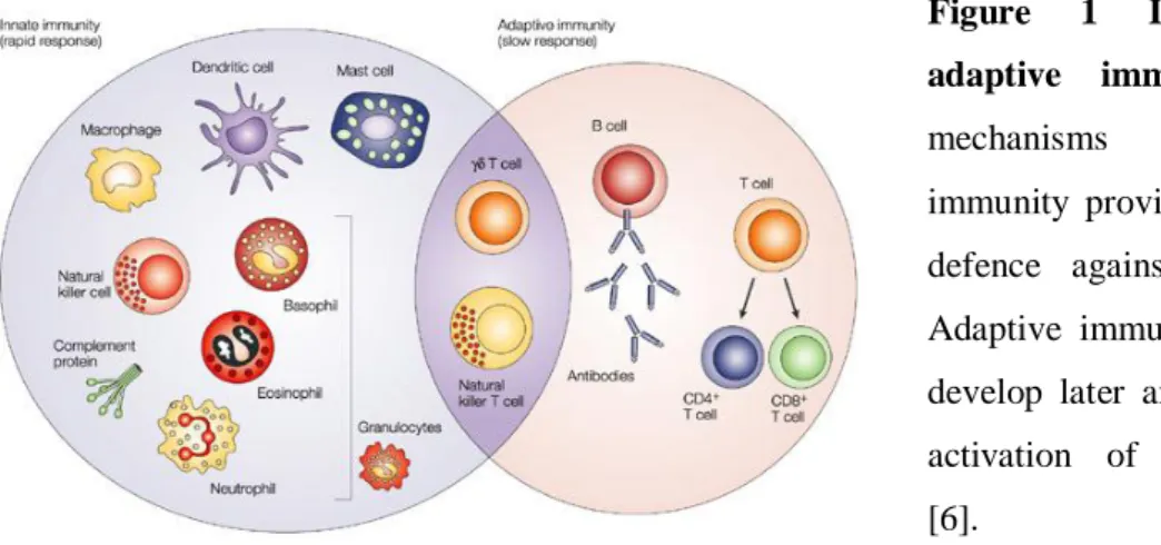
Two general systems of immunity to infectious agents have been selected during evolution of vertebrates: the early reactions of innate immunity and the later responses of adaptive immunity (Figure 1) [1, 3, 4]. Both types of responses depend on the ability of the body to distinguish between own and foreign. Two categories of pathogens can be distinguished: those that occur primarily inside a host cell (all viruses, some bacteria, and certain protozoan parasites) and those that occur primarily in the extracellular
2 compartments of the host (most bacteria and other cellular pathogens) [5]. The essential difference between the two systems is the way by which they recognize microorganisms [4]. Innate immunity (also called natural or native immunity) consists of cellular and biochemical defence mechanisms to respond rapidly to infections. These mechanisms react only to microbes and not to non-infectious substances, and they respond in essentially the same way to repeated infections. The principal components of innate immunity are (1) physical and chemical barriers, such as epithelia and antimicrobial substances produced at epithelial surfaces; (2) phagocytic cells (neutrophils, macrophages) and NK (natural killer) cells; (3) blood proteins, including members of the complement system and other mediators of inflammation; and (4) proteins called cytokines that regulate and coordinate many of the activities of the cells of innate immunity. The mechanisms of innate immunity are specific for structures that are common to groups of related microbes and may not distinguish fine differences between foreign substances. Innate immunity provides the early lines of defence against microbes [1].
The adaptive immunity has been intensively studied with artificial antigens, such as haptens and foreign serum proteins, leading to a precise understanding of many of the cells, proteins, and genes involved [2]. The defining characteristics of adaptive immunity are exquisite specificity for distinct molecules and an ability to "remember" and respond more vigorously to repeated exposures to the same microbe. The adaptive immune system is able to recognize and react to a large number of microbial and non-microbial substances. In addition, it has an extraordinary capacity to distinguish among different, even closely related, microbes and molecules, and for this reason it is also
Figure 1 Innate and adaptive immunity. The
mechanisms of innate immunity provide the initial defence against infections. Adaptive immune responses develop later and consist of activation of lymphocytes [6].
3 called specific immunity. Sometimes it is also called acquired immunity, to emphasize that potent protective responses are "acquired" by experience [1]. Adaptive immunity is mediated by T and B lymphocytes bearing clonally-distributed antigen receptors [2]. Foreign substances that induce specific immune responses and are the targets of such responses are called antigens [1]. The specificities of these receptors are generated by somatic mechanisms, and therefore are not products of natural selection directed by pathogens. Rather, several random processes contribute to the generation of the specificities of antigen receptors. Consequently, each lymphocyte can have a receptor with unpredicted specificity. In particular, a mature peripheral lymphocyte can have a receptor specific for a self-antigen. Therefore, a signal received through the antigen receptor is not sufficient on its own for the activation of naive lymphocytes. Indeed, it has been well documented that a second so-called co-stimulatory signal is required for lymphocyte stimulation [2].
Innate and adaptive immune responses are components of an integrated system of host defence in which numerous cells and molecules function cooperatively. The mechanisms of innate immunity provide effective defence against infections. However, many pathogenic microbes have evolved to resist innate immunity, and their elimination requires the powerful mechanisms of adaptive immunity. There are two important links between innate immunity and adaptive immunity. First, the innate immune response to microbes stimulates adaptive immune responses and influences the nature of the adaptive responses (Figure 2). Second, adaptive immune responses use many of the effector mechanisms of innate immunity to eliminate microbes, and they often function by enhancing the antimicrobial activities of the defence mechanisms of innate immunity [1]. The innate immune system is believed to have predated the adaptive immune response on several grounds [2].
Innate immunity is phylogenetically the oldest system of host defence, and the adaptive immune system evolved later. In invertebrates, host defence against foreign invaders is mediated largely by the mechanisms of innate immunity, including phagocytes and circulating molecules that resemble the plasma proteins of innate immunity in vertebrates. Adaptive immunity, consisting of lymphocytes and antibodies, first appeared in jawed vertebrates and became increasingly specialized with further evolution.
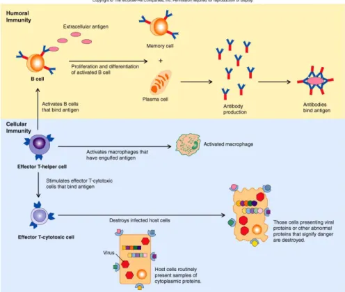
4 There are two types of adaptive immune responses, called humoral immunity and cell-mediated immunity, which are mediated by different components of the immune system and function to eliminate different types of microbes (Figure 3).
Figure 3 Types of adaptive immunity. In humoral immunity, B lymphocytes secrete
antibodies that prevent infections and eliminate extracellular microbes. In cell-mediated immunity, T lymphocytes either activate macrophages to kill phagocytosed microbes or CTLs directly destroy infected cells [7].
Figure 2 Innate and adaptive immunity. When a pathogen
enters the body, mammals initially attempt to eliminate it by innate immunity. If the pathogen has previously infected (not correct, also in case of the first infection you will have an adaptive immune response) the organism, adaptive immunity then operates to specifically exterminate the returning invader [6].
5 Humoral immunity is mediated by the secretion of antibodies by B lymphocytes (also called B cells). Antibodies recognize microbial antigens, neutralize the infectivity of the microbes, and target microbes for elimination by various effector mechanisms. Humoral immunity is the principal defence mechanism against extracellular microbes and their toxins because secreted antibodies can bind to these microbes and toxins and assist in their elimination. Therefore, immunization schemes that trigger pathogen- or toxin-specific antibodies belong to the most successful prophylactic treatments in medical history. Antibodies themselves are specialized, and different types of antibodies may activate different effector mechanisms. For example, some types of antibodies promote phagocytosis, and others trigger the release of inflammatory mediators from leukocytes such as mast cells. Cell-mediated immunity, also called cellular immunity, is mediated by T lymphocytes (also called T cells). Intracellular microbes and viruses survive and proliferate inside phagocytes and other host cells, where they are inaccessible to circulating antibodies. Defence against such infections is a function of cell-mediated immunity, which promotes the destruction of microbes residing in phagocytes or the killing of infected cells to eliminate reservoirs of infection [1].
1.2 Phagocytosis
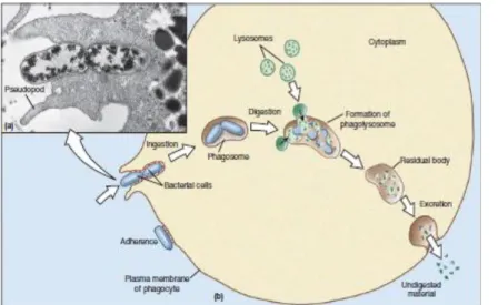
Phagocytosis, the process by which cells internalize particles larger than 0.5 μm in diameter, plays a critical role in innate immunity, both by facilitating the removal and killing of pathogens and by priming the adaptive immune response (Figure 4) [8, 9, 10, 11, 12, 13]. It is an essential physiological function, common to most eukaryotic cell types [14] and involves the activation of complex signalling networks within the host cell [15]. As critical effectors of the innate immune system, macrophages use signaling processes during adhesion to accomplish many essential cellular functions, including phagocytosis [16].
Despite the complexity associated with different phagocytic mechanisms, a number of shared features follow: Particle internalization is initiated by the interaction of specific receptors on the surface of the phagocyte (some capable of direct recognition, the so-called pattern recognition receptors (PRRs), and others that recognize opsonins on the pathogens) with ligands on the surface of the particle [8, 10]. This leads to the polymerization of actin at the site of ingestion, and the internalization
6 of the particle via an actin-based mechanism. After internalization actin is shed from the phagosome, and the phagosome matures by a series of fusion and fission events with components of the endocytic pathway, culminating in the formation of the mature phagolysosome. Since endosome-lysosome trafficking occurs primarily in association with microtubules, phagosome maturation requires the coordinated interaction of the actin and tubulin based cytoskeleton [10].
Numerous phagocytic receptors exist that can bind their target directly or indirectly through opsonins [13, 14]. Of these receptors, the opsonic Fcγ (FcγRs) and complements receptors (CRs) are the best described and exhibit different phagocytic mechanisms and subsequent cellular responses that reflect important differences in their signalling pathways [8, 17, 18, 19]. Ligand-bound receptors classically zipper around the phagocytic prey and induce intracellular signalling cascades that lead to the activation and recruitment of signalling and adaptor molecules at sites of particle binding. These locally assembled signalling complexes reorganize the actin cytoskeleton and regulate membrane dynamics underneath bound particles through the activation of Rho- and Arf-family GTP-binding proteins, respectively. According to the zipper model, phagocytosis of bound particles requires continuing ligation of phagocytic receptors around the whole phagocytic object, at least for spherical particles [10, 14]. The recognition mechanisms leading to phagocytosis occur either cellularly or humorally [10].
Fcreceptors (FcRs) mediate phagocytosis of IgG opsonized particles and complement receptors (CRs) recognize the particles opsonized with C3b/C3bi,
Figure 4 Phagocytosis of bacteria by a neutrophil.
a) Extensions of cytoplasm, called pseudopodia, surround the bacteria. b) Steps of the process to destroy the invading bacteria [5].
7 components present in serum. Interaction of complement-opsonized particles with CR3 triggers the engulfment of complement opsonized particles and plays important roles in cell adhesion, as well as in phagocytosis [20, 21].
1.2.1 Fc Receptor mediated phagocytosis
Most of our understanding of the signaling pathways leading to phagocytosis in macrophages comes from studies of the FcRs [9]. In mammals, binding of immunoglobulins (Igs) to foreign particles leads to the prompt clearance of these particles from the organism. Igs act as opsonins, molecules that render the particle they coat more susceptible to the engulfment by phagocytic cells. The conserved Fc domain of the Igs is recognized by Fc receptors present on professional phagocytes, such as neutrophils and macrophages, and the opsonized particle is rapidly internalized. This internalization is characterized by a dramatic, actin-dependent extension of the plasma membrane around the particle and is followed by secondary activity, such as the production of reactive oxygen species and the release of inflammatory cytokines from the phagocyte [17]. Leukocyte Fc receptors promote the phagocytosis of opsonized particles and deliver signals that stimulate the microbicidal activities of the leukocytes. Fc receptors for different Ig heavy chain isotypes are expressed on many leukocyte populations and serve diverse functions in immunity. Of these Fc receptors, the ones that are most important for phagocytosis of opsonized particles are receptors for the heavy chains of IgG antibodies, called Fcγ receptors. There are three types of Fcγ receptors, which have different affinities for the heavy chains of different IgG subclasses and are expressed on different cell types [1, 10, 17, 22].
Phagocytosis of IgG-coated particles is mediated by binding of the Fc parts of opsonizing antibodies to Fcγ receptors on phagocytes. Therefore, the IgG subtypes that bind best to these receptors (IgG1 and IgG3) are the most efficient opsonins for promoting phagocytosis. Antibody molecules attached to the surface of a microbe or macromolecular antigen form multivalent arrays and are bound by phagocyte Fc receptors with much higher avidity than free circulating antibodies. Binding of multiple Fc receptors to antibody-coated particles leads to engulfment of the particles and their internalization in phagocytic vesicles. These phagosomes fuse with lysosomes, and the phagocytosed particles are destroyed in the phagolysosomes [1].
8 Binding of opsonized particles to Fc receptors, particularly FcγRI, also activates phagocytes by virtue of signals transduced by the FcR γ chain. These signals result in the activation of several tyrosine kinases in the phagocytes, which stimulate production of various microbicidal molecules [1, 23].
1.2.2 Complement receptors mediated phagocytosis
Complement-receptor mediated phagocytosis is morphologically distinct from that by FcRs, although both processes require actin polymerization. Complement-opsonised particles „sink“ into the phagocyte. There is minimal membrane disturbance, and the internalization usually does not lead to an inflammatory response or oxidative burst [10]. Complement factors, present in serum, opsonize bacteria for phagocytosis by CRs on macrophages. Several receptors that participate in phagocytosis of complement-opsonized particles, including CR1, CR3 and CR4 are expressed on macrophages and neutrophils [10, 14]. CR3 represents a member of leukocyte β2-integrin family and contains multiple binding sites, which are able to interact with a range of ligands [22].
Microbes on which the complement system is activated by the alternative or classical pathway become coated with C3b, iC3b, or C4b and are phagocytosed by the binding of these proteins to specific receptors on macrophages and neutrophils. C3b and C4b (the latter generated by the classical pathway only) bind to CR1, and iC3b binds to CR3 (Mac-1) and CR4. By itself, CR1 is inefficient in inducing the phagocytosis of C3b-coated microbes, but its ability to do so is enhanced if the microbes are coated with IgG antibodies that simultaneously bind to Fcγ receptors. C3b-and iC3b-dependent phagocytosis of microorganisms is a major defense mechanism against infections in innate and adaptive immunity [1].
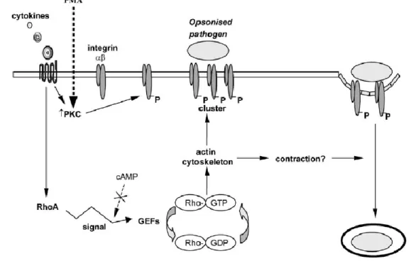
Integrins on the surface of macrophages are linked to actin filaments by actin-binding proteins such as vinculin and talin which are required for cytoskeletal rearrangement. While FcRs are constitutively active for phagocytosis, the CRs of residental peritoneal macrophages bind but do not internalize particles in the absence of additional stimuli [24, 25]. The activation of integrins is achieved via inside-out signaling, a process whereby the stimulation of cytokine and chemokine receptors activate intracellular signals (Figure 5). Those signals in turn regulate the avidity and/ or affinity of the integrins for the ligand, lateral mobility of integrins in the plasma membrane and subsequent binding of the cytoplasmic tails of integrins to cytoskeletal
9 proteins. Whilst the molecular details of inside-out signaling and the activation process of integrins are not precisely known, several potential elements in the mechanisms of activation have been identified. Cytokines (e.g. TNF-a and IFN-g) and phorbol esters (e.g. PMA) activate integrins via the activation of protein kinase C (PKC). PMA also induces an increase in phosphorylation of CD18. Examination of the cytoplasmic domains of CR3 and CR4 suggests several candidate sites for phosphorylation by PKC. PKC activation is associated with increased diffusion of the β2 chain of integrins. This suggests that phosphorylation of the β2 chain by PKC may serve as a regulatory mechanism for CR-mediated phagocytosis, and supports a role for β2 integrin phosphorylation by PKC that releases integrins from cytoskeletal constraints. Activation of integrins is known to be associated with a quantitative up-regulation of their functions. Upon activation, an integrin is thought to undergo a conformational change that enhances its ability to bind ligand. Following binding of the activated receptor to a Complement opsonized particle, CRs cluster and re-establish a firm link with the cytoskeleton. The few contact sites between the macrophage and Complement opsonized particle during the phagocytic process are rich in F-actin and cytoskeletal proteins such as vinculin, paxillin and a-actinin. CR-mediated phagocytosis results in the reorganization of polymerized actin at the site of ingestion and internalization of the opsonized particle. The signaling that induces actin polymerization and particle uptake by CR is not precisely known.
CR-mediated phagocytosis activates RhoA, a member of the Rho family of small GTPases (Figure 5). Rho and other GTPases are central in the regulation of the actin cytoskeleton, and in particular, RhoA stimulates contractile processes. Activation of RhoA results in the bundling of actin filaments and clustering of integrins. Rho-mediated signaling also promotes the assembly of stress fibres and focal adhesions, which regulate myosin filament formation and contractility. RhoA activation has been proposed to mediate integrin clustering at the site of ingestion, while regulating contractility of the actin cytoskeleton to allow the opsonized particle to sink inside the macrophage [25, 26].
The similarities in the roles of complement and Fc receptors in phagocytosis and inflammation raise the question of which is the more important effector mechanism of humoral immunity [1]. Also, IgM antibody-mediated protective immunity and pathologic reactions are dependent only on the complement system because IgM is an
10 efficient activator of the complement system but does not bind to leukocyte Fc receptors [1, 27]. To study complement mediated phagocytosis, there were used IgM coated particles which, therefore, were incubated with serum (which contains the complement components), leading to the recognition of these coated particles by complement receptors [28, 29, 30, 31].
Figure 5 A model for complement-receptor mediated phagocytosis. CRs are inactive at the
cell surface. Cytokines released at a site of infection activate macrophages through cytokine receptors. This activates PKC and RhoA. PKC phosphorylates the β chain and activates integrins. RhoA, under the regulation of GEFs, stimulates downstream effectors to promote reorganization of the actin based cytoskeleton. This results in the bundling of actin filaments and the clustering of integrins. It is also possible that RhoA activation stimulates contractility, allowing the opsonized particle to sink into the cytosol of the macrophage [25].
1.3 Tyrosine Kinases
Proteins are the most diverse of all macromolecules, and each cell contains several thousand different proteins, performing a wide variety of functions [32], where each protein has a unique and defined structure that enables it to carry out a particular function [13].
Protein kinases catalyze the transfer of the γ-phosphate group from ATP to the side chains of serine and threonine (protein-serine/threonine kinases) or tyrosine (protein-tyrosine kinases) residues. By contrast, protein phosphatases catalyze the
11 removal of phosphate groups from the same amino acids by hydrolysis. The combined action of protein kinases and protein phosphatases mediates the reversible phosphorylation of many cellular proteins. Frequently, protein kinases function as components of signal transduction pathways in which one kinase activates a second kinase, which may act on yet another kinase. The sequential action of a series of protein kinases can transmit a signal received at the cell surface to target proteins within the cell, resulting in changes in cell behaviour in response to environmental stimuli [32]. Protein-tyrosine phosphorylation is a mechanism for signal transduction that appeared with the evolution of multicellular organisms. Over 90 different protein-tyrosine kinases are encoded by the human genome. These kinases are involved in the regulation of growth, division, differentiation, survival, attachment to the extracellular matrix and migration of cells. Expression of mutant protein-tyrosine kinases that cannot be regulated and are constitutively active can lead to uncontrolled cell division and development of cancer.
Protein tyrosine kinases can be divided into receptor and non-receptor classes by virtue of whether they possess or lack extracellular ligand-binding and transmembrane domains. On the basis of sequence similarity in the catalytic kinase domain and the presence of common structural motifs, numerous families of non-receptor tyrosine kinases have been defined. Non-receptor tyrosine kinases may be recruited to the plasma membrane, where they mediate cellular signaling by cell surface receptors lacking intrinsic protein tyrosine kinase activities, controlling processes as diverse as the immune response, cells adhesion and neuronal cell migration. For instance, members of the Src family of protein tyrosine kinases are activated in response to stimulation of growth factor receptors, different G-protein-coupled receptors, and many other extracellular stimuli [13, 33].
One member of this group of kinases is the proline-rich tyrosine kinase (Pyk2, also designated RAFTK, CAK and CADTK). Pyk2 has an N-terminus with similarity to band 4.1 homology domain, a centrally located protein tyrosine kinase domain and two proline-rich regions at the C-terminus (Figure 6).
12 Figure 6 Non-receptor tyrosine kinase Pyk2. Proline-rich tyrosine kinase (Pyk2) has a central
kinase domain, two proline-rich-regions (PRRs) and four tyrosine phosphorylation sites whereas the phosphorylation of Tyr402 and Tyr881 create binding sites for Src and growth factor receptor-bound 2 (GRB2), respectively. Pyk2 contains also a C-terminal focal adhesion targeting (FAT) domain that binds to paxillin. However, Pyk2 shows perinuclear distribution and is not strongly localized to focal contacts in many cells [34].
Proline-rich tyrosine kinase 2 is a non-receptor tyrosine kinase that is expressed in cells from neural, epithelial, and hematopoietic origin [35, 36, 37]. Pyk2 does not harbor SH2 or SH3 domains but contains several binding sites for different SH2/SH3-containing signaling proteins. Following activation/autophosphorylation of this enzyme at Tyr402, Pyk2 may bind cytoskeletal proteins (like paxillin), linkers (like p130Cas), or other tyrosine kinases (including Src family members), which may potentially impinge Pyk2 in a variety of signal transduction pathways [35]. Also bind to Rho-GAP protein Graf and a LIM domain containing proteins, suggesting a role in regulation of the cytoskeleton and cellular morphology in response to extracellular stimuli [38, 39, 40]. The autophosphorylation site of Pyk2 serves as a docking site for the SH2 domain of Src [33], allowing it to achieve an active conformation. Following activation, both Pyk2 and the Src kinases function in tandem to activate downstream signaling molecules [41]. It has been shown that, together with Src, Pyk2 functions as a link between heterotrimeric G-protein-coupled receptors and the mitogen-activated protein (MAP) kinase signaling pathway [33].
The function of Pyk2 in innate immune cells is not clear. Previous work has shown that Pyk2 is important in macrophage activation. Pyk2-deficient macrophages exhibit impaired adhesion and migration owing to decreased Rho GTPase, and PI3K activation after integrin ligation. Previous studies using Pyk2 inhibitors suggested that this enzyme is critical for superoxide production in human PMNs stimulated with TNF-α. Furthermore, it was found that Pyk2 functions primarily in the integrin mediated signaling pathway and that Pyk2 deficiency results in reduced adhesion-mediated degranulation, which results in impaired host defense [41]. Also, studies with Yersinia (a genus of bacteria in the family Enterobacteriaceae) shown that Pyk2 function in Yersinia uptake by macrophages and, therefore, Pyk2 may be an integral part of the host response to Yersinia infections. Also in the same work, was found that Pyk2 autophosphorylation becomes elevated upon Yersinia infection in FAK-/- cells [42].
13 Pyk2 is also shown to be a critical intracellular signaling molecule, integrating chemokine and growth factor receptor stimulation with a variety of downstream pathways, including Ras, mitogen-activated protein (MAP) kinase, protein kinase C and inositol phosphate metabolism [36, 43]. Previously, Pyk2 has been linked to migration and adhesion processes as shown by its activation in response to different chemokines and integrins. Pyk2–deficient mice have been generated and analysis shows they develop normally except for exhibiting defective macrophage migration. Similarly, B lymphocytes derived from Pyk2–deficient mice displayed decreased motility (background migration in the absence of stimuli) that was accompanied with decreased responsiveness to a variety of chemokines [43].
Pyk2 can be activated by a variety of extracellular signals that elevate the intracellular calcium concentration. In addition, treatment with phorbol esters or agonists of G-protein-coupled receptors leads to Pyk2 tyrosine phosphorylation. Moreover, Pyk2, like FAK, can be regulated by the activation of integrin receptors. However, Pyk2 is not localized in focal contacts but rather concentrated in the perinuclear region of cells [33].
1.4 Recombinant protein expression in insect cells
The techniques developed in recombinant DNA technology have had an impact on every area of study in immunology. Genes can be cloned, DNA can be sequenced and recombinant protein products can be produced, providing components with which the structure and function of the immune system is studied [44]. These cloned genes can be transfected into cultured cells by several methods, expressing the recombinant proteins and purify them, afterwards [45]. It is known that insect cells, unlike bacteria, are capable of performing many of the processing events that are required for forming biologically active, foreign proteins [46]. Successful culture of insect cells requires a basic familiarity with insect cell physiology and general cell culture methods. The materials and methods for use with insect cell culture have evolved and contributed to the advancement of BEVS technology. The most common cell lines used for BEVS applications are Sf9, Sf-21, from Spodoptera frugiperda insect species, Tn-368 and BTI-TN-5B1-4, from Trichoplusia ni insect species. Of these, Sf9, a clonal isolate of the Spodoptera frugiperda cell line IPLB-Sf21-AE, is probably the most widely used. Sf9 was originally established from ovarian tissue of the fall armyworm. Although there
14 is significant scientific data on the characteristics of this Lepidopteran cell line, it remains to be confirmed whether it is the best line for virus or recombinant protein production. Ongoing research suggests that different insect cell lines may support varying levels of expression and differential glycosylation with the same recombinant protein [47].
1.4.1 The Baculovirus Expression Vector System
The Baculovirus Expression Vector System (BEVS) is one of the most powerful and versatile eukaryotic expression systems available. In this system, several Baculovirus genes non-essential in tissue culture life cycle (polyhedrin, p10, basic) may be replaced by heterologous genes [46, 48, 49, 50].
Baculoviruses (family Baculoviridae) belong to a diverse group of large double stranded DNA viruses that infect many different species of insects as their natural hosts.
Baculovirus strains are highly species-specific and are not known to propagate in any non-invertebrate host. The Baculovirus genome is replicated and transcribed in the nuclei of infected host cells where the large Baculovirus DNA (between 80 kb and 200 kb) is packaged into rod-shaped nucleocapsids.Since the size of these nucleocapsids is flexible, recombinant Baculovirus particles can accommodate large amounts of foreign DNA. Autographa californica nuclear polyhedrosis virus (AcNPV) is the most extensively studied Baculovirus strain. Its entire genome has been mapped and fully sequenced. Infectious AcNPV particles enter susceptible insect cells by facilitated
endocytosis or fusion, and viral DNA is uncoated in the nucleus [46].
Since the Baculovirus genome is generally too large to easily insert foreign genes, heterologous genes are cloned into transfer vectors. Co-transfection of the transfer vector and AcNPV DNA into Spodoptera frugiperda (Sf) cells, after growing the cells from mid- to late-exponential growth phase, allows recombination between homologous sites, transferring the heterologous gene from the transfer vector to the AcNPV DNA [48, 51] (Figure 7). AcNPV infection of Sf cells results in the shut-off of host gene expression allowing a high rate of recombinant mRNA and protein production. Recombinant virus induces recombinant target-protein expression and also infects additional insect cells thereby resulting in additional recombinant virus [46, 51]. The recombinant Baculovirus system for expression of heterologous proteins in insect cells was developed in the laboratories of Max Summers and Lois Miller. Development
15 of this expression system was based on observations of the wild-type baculovirus life cycle [51].
Choosing the right system for foreign gene expression can be particularly important in obtaining biologically active recombinant protein [46, 48]. Several unique features of the BEVS have made it the system of choice for many applications. Some advantages of using this system are: the use of insect cell, functional activity of the recombinant protein, high expression levels, capacity of large inserts, ease of purification, between others [46, 52]. The MOI (multiplicity of infection) and the time of infection are two parameters that are easily manipulated and that may be important in optimizing heterologous protein yields [50].
1.5 Aims of the project
Several studies have shown that Pyk2 may bind cytoskeletal proteins (like paxillin), linker proteins (like p130Cas), or other tyrosine kinases (including Src family members), which may potentially impinge Pyk2 in a variety of signal transduction pathways. Previous work has shown that Pyk2 is important in macrophage activation, Figure 7 Generation of recombinant proteins with Baculovirus Expression Vector System. The cDNA to be expressed
is cloned into a transfer vector (pVL1392) under the control of the polyhedrin promoter. This vector is co-transfected with linearized baculovirus DNA into the host insect cells. Through homologous recombination, the cDNA of interest replaces the polyhedrin cDNA [51].
16 but the overall function of this protein in innate immune cells is still unclear. Pyk2 was also shown to be a critical intracellular signaling molecule, integrating chemokine and growth factor receptor stimulation with a variety of downstream pathways, including Ras, mitogen-activated protein (MAP) kinase, protein kinase C and inositol phosphate metabolism. As this protein is expressed in macrophages, and can be activated during phagocytosis, in the present project the aim was to know if this protein is recruited during complement receptor-mediated phagocytosis. First of all, the conditions should be improved, to further have a reliable Pyk2 staining and an efficient uptake of complement-opsonized particles. Pyk2 staining in macrophages, after activation of complement-mediated phagocytosis can show afterwards, if this protein is recruited or not.
In between, another project took place. The major aim was to express recombinant proteins with a Baculovirus expression vector system in insect cells Sf9. This way, active proteins could be produced, which can be further used in in vitro as well as in vivo studies. The TAT sequence was already shown to induce the uptake of proteins in vivo and FRNK (FAK-related non-kinase), can be used for studies of FAK, which is implicated in numerous cellular processes, including cell migration, differentiation, survival and proliferation. Thus, a recombinant protein bearing the TAT sequence, GST and FRNK wanted to be produced, to further investigate if the TAT sequence allows the uptake of FRNK and, if it this happens, this recombinant protein could be used for studies in vivo. Additionally, the kinase domain of Hck should be expressed as a GST-fusion protein in Sf9 and the kinase activity of the purified protein should be verified using an in vitro kinase assay.
17 2. Results
In the present work is separated in two different projects. Firstly, the analysis of the recruitment of Pyk2 in macrophages during complement receptor-mediated phagocytosis is presented. Secondly, the recombinant expression of different proteins in Sf9 insect using the baculovirus expression vector system is shown. The proteins were afterwards purified for further use in in vitro and in vivo assays.
2.1 Analysis of the recruitment of Pyk2 in macrophages after establishing IgG-versus complement-mediated opsono-phagocytosis assay
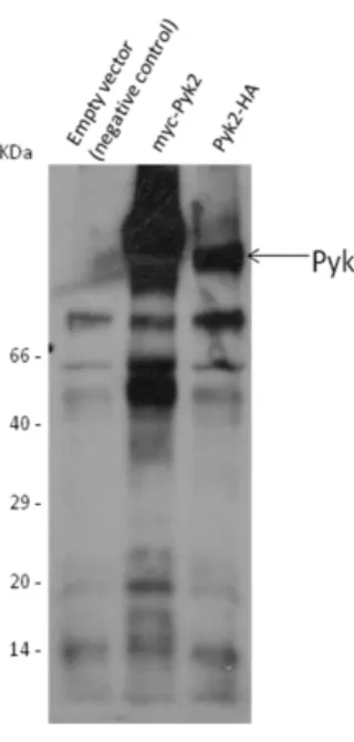
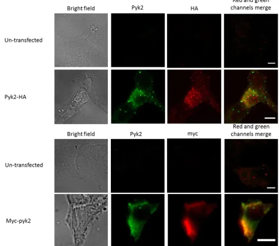
To analyze the recruitment of Pyk2 in during complement receptor-mediated phagocytosis, first of all the conditions had to be optimized to have a reliable Pyk2 staining and an efficient uptake of complement-opsonized particles. As macrophages are hard to transfect, HEK 293T cells were used. HEK 293T cells were transfected with Pyk2-HA, Pyk2-myc or the empty vector as a negative control. A western blot analysis of the transfected cells was made and it shows positive transfection for the two constructs (Figure 8). After staining, the cells were analyzed at the microscope. The results shows that the transfection worked, and as the cells had the same sites stained can be concluded that the Pyk2 antibody is specific for the protein (Figure 9).
Figure 8 Transfection of 293T cells with Pyk2 constructs. 293T cells
were transfected with the indicated constructs and were lysed 48 h later. Expression of the constructs was analyzed by western blotting using a Pyk2 AB 1:400 and an appropriate secondary antibody. Normally the size of the constructs should be nearly the same, as the single HA-tag compared to single myc-HA-tag are similar in size but some difference is detected. This can be explained by the 6xmyc tag at the N-terminus of Pyk2 compared to the 2xHA tag of the HA-Pyk2 construct.
18 After this assay, the best procedure to fix and permeabilize the cells was determined. During this assay, the cells were stained for Pyk2 (1:50 in blocking solution) and four approaches were made:
1. 4% PFA 15 min at RT to fix the cells and ice-cold Acetone 10 min at -20ºC to permeabilize.
2. 4% PFA 15 min at RT to fix the cells and 0.4% Triton X-100 in PBS ++ 3 min at RT to permeabilize.
3. 4% PFA 15 min at RT to fix the cells and 0.4% Saponin 10 min at RT to permeabilize.
Figure 9 Analysis of Pyk2 expression by transfected cells using immunofluorescence. 293T
cells were stained after fixation with 4% PFA and permeabilization with 10% FCS and 0.2% Saponin in PBS++. The Pyk2, myc and HA AB were diluted 1:200 in blocking solution. The secondary antibodies used were a mouse Cy3 antibody for HA and myc and a goat-anti-mouse Cy2 antibody for Pyk2. Thus, the Pyk2 can be distinguished from the HA and myc as the Cy3 fluorescence is detected by a TRITC channel and the Cy2 fluorescence is green detected by a FITC channel. Scale bar 10 μm.
19 4. MeOH ice-cold 5 min at -20ºC to fix the cells and 0.4% Triton X-100 in PBS ++
3 min at RT to permeabilize.
After staining, the cells were analyzed using a fluorescence microscope (Figure 10). For all samples the same exposure times were used to allow comparison of the staining. It can be seen that the best way to fix and permeabilize the cells was the second approach, as with the same conditions for all approaches, this one has the highest signal in the green channel. The staining procedure is described in the methods section.
It is described in the literature that macrophages need to be pre-activated before infection, and only this way bound particles will be phagocytosed by the complement receptors [14, 19]. To pre-activate the macrophages normally PMA or LPS are used [53]. Beside this, it was unclear which coating procedure assists maximal stimulation of the cells. In thus, Raw macrophages were seeded on fibronectin (1 µg/mL) or 0.1% of gelatine and maintained 2 hours at 37ºC. After this time, the cells were washed once with DMEM with 0.5% h.i. FCS and stored in this medium at 37ºC. The next day, the medium was supplemented with 150 ng/mL of PMA or 300 ng/mL of PMA in different wells and with 1 mM HEPES. The range of PMA concentration was as in the literature Figure 10 Analysis of fixation and permeabilization parameters for Pyk2 immunostaining. Raw macrophages were seeded on Poly-L Lysin/Fibronectin, get fixed and permeabilized as indicated and stained for Pyk2 using appropriate antibodies. Scale bar 10 μm.
20 [6, 11, 19, 53]. The cells were then incubated for 15 min at 37ºC and then analyzed at the microscope, taking pictures every 5 minutes for one hour. The movement of the cells and the spreading was analyzed by ImageJ (data not shown). It was seen that the cells spread better on Fibronectin, however, on Gelatine the cells seems to be more activated as they displayed increased migration. The cells not pre-activated showed only marginal movement. Therefore, to analyze the recruitment of Pyk2 in the cells during phagocytosis it was chosen to use Gelatine for coating and 200 ng/mL of PMA to pre-activate the cells, before infection.
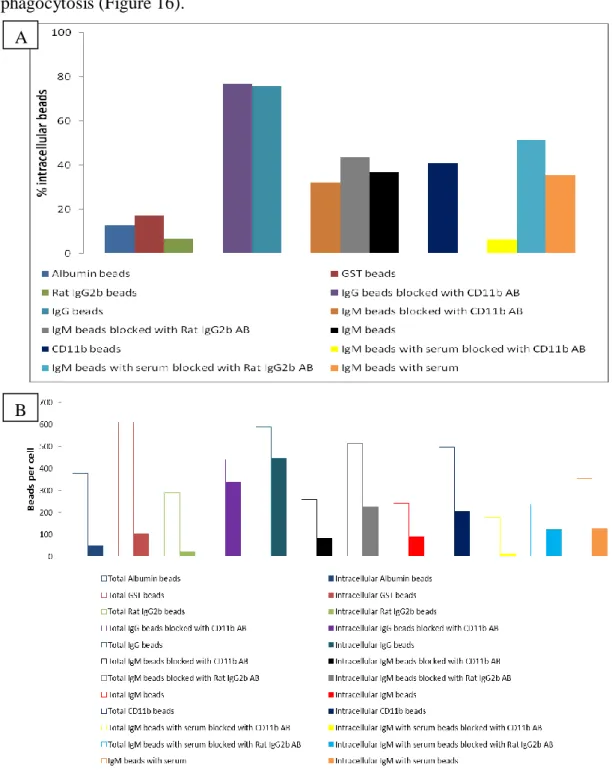
It is well known that Actin plays an important role during phagocytosis [17, 54, 55] and there are made recent advances in this field, mostly related to FcR and CR3 [55]. Thus, the cells were infected with coupled beads and filamentous actin was stained to see if these beads were in fact phagocytosed by the cells (Figure 11). The beads were coupled with Albumin and GST, negative controls for phagocytosis as they don´t bind to receptors present in macrophages, IgG that, as described before, are internalized by the FcRs which allows have a positive control for phagocytosis, IgM that is showed to be internalized by the CRs after incubation with serum that contains the complement components, and iC3b that is a complement component and the macrophages has a iC3b receptor which recognize opsonized particles with this component. It was seen in all samples (except for Albumin as expected), that Actin was recruited to sites of infection. These results indicate that the beads are in fact being phagocytosed by the macrophages, which allows us to proceed to the analysis of Pyk2.
The Pyk2 staining did not showed any clear recruitment of this protein during phagocytosis (Figure 11). The analysis of Pyk2 continued with all conditions described before, and after three unsuccessful attempts was made an assay to see if the beads were in fact internalized by the cells. To this, the coupled beads were stained before infection with CFSE 1:1000 in PBS and after infection with antibodies against the coupled proteins to distinguish between intracellular beads from adhered, extracellular beads (Figure 12) (This procedure is described in the methods section).
21 Figure 11 Actin recruitment during phagocytosis. Raw macrophages were incubated for 20
min with the indicated beads and fixed. Samples were stained for actin and Pyk2 staining. The intensity, exposure time and signal amplification in both channels were adjusted so that in the sample without staining there was no signal. Scale bars 10 μm.
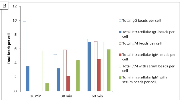
The IgM beads were opsonized with complement components by incubating with mice serum at 37ºC for 1 h. The quantification of the first intra- and extracellular assay revealed the internalization of IgG and IgM with serum beads, as well as the IgM beads alone (Figure 13). The IgG beads are internalized by the Fc receptors [1, 10, 11, 31, 56] and the IgM with serum beads should be internalized by the CRs. But as we can see an IgM beads internalization it was not sure that the CRs are active for IgM with serum beads.
22 Figure 12 Internalization of coated latex beads by mouse macrophages. All the beads were
stained with CFSE before infection and after infection and fixation the beads were stained again, and as there was no permeabilization only the extracellular beads should be stained. A streptavidin-Cy3 or Cy3 antibody was used, depending if the beads were biotinylated or not during CFSE staining (only the beads without antibody against the protein coupled were biotinylated, as the Albumin beads). The intra- and extracellular beads were distinguished afterwards. The beads in the cells were counted after analysis at the microscope, where the CFSE stained beads were detected with a FITC channel and the Cy3 stained beads with a TRITC channel. In this case the time of infection was 60 min. Scale bar 20 μm.