Anatomical variants of the ostiomeatal complex:
tomographic findings in 200 patients*
Variações anatômicas do complexo ostiomeatal: achados tomográficos em 200 pacientes
Anna Patricia de Freitas Linhares Riello1, Edson Mendes Boasquevisque2
OBJECTIVE: The present study was aimed at evaluating the frequency and types of anatomical variants of the ostiomeatal complex. MATERIALS AND METHODS: Computed tomography studies of 200 patients evaluated for clinical suspicion of sinusopathy were reviewed. RESULTS: Two or more types of anatomical variants were observed in 83.5% of the patients. The most frequent anatomical variants involved the middle turbinate (84%) and the nasal septum (34%). Other common variants found were the presence of pneumatized agger nasi cells and infraorbital ethmoid cells (Haller cells). CONCLUSION: The prevalence of anatomical variants in the ostiomeatal complex is high, the most frequent ones involving the middle turbinate and the nasal septum.
Keywords: Computed tomography; Ostiomeatal complex; Paranasal cavities; Anatomy; Drainage.
OBJETIVO: O objetivo deste trabalho foi verificar a freqüência e os tipos de variações anatômicas do com-plexo ostiomeatal. MATERIAIS E MÉTODOS: Foram revisadas 200 tomografias computadorizadas de pa-cientes com suspeita clínica de sinusopatia. RESULTADOS: Observamos um ou mais tipos de variações ana-tômicas em 83,5% dos pacientes, as mais freqüentes envolvendo o corneto médio (84%) e o septo nasal (34%). Outras variantes anatômicas comuns encontradas foram a presença de aeração do agger nasi e células etmoidais infra-orbitárias (células de Haller). CONCLUSÃO: As variações anatômicas do complexo ostio-meatal anterior são muito corriqueiras. As mais freqüentes envolvem o corneto médio e o septo nasal.
Unitermos: Tomografia computadorizada; Complexo ostiomeatal; Cavidades paranasais; Anatomia; Drena-gem.
Abstract
Resumo
* Study developed at Labs D’Or Laboratório e Imagem, Rio de Janeiro, RJ, and Medimagem, Niterói, RJ, Brazil.
1. Master in Radiology, MD, Radiologist at Medimagem, Niterói, RJ, and Labs D’Or Laboratório e Imagem, Rio de Janeiro, RJ, Brazil.
2. PhD, Associate Professor, Department of Radiology – Uni-versidade Estadual do Rio de Janeiro (UERJ), Niterói, RJ, MD, Radiologist, Instituto Nacional de Câncer (INCA), Rio de Janeiro, RJ, Brazil.
Mailing address: Dra. Anna Patricia Riello. Avenida Visconde de Albuquerque, 1102/201, Leblon. Rio de Janeiro, RJ, Brazil, 22450-000. E-mail: [email protected]
Received July 13, 2006. Accepted after revision September 14, 2007
The protocol consisted of coronal and axial slices, respectively, perpendicular and parallel to the palate, with 2–3 mm in thick-ness. For patients who could not tolerate the prone position (hyperextended neck) required for coronal images acquisition, helical acquisition was performed with 2–3 mm collimation and computer-gener-ated reconstructed coronal views.
In all of the cases, bone algorithm was utilized for documentation, and soft tissues window was utilized in some selected cases.
RESULTS
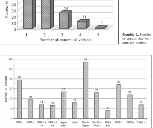
One or more anatomical variants (Graphic 1) were identified in 167 of the 200 cases evaluated (83.5%). The majority of patients presented with up to two ana-tomical variants (38% with one; 27% with two variants).
The frequency of anatomical variants of the ostiomeatal complex is shown on
Riello APFL, Boasquevisque EM. Anatomical variants of the ostiomeatal complex: tomographic findings in 200 patients. Radiol Bras. 2008;41(3):149–154.
INTRODUCTION
Sinusopathy is a common clinical prob-lem that, sometimes, does not present an adequate response to medicamentous therapy. Computed tomography (CT) is the method of choice for evaluating these cases, particularly in the setting of a prob-able surgical intervention(1). Endoscopic surgery has been increasingly utilized, re-quiring a meticulous assessment and a de-tailed description of both nasal and paranasal cavities structures(2). Considering
that the main objective of this type of sur-gery is to reopen the natural ways of drain-age of paranasal cavities, it is very impor-tant that the radiologist is aware of the ostiomeatal complex variants, describing them in a comprehensible way for the otorhinolaryngologist(3–8).
The present study was aimed at evalu-ating the frequency and types of anatomi-cal variants of the ostiomeatal complex.
MATERIALS AND METHODS
Graphic 2. Most of these anatomical variants involved the middle turbinate (84%) and the nasal septum (34%) (Figure 2), includ-ing: unilateral middle turbinate pneumati-zation in 39 cases (23%), bilateral pneuma-tization in 19 cases (11%), unilateral para-doxical middle turbinate curvature in 34 cases (20%) and bilateral paradoxical middle turbinate curvature in 24 cases (14%).
Other pneumatized structures were: agger nasi cells (Figure 3) in 27 cases (16%), unilateral or bilateral pneumatiza-tion of the uncinate process in 26 cases (16%) (Figure 5), and infraorbital (Haller) cells in 16 cases (10%) (Figure 4).
Among the variants of the uncinate pro-cess insertion which were found in 10 cases, seven (3.5%) presented the uncinate process inserted into the middle turbinate,
two (1%) into the ethmoid cribriform plate, and one (0.5%) into the middle orbital wall. Other less frequent variants found were: aberrant ethmoidal bulla in eight cases (5%), hypoplastic middle turbinate in 14 cases (8%), paradoxical inferior turbinate, medial deviation of the uncinate process, hypoplastic maxillary sinus, and nasal sep-tum spur without associated deviation in two cases each (1%), and only one case (0.6%) with each of the following variants: bilateral paradoxical inferior turbinate, spi-ral-shaped middle turbinate, pneumatized superior turbinate, hypoplastic middle tur-binate attached to the uncinate process, enlarged inferior turbinate, double maxil-lary infundibulum, choanal atresia, unci-nate process attached to the infraorbital ethmoid cell, accessory middle turbinate and duplicate middle turbinate.
DISCUSSION
The ostiomeatal complex is differently defined by several authors.
Scribano et al.(9) have defined the ostio-meatal complex as a complex including the maxillary sinus ostium, ethmoid infundibu-lum and middle meatus; in other words, as the final site of drainage from the frontal and maxillary sinuses and anterior ethmoi-dal cells.
Casiano(10) has defined the ostiomeatal complex as the ethmoid bulla, uncinate process and adjacent spaces and ostia draining the anterior sinuses (anterior eth-moid sinus, frontal and maxillary sinuses). Zinreich et al.(11) have defined the ostio-meatal complex as the group of bony struc-tures and aerated channels into which the paranasal cavities drain, and have subdi-vided the complex into three parts. The first most anterior portion of the complex in-cludes structures surrounding the frontal recess; the second one corresponds to the structures including the maxillary sinus and middle meatus; and the third and most pos-terior portion includes the structures sur-rounding the sphenoethmoidal recess. The ostiomeatal complex would be formed by the two first portions(11). Mafee et al.(12) and Mafee(13) have described the ostiomeatal complex similarly to the definition by Zinreich et al.(11).
Laine & Smoker(14) have defined the ostiomeatal complex as an aerated channel of the middle meatus representing the final common pathway for drainage of the max-illary and frontal sinuses and anterior eth-moid cells, delimited by the uncinate pro-cess, ethmoidal bulla and middle turbinate. Shankar et al.(6) have defined ostio-meatal complex as a complex including the maxillary ostium, ethmoid infundibulum, hiatus semilunaris, middle meatus, frontal recess, ethmoid bulla and uncinate process. In the present study, the concept devel-oped by Stammberger & Kennedy(7) was adopted, defining ostiomeatal complex as a functional unit of the anterior ethmoid complex representing the final common pathway for drainage and ventilation of the frontal, maxillary and anterior ethmoid cells. Any of these cells, clefts, ostia, re-cesses or cavities may be affected by a pathological process, thereby contributing
Graphic 1. Number of anatomical vari-ants per patient. Number of anatomical variants
Number of patients
Graphic 2. Main anatomical variants found. CMAu, unilateral pneumatized middle turbinate; CMAb, bilateral pneumatized middle turbinate; CMAu + Hr, bilateral, pneumatized and hypertrophic middle turbinate; Pcs Unc Pneu, pneumatized uncinate process; Bulla Etm Aberr, aberrant ethmoid bulla; CMPu, unilateral paradoxical middle turbinate; CMPb, bilateral paradoxical middle turbinate; CMHo u, unilat-eral, hypoplastic middle turbinate.
N
u
m
b
e
r
o
f
p
a
ti
e
n
to the symptoms and pathophysiology of sinusitis (Figure 1).
Anatomical variants of the ostiomeatal complex were found in 83.5% of cases in the present casuistic, a rate within the fre-quency interval observed by other authors — Bolger et al.(3) have reported a frequency of 64.9%; Pérez-Piñas et al.(5), 67%; Tonai & Baba(15), 75%; and Earwaker(16), 93%.
The groups evaluated, as well as pedi-atric groups, ranged considerably in size and distribution (Graphic 1)(4). Kinsui et al. have compared symptomatic and asymp-tomatic groups(17), and other authors have evaluated patients with suspected or con-firmed sinusopathy(5,9,15,16,18,19). Earwaker(16) has studied 800 individuals, and the major-ity of authors have studied groups ranging from 71 to 200 patients(3–5,7,15,19–21), simi-larly to the number of patients included in the present study (200 patients).
The authors have reported quite differ-ent frequencies of anatomical variants (Graphic 1). Maybe, these divergences could be explained either by populational differences, by the definition adopted for
gree, and lower rates suggest that only large turbinates may have been taken into con-sideration. This finding may have been in-fluenced by the sensitivity of the analysis method. There may be inherent differences among macroscopic anatomical study, analysis of conventional radiographs, to-mographic studies and techniques of mi-croscopic dissection(3). For Pérez-Piñas et
al.(5), the middle turbinate was aerated only in cases where both the vertical cribriform plate and inferior bulbar portion were pneumatized. The present study has adopted the definition by Zinreich et al.(23), who have considered any pneumatization degree as concha bullosa.
Middle turbinate aeration was found in 42.5% of patients in the present study (Fig-ure 2). Other studies have found prevalence of 21%(19), 28%(15,20), 30%(18), 33%(17) and 35%(16), all of them ranging between 21% and 73%(5), excepting Dutra and Mar-chiori(4) whose study has included only pediatric patients (4%).
Typically, the curved portion of the middle turbinate points toward the nasal anatomical variants (Graphic 1), or by the
methodology of analysis (3).
Nasal septum
Deviation of the nasal septum can be defined as any midline deviation(16,20) and may be cartilaginous, osteocartilaginous or osseous. Severe nasal septal deviation may result in compression of the inferior or middle turbinate, causing obstruction of the normal mucus flow and, consequently, sec-ondary inflammation and infection(14,20).
Deviation of the nasal septum was found in 28.5% of cases in the present study (Figure 2). In other studies, this find-ing ranged from 14.1% to 80%: Dutra & Marchiori(4), 14.1%; Kinsui et al.(17), 23.3%; Arslan et al.(18), 36%; Earwaker(16), 44%; and Pérez-Piñas et al.(5), 80%.
Middle turbinate or middle concha Concha bullosa is one of the most fre-quently found anatomical variants(22,23). The differences reported in the prevalence of middle turbinate pneumatization may have been influenced by the aeration
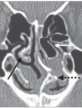
de-Figure 1. Normal anatomy of the anterior ostiomeatal complex. Frontal sinus (SF); Maxillary sinus (SM); maxillary sinus infundibulum (inf); maxillary sinus ostium (o); eth-moid bulla (BE); nasal septum (SN); inferior turbinate (CI); middle turbinate (CM); fron-tal recess (arrow head); uncinate process (arrow); middle meatus (dashed arrow); hia-tus semilunaris (white arrow).
septum. In cases where the curvature un-usually occurs toward the opposite side, they are called paradoxical turbinates(14,20). This variation can be observed either in superior, middle or inferior turbinates, al-though this is much more frequent in middle turbinates. Again, the prevalence reported by different authors may diverge because some of them, like the authors of the present study, consider any involved portion of the turbinate as paradoxical cur-vature, whereas others may consider this variation only in cases where the whole turbinate is unusually curved towards the opposite side.
In the present study, 58 patients were found with paradoxical middle turbinates (29%). Arslan et al.(18) have found this vari-ant in 3% of cases, Bolger et al.(3) in 26,1%, Earwaker(16) in 43%, Pérez-Piñas et al.(5) in 73% and Tonai & Baba(15) in 28%.
Agger nasi
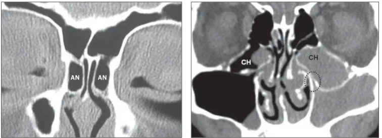
The agger nasi is the most superior rem-nant of the first ethmoturbinate, which per-sists as a mound or tuberosity immediately anterior and superior to the insertion of the middle turbinate(7,12). An agger nasi cell results when this area becomes pneuma-tized. Agger nasi cells present a close rela-tionship with five different cranial bones: lacrimal bone, maxillary bone, ethmoid bone, frontal bone and nasal bone(3). Very small agger nasi cells may be missed dur-ing anatomical dissections but not at CT.
Zinreich et al.(23), based on their expe-rience with CT of paranasal sinuses, nasal endoscopy and functional endoscopic sur-gery, have demonstrated that agger nasi cells are air cells under the frontal sinus extending anterosuperiorly toward the frontal recess, reaching the lacrimal fossa inferolaterally, and laterally adjacent to the nasal bones. Frequently, agger nasi is the antero-inferior border of the frontal re-cess(3,24), and agger nasi aeration may be implied in cases of chronic frontal sinusi-tis. In the present study, the Zinreich et al.(23)agger nasi concept was adopted.
Agger nasi cells were found in 13.5% of patients in the present casuistic (Figure 3), but other authors have reported preva-lence rates of 7%(20), 86%(15), 96%(16) and 98%(3).
Infraorbital ethmoid cells or Haller cells Arslan et al.(18) and Meloni et al.(19) have defined infraorbital ethmoid cell or Haller cell as a pneumatized ethmoid cell between the orbit and the maxillary sinus. Zinreich et al.(8,23) have described infraorbital eth-moid cell or Haller cell as an etheth-moid cell that is found below the ethmoid bulla, at-tached to the floor of the maxillary sinus, adjacent to the maxillary infundibulum, as part of the lateral wall of the infundibulum. Stammberger & Kennedy(7) and Bolger et al.(3) have added that these cells are found precisely in the region of the maxillary si-nus ostia. It is postulated that infraorbital
ethmoid cells might constitute an etiologi-cal factor in the recurrent maxillary sinusi-tis(8), but, according to Bolger et al.(3) they should be analyzed on a case-by-case ba-sis, considering that other authors have not observed statistically significant differ-ences between patients with and without inflammatory sinus disease. In the present study, the definition coined by Stammber-ger & Kennedy(7) and Bolger et al.(3) was adopted.
In the present study, infraorbital eth-moid cells were found in 8% of cases (Fig-ure 4), but other authors have reported a wide variation in the prevalence rates such as 1%(4), 5.5%(20), 6%(18), 9%(17), 20%(5), 36%(15) and 45%(3).
The uncinate process
The most common anatomical variants of the uncinate process are represented by insertion into an unusual topography and pneumatization.
The uncinate process is a superior ex-tension from the lateral wall of the nasal cavity and generally is inserted into the postero-medial portion of the agger nasi(11,16).
Uncinate process insertion into other structures may result in a blind-end ob-struction. In cases where the uncinate pro-cess inserts onto the lamina papyracea, the maxillary sinus drainage may be im-paired(16,25). In the present study, this ana-tomical variation was found in one case
Figure 3. Agger nasi(AN) cells. Figure 4. Infraorbital ethmoid cells or Haller cells (HC). Note the obliteration of the left maxillary infundibulum (dashed circle) and completely obscured maxil-lary sinus and infraorbital cell. Also, a less exuberant mucous thickening is ob-served at right.
Table 1 Comparison of data from different authors.
Authors
Riello & Boasquevisque Arslan et al.(18) Bolger et al.(3) Dutra & Marchiori(4) Earwaker(16) Kayalioglu et al.(20) Kinsui et al.(17) Meloni et al.(19) Pérez-Piñas et al.(5) Tonai & Baba(15)
n 200 200 202 71 800 172 150 100 110 75 Middle turbinate pneumatization 42.5% 30% 53% 4% 35% 28% 33% 21% 73% 28% Paradoxical middle turbinate curvature 29% 3% 26.1% UD 43% UD UD UD 27% 25% Infraorbital ethmoid (Haller) cells 8% 6% 45% 1% UD 5.5% 9% 10% 20% 36% Agger nasi cells 13.5% UD 98% ND 96% 7% UD UD UD 86% Septum deviation 28.5% 36% UD 14.1% 44% UD 23.3% UD 80% UD Uncinate process pneumatization 13% 4% 2.5% UD 9.1% UD UD UD UD UD
n, number of patients; UD, unavailable data.
(0.5%). If the uncinate process attaches to the middle turbinate or cribriform plate, the frontal and homolateral maxillary sinuses drainage may be impaired, resulting in a mechanism of sinus mucus recirculation. In the present study, these variants were found, respectively, in seven cases (3.5%) and in two cases (1%). Earwaker(16), in a study of 800 cases, have described variants of the uncinate process in detail, classify-ing them in association with other variants of the ostiomeatal complex and subdivid-ing them differently from the present study. Other authors included in Table 1 have not approached anatomical variants of the uncinate process.
Pneumatized uncinate process (or unci-nate bulla) also has been associated with poor sinus ventilation(11,14), specifically of the anterior ethmoid, frontal recess and of the infundibulum region. A careful analy-sis of consecutive CT images suggest that the pneumatization of the uncinate process occurs because of agger nasi excavation in the most anterosuperior region of the unci-nate process(3).
In the present study, pneumatization of the uncinate process was found in 13% of cases (Figure 5), whereas other authors re-port prevalence rates of 2.5%(3), 4%(18) and
9.1%(16).
CONCLUSION
Different and frequent anatomical vari-ants may be found in the anterior ostio-meatal complex, and a single individual may present with different variants. In the
present study, the most frequent variants were those involving the middle turbinates, particularly their pneumatization and para-doxical curvature, deviation of the nasal septum and pneumatized agger nasi cells, infraorbital ethmoid cells and uncinate pro-cess.
REFERENCES
1. Melhem ER, Oliverio PJ, Benson ML, et al. Op-timal CT evaluation for functional endoscopic si-nus surgery. AJNR Am J Neuroradiol. 1996;17: 181–8.
2. Ludwick JJ, Taber KH, Manolidis S, et al. A com-puted tomographic guide to endoscopic sinus surgery: axial and coronal views. J Comput Assist Tomogr. 2002;26:317–22.
3. Bolger WE, Butzin CA, Parsons DS. Paranasal si-nus bony anatomic variations and mucosal abnor-malities: CT analysis for endoscopic sinus sur-gery. Laryngoscope. 1991;101(1 Pt 1):56–64. 4. Dutra LD, Marchiori E. Tomografia
computado-rizada helicoidal dos seios paranasais na criança: avaliação das sinusopatias inflamatórias. Radiol Bras. 2002;35:161–9.
5. Pérez-Piñas, Sabaté J, Carmona A, et al. Anatomi-cal variations in the human paranasal sinus region studied by CT. J Anat. 2000;197:221–7.
6. Shankar L, Evans K, Hawke M, et al. An atlas of imaging of the paranasal sinuses. London: Martin Dunitz; 1994.
7. Stammberger HR, Kennedy DW. Paranasal si-nuses: anatomic terminology and nomenclature. The Anatomic Terminology Group. Ann Otol Rhinol Laryngol Suppl. 1995;167:7–16. 8. Zinreich SJ, Kennedy DW, Rosenbaum AE, et al.
Paranasal sinuses: CT imaging requirements for endoscopic surgery. Radiology. 1987;163:769–75. 9. Scribano E, Ascenti G, Loria G, et al. The role of the ostiomeatal unit anatomic variations in in-flammatory disease of the maxillary sinuses. Eur J Radiol. 1997;24:172–4.
10. Casiano RR. Correlation of clinical examination with computer tomography in paranasal sinus disease. Am J Rhinol. 1997;11:193–6. 11. Zinreich SJ, Albayram S, Benson M, et al. The
ostiomeatal complex and functional endoscopic surgery. In: Som PM, Curtin HD, editors. Head and neck imaging. 4th ed. St. Louis: Mosby; 2003. p. 149–74.
12. Mafee MF, Chow JM, Meyers R. Functional
en-doscopic sinus surgery: anatomy, CT screening, indications, and complications. AJR Am J Roentgenol. 1993;160:735–44.
13. Mafee MF. Preoperative imaging anatomy of nasal-ethmoid complex for functional endoscopic sinus surgery. Radiol Clin North Am. 1993;31:1–20. 14. Laine FJ, Smoker WR. The ostiomeatal unit and
endoscopic surgery: anatomy, variations and im-aging findings in inflammatory diseases. AJR Am J Roentgenol. 1992;159:849–57.
15. Tonai A, Baba S. Anatomic variations of the bone in sinonasal CT. Acta Otolaryngol Suppl. 1996; 525:9–13.
16. Earwaker J. Anatomic variants in sinonasal CT. Radiographics. 1993;13:381–415.
17. Kinsui MM, Guilherme A, Yamashita HK. Varia-ções anatômicas e sinusopatias: estudo por tomo-grafia computadorizada. Rev Bras Otorrinolarin-gol. 2002;68:645–52.
18. Arslan H, Aydinlioglu A, Bozkurt M, et al. Ana-tomic variations of the paranasal sinuses: CT examination for endoscopic sinus surgery. Auris Nasus Larynx. 1999;26:39–48.
19. Meloni F, Mini R, Rovasio S, et al. Anatomic variations of surgical importance in ethmoid laby-rinth and sphenoid sinus. A study of radiological anatomy. Surg Radiol Anat. 1992;14:65–70. 20. Kayalioglu G, Oyar O, Govsa F. Nasal cavity and
paranasal sinus bony variations: a computed to-mographic study. Rhinology. 2000;38:108–13. 21. Ünlü HH, Akyar S, Caylan R, et al. Concha
bullosa. J Otolaryngol. 1994;23:23–7. 22. Zinreich SJ, Mattox DE, Kennedy DW, et al.
Con-cha bullosa: CT evaluation. J Comput Assist Tomogr. 1988;12:778–84.
23. Zinreich SJ, Kennedy DW, Gayler BW. Com-puted tomography of nasal cavity and paranasal sinuses: an evaluation of anatomy for endoscopic sinus surgery. Clear Images. 1988;1:2–10. 24. Daniels DL, Mafee MF, Smith MM, et al. The
frontal sinus drainage pathway and related struc-tures. AJNR Am J Neuroradiol. 2003;24:1618– 27.