IX
Radiol Bras. 2008 Mai/Jun;41(3):IX–X Sérgio Daher1
, Murilo Tavares Daher2
, Wilson Eloy Pimenta Júnior3
, Márcio Martins Machado4
,
Renato Tavares Daher5
, Ricardo Tavares Daher6
Study developed in the Unit of Surgery of the Spine – Department of Orthopedics and Traumatology at Faculdade de Medicina da Universidade Federal de Goiás (DOT-FM/UFG), Goiânia, GO, Brazil. 1. MD, Orthopedist, Surgeon-in-Chief, Unit of Surgery of Spine – Department of Orthopedics and Traumatology at Faculdade de Medicina da Universidade Federal de Goiás (DOT-FM/UFG), Goiânia, GO, Brazil. 2. Titular Member of Sociedade Brasileira de Ortopedia e Traumatologia (SBOT) (Brazilian Society of Othopedics and Traumatology), Trainee in the Group of Scoliosis at Associação de Assistência à Criança Deficiente (AACD) (Asso-ciation for Assistance of Disabled Children), São Paulo, SP, Brazil. 3. MD, Neurosurgeon, Assistant for the Unit of Surgery of the Spine – Department of Ortho-pedics and Traumatology at Faculdade de Medicina da Universidade Federal de Goiás (DOT-FM/UFG), Goiânia, GO, Brazil. 4. PhD, Radiologist at Hospital de Acidentados de Goiânia, Goiânia, GO, Brazil. 5. MD, Resident in Radiology, Faculdade de Medicina da Universidade Federal de Goiás (FM/UFG), Goiânia, GO, Brazil. 6. Graduate Student of Medicine at Escola Superior de Ciências da Saúde (ESCS), Brasília, DF, Brazil. Mailing address: Dr. Sérgio Daher. Primeira Ave-nida, s/nº, Setor Universitário. Goiânia, GO, Brasil, 74065-050. E-mail: daher.sergio@uol.com.br; murilodaher@uol.com.br
0100-3984 © Colégio Brasileiro de Radiologia e Diagnóstico por Imagem
Which is your diagnosis?
•
Qual o seu diagnóstico?
A 30-year-old, male patient, driver, has presented with lumbar pain irradiating especially to his lower limbs, and worsening with deambulation, for six months. At clinical examination, the patient presented pain and significantly limited flexion of the lumbar spine. No apparent deformity was observed. No alteration was found at neurological examination.
Daher S, Daher MT, Pimenta Júnior WE, Machado MM, Daher RT, Daher RT. Which is your diagnosis? Radiol Bras. 2008;41(3):IX–X.
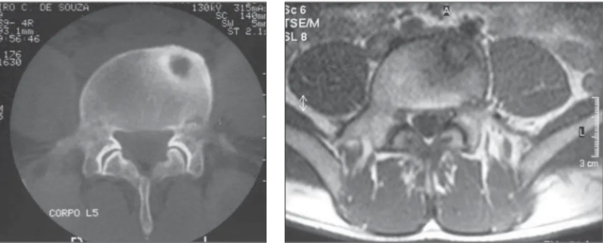
Figure 3. MRI, axial, T2-weighted image. Figure 4. Post-resection, axial CT image of the lesion.
X Radiol Bras. 2008 Mai/Jun;41(3):IX–X
Imaging findings
A radiograph of the lumbar spine was requested and did not demonstrate any bone alteration. Technetium-99 bone scin-tigraphy was subsequently performed, demonstrating increased uptake in the fifth lumbar vertebra, compatible with a mild osteogenic process. At computed tomogra-phy (CT), a lytic lesion was observed in the anterior and lateral portions of L5, with an adjacent sclerotic area (Figure 1), which at magnetic resonance imaging (MRI) pre-sented low-intensity signal on T1-weighted, and a slightly high-intensity signal on T2-weighted sequences (Figures 2 and 3).
The patient underwent surgery for com-plete excision of the lesion by anterior, ret-roperitoneal approach, with insertion of an iliac bone autograft. Histopathologic analy-sis of the excised material demonstrated a tumor nidus constituted by thick vascular bars of osteoblastic tissue surrounded by vascular fibrous tissue and presence of adjacent sclerotic cortical bone. The patient progressed with pain relief, and a new CT study confirmed the complete excision of the lesion (Figure 4).
Diagnosis: Osteoid osteoma of the L5 vertebral body.
COMMENTS
Osteoid osteoma was firstly described in 1935 by Jaffe(1), and represents about 10% of all benign bone tumors, affecting mainly the metaphysis and diaphysis of lower limbs long bone. In 10–20% of cases, osteoid osteomas affect the spine, particu-larly in the lumbar and thoracic regions(2–4). The prevalence is higher in the male population, especially in the age range be-tween five and 25 years, the differential diagnosis being important in dorsodynia, particularly in children and adolescents(5). Microscopically, this lesion consists of a well delimited nidus with a bone growth pattern, containing numerous osteoblasts producing osteoid and bone tissue, very simi-lar to osteoblastoma, although this later is a typically more aggressive lesion, without sclerotic ring and with a nidus > 1.5 cm in its largest axis(4,6). Other differential diag-noses are osteosarcoma and osteomyelitis. In cases where the axial skeleton is in-volved, the posterior spinal elements are affected. Presentation in the vertebral body, like in the present case, is unusual, (2–4,7).
Clinically, the main symptom is pain which classically worsens at night and is well-relieved with non-steroidal anti-in-flammatory and salicylate drugs. Radicu-lar symptoms may be or not be present, and neurological deficit is rarely detected(2–4). Frequently, scoliosis is associated, with non-rotational curves, typically with the lesion localized on the apex of the defor-mity, in the concavity of the curve(2,6). Usu-ally, in cases where the lumbosacral tran-sition is involved, the deformity apex is cranial to the lesion, with presence of pel-vic obliquity(3). In the present case, such deformities were not found, despite the localization of the lesion in the L5.
Radiographically, the lesion shows up like a sclerotic area, and a radiolucent ni-dus may be or not be observed(6). Because of the complex anatomy of the vertebrae, this lesion is hardly or even not visual-ized(4). Bone scintigraphy shows hyperup-take, and is highly sensitive for early as-sessment of patients with back pain and suspicion of osteoid osteoma(3). Computed tomography shows a lytic lesion sur-rounded by sclerosis, with or without cen-tral calcification. Classically, this is consid-ered as the best method for diagnosis and localization of this type of lesion. At MRI, a heterogeneous lesion can be observed, with hyperintense signal on T2-weighted images, and calcification within the nidus (if present) and reactive sclerosis with hypointense signal on all sequences. Bone marrow and soft tissues edema may be present, tending to simulate more aggres-sive lesions such as osteomyelitis and ma-lignant neoplasms(4,8).
The treatment for osteoid osteoma is surgical; nidus ablation must be complete to avoid the lesion recidivation (9–11). Open resection(9–11) or CT-guided percutaneous procedures may be utilized in the lesion ablation(12–14). In the present case, because of the anterior localization of the lesion in the vertebral body, the anterior retroperito-neal approach was adopted, with curettage and implantation of iliac bone autograft.
Arthrodesis must be performed in cases where the lesion is localized in the poste-rior spinal elements and the spinal stabil-ity may be affected as a result of the lesion excision(11).
Several methods are available for an accurate intraoperative localization of the
lesion: preoperative radioactive isotope injection, specimen CT, preoperative iden-tification with Kirschner wire or simply intraoperative radiography(3). None of these methods were utilized in the present case, however, during the clinical evaluation, a complete curettage of the nidus was ob-served, and later confirmed by CT at the postoperative follow-up.
The vertebral involvement is relatively unusual in cases of osteoid osteomas typi-cally localized in posterior spinal elements. Localization in the vertebral body is rare. This lesion is a relevant cause of back pain, particularly in children and adolescents and especially in the presence of secondary scoliosis.
REFERENCES
1. Jaffe HL. Osteoid osteoma: a benign osteoblastic tumor composed of osteoid and atypical bone. Arch Surg. 1935;31:709–28.
2. Domans JP, Moroz L. Infection and tumors of the spine in children. J Bone Joint Surg Am. 2007;89 Suppl 1:S79–97.
3. Crist BD, Lenke LG, Lewis S. Osteoid osteoma of the lumbar spine: a case report highlighting a novel reconstruction technique. J Bone Joint Surg Am. 2005;87:414–5.
4. Defino HLA, Pereira CU, Barbosa CSP. Tumores benignos e lesões pseudotumorais da coluna ver-tebral. Rio de Janeiro: Revinter; 2002. 5. Basile Jr R, Barros Filho TEP, Bonetti VL, et al.
Dor nas costas em crianças e adolescentes. Rev Bras Ortop. 1994;29:144–8.
6. Ozaki T, Liljenqvist U, Hillmann A, et al. Osteoid osteoma and osteoblastoma of the spine: experi-ences with 22 patients. Clin Orthop Relat Res. 2002;(397):394–402.
7. MacLellan DI, Wilson FC Jr. Osteoid osteoma of the spine. A review of the literature and report of six new cases. J Bone Joint Surg Am. 1967;49: 111–21.
8. Assoun J, Richardi G, Railhac JJ, et al. Osteoid osteoma: MR imaging versus CT. Radiology. 1994;191:217–23.
9. Maiuri F, Signorelli C, Lavano A, et al. Osteoid osteomas of the spine. Surg Neurol. 1986;25: 375–80.
10. Pettine KA, Klassen RA. Osteoid osteoma and osteoblastoma of the spine. J Bone Joint Surg Am. 1986;68:354–61.
11. Avanzi O, Joilda FG, Dezen EL, et al. Tumores benignos e lesões pseudotumorais da coluna ver-tebral. Análise de 60 pacientes. Rev Bras Ortop. 1996;31:131–42.
12. Labbé JL, Clement JL, Duparc B, et al. Percuta-neous extraction of vertebral osteoid osteoma under computed tomography guidance. Eur Spine J. 1995;4:368–71.
13. Mazoyer JF, Kohler R, Bossard D. Osteoid osteoma: CT-guided percutaneous treatment. Radiology. 1991;181:269–71.
