ABSTRACT
treatment on bond strength to enamel after tooth
bleaching: an
in situ
study
!"#1, Sandra Kiss MOURA2$%&'()(!3$!*'+-+'0!#1,
Renata Corrêa PASCOTTO1
1- Department of Dentistry, State University of Maringá, Maringá, PR, Brazil.
2- Department of Dentistry, University North of Paraná (UNOPAR), Londrina, PR, Brazil. 3- Private practice, Maringá, PR, Brazil
4-- Renata Corrêa Pascotto - Av. Mandacaru, 1550 - bloco S/08 - 87.080-000 - Maringá - PR - Brasil - Phone: +55 44 9982-0215 - +55
+'89:-';<$=>-:'-<$>>+98$
O
insitu
human enamel restored after tooth bleaching. Material and Methods: Forty human teeth 8 specimens each: Gct (control group, restored on unbleached enamel); Gbl (restored !"#$%&' *& min and restored); G7d (bleached, exposed to saliva in situ for 7 days and restored); and G14d (bleached, exposed to saliva in situ %+ -/
cut into 0.8 mm23 -5
6 9$ $ -< $ =>?@A>3Bpost-hoc test (αC&-&$
as the experimental unit. Results: Mean bond strength results found for Gbl in comparison " 9 #<D&-&%-E $ 9 "$ " "H #<I&-&-J "%+9 "#<D&-&%-K - L J and exposure to human saliva in situ H-J exposure to human saliva in situ %+- *& H techniques to be performed.
Key words: Tooth bleaching. Dental enamel. Saliva. Antioxidants. Adhesives.
INTRODUCTION
3 9 for tooth-bleaching treatments14. Despite the
9 $ demonstrated that bleaching agents might have enamel structures1,20. Mineral loss, increased
surface roughness, decreased microhardness,
decreased fracture toughness, and reduced enamel $ H' after bleaching, have already been reported1,4,16,20,26.
Changes in the amount, length, and morphology of resin tags observed in studies using scanning electron microscopy (SEM) suggest decreased penetration of resin materials into bleached enamel29. Clinically, this decrease is important
Y17.
Enamel surface alterations after tooth bleaching, such as those mentioned above, have been found to be more pronounced under in vitro than in in
situ settings1,11- 9
ability of saliva to prevent such alterations in the dental structure1, and suggests the reversibility of
bleaching-related changes. By placing bleached $ the effects of salivary antioxidants on enamel can be better observed26. Hence, in situ methodologies
have been used to estimate the time needed to reverse bleaching-related effects on enamel $ bleaching teeth can be restored1. Some authors
\+ ] 3 procedures after bleaching5,23,29$
H 28
and 14 days for dentin2. Nevertheless, in some
$ to have their teeth rehabilitated. In order to permit immediate restorations after bleaching, several researchers have attempted the use of different antioxidant agents prior to performing restorative $ 12,14,16,27.
$ Y
= - shed some more light on the issue, the objective
in situ%&'
gel on composite bond strength to human enamel after bleaching.
MATERIAL AND METHODS
5w of Maringá Research Ethics Committee (protocol *&z{\&&z- A informed consent before participating in the study.
Dental specimen preparation
Fifty sound extracted human teeth (premolars $
18$ \'
#EH-&]& &-> 3 # face for premolars and vestibular or proximal face
#Diamond Wheel
&%\}9$5 J $L>$w5>)
adapted to a sectioning machine (Isomet 1000, J$~3J$~$w5> $ - Y $ 3 minimum height of 3.1 mm. After inspection under optical microscopy 20X (BEL MicroImage Analyzer, Bel Photonics, Monza, Milano, Italy), 10 specimens 3$ from the study.
Before their use in the experiment, specimens = %& $ $ "#
resin); Gbl (specimens restored immediately after !"# sodium ascorbate gel for 60 min before restoration); G7d (bleached specimens restored after being exposed to saliva in situ for 7 days); and G14d (bleached specimens restored after being exposed to saliva in situ for 14 days). Figure 1 illustrates the steps involved in the different experimental
-Bleaching procedure
Specimens belonging to Gbl, Gsa, G7d and "%+ 9 3 (Cera utilidade rosa Epoxiglass, Epoxiglass Ind. Com. de Produtos Químicos Ltda., Diadema, SP, J6 expose the enamel surface. A thin layer (1 mm) ]H-' =
#< 9 $ 5~$ J$ A $
Australia
->$ 9 tipped suction device (Sug-Plast, DFL Indústria e Comércio S.A., Rio de Janeiro, RJ, Brazil) and - + $ -5 +&$ ]& $
-Treatment with sodium ascorbate
5 9 3 $ "%&' sodium ascorbate gel (pH 7) manipulated according to Kimyai and Valizadeh14 (2006) shortly before its
$ %&&' humidity for 60 minutes. After this period specimens ]& $
-Exposure to saliva in situ
To evaluate the effect of exposure time to human saliva on composite bond strength to bleached $] # - $
diseases, n = $
systemic disorders!
smokers and current use of orthodontic appliances 9 -> (Jeltrate Dustless, Dentsply, York, PA, USA) of the 3$ # A$$ 3$<>$w5> for each volunteer. Polyvinyl siloxane blocks (Elite HD+, Zhermack Clinical, Badia Polesine, Rovigo, --- cyanoacrylate-based glue (Super Bonder, Loctite,
São Paulo, SP, Brazil)-
fabricated in acrylic resin (JET, Clássico Artigos Odontológicos Ltda., Campo Limpo Paulista, SP, Brazil). The 16 specimens to be assessed in situ
#"H"%+93#L utilidade Asfer, ASFER – Indústria Química Ltda., São Caetano do Sul, SP, Brazil) to the cavities left by the polyvinyl siloxane blocks in the intraoral # * - 3 $9
-Each volunteer received their palatal device day and night. It could only be removed during $ Y # $ - $ 6 - A
$ = -@ $#"H removed from the palatal devices (2, 3 and 3), $ the palatal devices until the morning of the 15th
$ specimens (G14d) removed. After exposure to $ ]& $
-Restoration with composite resin
J $ 3 # Otto inox, Arminger & Cia Ltda, São Leopoldo, /5$J6- ]H' ]& ]& ->$ #> Single Bond 2, 3M-ESPE, 3M do Brasil, Campinas, 5<$ J6 $ \& jets of air for 5 seconds. Composite resin (Filtek Z350, 3M-ESPE, 3M do Brasil, Campinas, SP, J6 % light activated (600 mW/cm2 =
\+-Microtensile testing
$9 brackets of a precision sectioning machine (Isomet %&&&$J$~3J$~$w5>3-5 # Corp. 12205) perpendicularly through the tooth/ restoration interface into 0.9 mm slices. Each slice 9 be further sectioned as to obtain tooth/restoration 3} =
0.8 mm2-
digital caliper (Zaas Precision, Amatools, Piracicaba, 5<$J6-> $3 %&&' testing.
3 jig (Odeme Biotechnology, Joaçaba, SC, Brazil) cyanoacrylate glue (adhesive ZAP and Zip Kicker accelerator, Colas, Pacer Technology, USA), leaving just the enamel/resin interface exposed. This 3 3 centralized on the device and tension distribution
-53
tensile-tested at 0.5 mm/min in a universal testing machine (EMIC DL 2000, São José dos Pinhais, </$ J6 - ? divided by the cross-sectional area in mm2 and
<-
$ under an optical microscope 40X (Bel MicroImage Analyzer, Bel Photonics, Monza, Italy), and fracture
9 7:
> $ fracture at the interface;
Cohesive fracture – enamel substrate failure only;
Cohesive fracture – restorative material failure only; or
Mixed fracture – enamel substrate and resin materials fractures (adhesive or resin composite) in the same test specimen.
Sticks that presented pretest failures, i.e., broke $
from the analysis21- $ 9
9 $]&' analysis under scanning electron microscopy (SEM). 53 $ = = # L $L= 50, Shimadzu Biotech), and observed under SEM (SS-550 Superscan, Shimadzu Biotech, Japan). @ 3 } } $ %&' $ statistical analysis10,25.
Statistical analysis
To guarantee independence of data, and at the same time reduce the variability, average 3 originating from each dental specimen21. As the
$ 6 = variance (ANOVA) and Tukey’s post-hoc$'
9-RESULTS
= 3 statistically homogenous (0.70–0.92 mm2, P=0.52),
indicating that differences in bond strength among = sectional area.
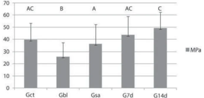
#5 " 39.61 (±13.71), Gbl 25.89 (±11.38), Gsa 36.27 (±16.04), G7d 43.60 (±15.24), and G14d 49.26 (±12.97) MPa (Figure 2). Results found for Gbl in comparison to Gct indicate that bleaching significantly reduced enamel adhesiveness #<D&-&%- E $ "$""H #<I&-&$ sodium ascorbate and exposure to saliva for 7 days recovered bond strength to enamel to the same level of unbleached specimens. Although bond "%+ ""H$9 that found for Gsa (P<0.01).
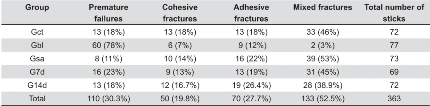
Table 1 illustrates premature failures and fracture mode15 observed in the different experimental
groups. Qualitative microscopic analysis (optical and SEM) revealed that fractures modes (Figures ]+ #\'$ #\' #\&'-Y
I' Interface of a test specimen from G7d seen with scanning electron microscopy. Typical fracture mode was !"#$%&'"*<=$
I' \ ?' ' & @ E F
'"*< =$
Group Premature
failures
Cohesive fractures
Adhesive fractures
Mixed fractures Total number of sticks
Gct 13 (18%) 13 (18%) 13 (18%) 33 (46%) 72
Gbl 60 (78%) 6 (7%) 9 (12%) 2 (3%) 77
Gsa 8 (11%) 10 (14%) 16 (22%) 39 (53%) 73
G7d 16 (23%) 9 (13%) 13 (19%) 31 (45%) 69
G14d 13 (18%) 12 (16.7%) 19 (26.4%) 28 (38.9%) 72
Total 110 (30.3%) 50 (19.8%) 70 (27.7%) 133 (52.5%) 363
" #H'$ "H#\]'$" "%+#
%'$"#%%'-DISCUSSION
3 $ 9 report to evaluate the influence of exposure time to saliva in situ (natural antioxidant effect) %&' ascorbate gel (artificial antioxidant effect) on composite resin bond strength to human enamel after bleaching.
The results found for Gbl in comparison " hydrogen peroxide reduced enamel adhesiveness ]'$ previous results1,4,16,20,26. This finding may be
explained by the delayed release of oxygen due to the presence of peroxides and their subproducts, $ produced during the photoactivation17, affecting the
polymerization of resin components15-17,29.
$ ascorbate increased composite resin bonding to -9 antioxidants to be able to immediately restore bond strength to bleached enamel12,14,27-E $$
et al.12 (2008) suggested that to obtain effective
results the exposure time to the antioxidant agent *&-> #"$ results. In another study, Torres, et al.26 (2006)
observed that the treatment of dental specimens \& 9 - B9 9 $ -$ $ $ to some studies12,27, more effective than solutions,
as the active compounds in the gel are released
-Although an in vitro study has demonstrated that the amount of sodium ascorbate required to reduce hydrogen peroxide is directly related to the concentration of the latter, and the reaction kinetics 9 to exert an effect8, the exact time required for the
antioxidant to reestablish bond capacity to bleached enamel in vivo has yet to be established. Lai, et
al.16#\&&\ %&'
ascorbate for at least a third of the bleaching agent - K $ Kimyai and Valizadeh14 (2006) left the antioxidant
] $
-Studies have also reported differences in the -5%&'
%& %&13
or 10 to 480 minutes12 after the application of a
sodium ascorbate gel and solution, respectively. $ ]' peroxide, the effective reversal of reduced bonding
%&\&12,26 of exposure
%&'
-Sazaki, et al.24 (2009) attempted to simulate
= 9 -J %&' solution, perhaps due to the fact that the antioxidant 9 incubated for 2 hours. In the present study, dental 3 %&&' 9$ -$ $ restore bond strength to enamel more effectively.
Therefore, in the present study, the treatment %&' *& be an effective clinical option for patients in need of
= "$-9
(2012), the authors reported on restorations carried %&' -demonstrated that no alterations in restoration $ 9$= little time-consuming. One of the limitations of $ $ 6 $ less reductive19. To circumvent this problem in
the present study, the sodium ascorbate gel used -Nevertheless, further investigations are needed to determine its shelf life.
? "$ "H$ "%+ $ exposure to saliva for 7 days in situ (Figure 2). Although several authors have recommended that H 14 days after bleaching3,12,15,17,23,26,29, 9
hydrogen peroxide concentrations can also Y end of the bleaching treatment and the performance of bonding procedures3. Studies using hydrogen
%+ procedure. The results of the present study support this assumption, as bond strength to bleached enamel after being exposed for 14 days (G14d) 9 found for sodium ascorbate-treated enamel (Gsa).
Results obtained in laboratory studies may differ from the clinical situation, because they commonly
9 3. In situ
studies have the advantage of being nearest to the clinical situation, as bleached dental specimens are placed under the direct effect of human saliva5. Human saliva acts on the oxidative stress
mediated by free radicals, and presents alternative antioxidants, including ascorbic acid and vitamin E10. A previous in situ and in vitro
that although bleaching agents are capable of altering dental enamel surface’s microhardness, $ $ for enamel mineral reposition20. This process helps
$ is an important component in the adhesive strength of enamel22. In an attempt to simulate the clinical
$ antioxidant effect of saliva could be assessed in the reestablishment of adhesive bond strength after bleaching, volunteers in the in situ experiment 3 = containing products. These products have been
6,
B remineralization process.
$ \' $ \' - = tensile as expected. The frequency of premature " #H' than that observed in other groups. During the specimen preparation procedures, microcracks might have been inadvertently produced mainly by the vibrations of the cutting instruments. 5 $ enamel, are responsible for the premature failure of microtensile bond specimens7. The high number
of pretest failures seen in the Gbl group emphasizes the fragility of bonding immediately after bleaching. In order to standardize procedures and avoid $ recommendations set out by Roulet and Van Meerbeek21 #\&&H- <
-5 used to analyze in detail the type of failure, and only those sticks presenting adhesive or predominantly
$ - microtensile tests mentioned by El Zohairy, et al.7
(2010), concerning the high presence of cohesive fractures. Moreover, to minimize the differences in adhesion that could occur in different parts of the $ bond strength for the sticks originating from the same dental specimen.
In conclusion, based on the results found in this study, the use of sodium ascorbate after = 9 Y - E $ $ for at least 7 days after tooth bleaching. From a chemical-molecular perspective, the alterations in the bleached enamel caused by the antioxidant 3
-CONCLUSIONS
%-L reduced by bleaching.
\-%&' to the bleached enamel for 60 minutes, bond unbleached enamel.
]- H$ %&' sodium ascorbate gel and unbleached enamel.
+-
human saliva in situ %+$
9 %&'
-REFERENCES
%=>$5</$K$>- study design on the impact of bleaching agents on dental enamel --\&&z!\%+]=H-\=J L$53/$K K$J/- ]' - Contemp Dent Prac. 2008;9:81-8.
3- Bittencourt ME, Trentin MS, Linden MS, Oliveira Lima Arsati YB, K¡K$K¢ K$- on shear bond strength of resin-based composite restorations. J Am Dent Assoc. 2010;141:300-6.
4- Cavalli V, Giannini M, Carvalho RM. Effect of carbamide peroxide bleaching agents on tensile strength of human enamel. Dent Mater. 2004;20:733-9.
5- Dishman MV, Covey DA, Baughan LW. The effects of peroxide bleaching on composite to enamel bond strength. Dent Mater. 1994;10:33-6.
H= £ >>$ 5 E$ > >$ K6 > - 9 microtensile versus microshear bond testing for evaluation of bond strength of dental adhesive systems to enamel. Dent Mater. 2010;26:848-54.
8- Freire A, Souza EM, Menezes Caldas DB, Rosa EA, Bordin CF, Carvalho RM, et al. Reaction kinetics of sodium ascorbate and dental bleaching gel. J Dent. 2009;37:932-6.
9- Garcia EJ, Mena-Serrano A, Andrade AM, Reis A, Grande RH, ~ >- %&' = =-Eur J Esthet Dent. 2012;7(2):154-62.
%&= "¤¤¥ <$ J ?$ L3 5$ E3 5$ / $ K$-5 % \ = control study. J Periodontol. 2009;80:1440-6.
11- Justino LM, Tames DR, Demarco FF. In situ and in vitro effects - @ Dent. 2004;29:219-25.
12- Kaya AD, Türkün M, Arici M. Reversal of compromised bonding in bleached enamel using antioxidant gel. Oper Dent. 2008;33:441-7.
%]=5$@3 55$/9>$A6E$>>>$E ZN. Comparison of the effect of hydrogel and solution forms of sodium ascorbate on orthodontic bracket-enamel shear bond strength immediately after bleaching: an in vitro study. Indian J Dent Res. 2010;21:54-8.
14- Kimyai S, Valizadeh H. The effect of hydrogel and solution of sodium ascorbate on bond strength in bleached enamel. Oper Dent. 2006;31:496-9.
15- Lago AD, Freitas PM, Netto NG. Evaluation of the bond strength >"-< ~5-2011;29:91-5.
16- Lai SC, Tay, FR, Cheung GS, Mak YF, Carvalho RM, Wei SH, et al. Reversal of compromised bonding in bleached enamel. J Dent Res. 2002;81:477-81.
17- Miguel LC, Baratieri LN, Monteiro S Jr, Ritter AV. In situ effect %&' = novel pilot study. J Esthet Restor Dent. 2004;16:235-42.
18- Moura SK, Pelizzaro A, Dal Bianco K, Goes MF, Loguercio AD, Reis A, et al. Does the acidity of self-etching primers affect bond strength and surface morphology of enamel? J Adhes Dent. 2006;8(2):75-83.
19- Muraguchi K, Shigenobu, S. Suzuki S, Tanaka T. Improvement of bonding to bleached bovine tooth surfaces by ascorbic acid treatment. Dent Mater J. 2007;26:875-81.
20- Pinto CF, Oliveira R, Cavalli V, Gianninni M. Peroxide bleaching agent effects on enamel surface microhardness, roughness and morphology. Braz Oral Res. 2004;18:306-11.
21- Roulet JF, Van Meerbeek B. Editorial: Statistics: a nuisance, a tool, or a must? J Adhes Dent. 2007;9:287-8.
22- Sa Y, Sun L, Wang Z, Ma X, Liang S, Xing W, et al. Effects of = 9E of human enamel: an in situ and in vitro study. Oper Dent. 2013;38(1):100-10.
23- Santana FR, Pereira JC, Pereira CA, Fernandes Neto AJ, Soares L - bond strength of indirect composite resin restorations to dentine. Braz Oral Res. 2008;22:352-7.
\+= 53 /$ K¢ K$ J /- %&' %&' = on the shear bond strength of enamel and dentin submitted to a home-use bleaching treatment. Oper Dent. 2009;34:746-52. \=555$L<K$5A- strength results of the different test methods: a critical literature
--\&%&!\*H=z]-26- Torres CR, Koga AF, Borges AB. The effects of anti-oxidant agents as neutralizers of bleaching agents on enamel bond strength. Braz J Oral Sci. 2006;5:971-6.
27- Türkün M, Çelik EU, Kaya AD, Arici M. Can the hydrogel form of sodium ascorbate be used to reverse compromised bond strength after bleaching? J Adhes Dent. 2009;11:35-40.
\=w?$L 3K$@6K- bleaching on the shear bond strength of composite to enamel. J Biomed Mater Res B Appl Biomater. 2008;84:363-8.