Case report
A 50-year-old never-smoking female, born and raised in the city of Goiânia, Brazil, presented with a two-year history of productive cough. She had no other complaints. The only finding during the physical examination was scattered fine rales on pulmonary auscultation.
After the initial investigation, the patient was diagnosed with bronchial hyperresponsi-veness associated with gastroesophageal reflux
Introduction
Microaspiration of lipid substances can lead to lipoid pneumonia.(1) The reported cases have been associated with the therapeutic use of mineral oil as a laxative by individuals with chronic constipation. However, other forms of treatment with lipid preparations might be implicated. We report the case of a female patient with chronic exogenous lipoid pneumonia secondary to the use of evening primrose oil.
Lipoid pneumonia secondary to long-term
use of evening primrose oil*
Pneumonia lipoide secundária ao uso prolongado de óleo de prímula
Marcelo Fouad Rabahi, Andreia Alves Ferreira, João Gabriel Piccirilli Madeira, Paulo Menzel Galvao, Sebastião Alves Pinto
Abstract
Lipoid pneumonia is an underdiagnosed disease that is caused by the aspiration of lipid particles into the lungs. Although most of the reported cases have been associated with the use of mineral oil as a laxative, other lipid substances can also cause the disease. We report the case of a 50-year-old female patient with a complaint of productive cough who was initially diagnosed with bronchial hyperresponsiveness and gastroesophageal reflux disease (GERD). The patient was treated for GERD. Because the productive cough persisted, the patient underwent chest CT, fiberoptic bronchoscopy, and open lung biopsy. She was diagnosed with lipoid pneumonia. The patient was questioned regarding the use of lipid substances, and she reported the chronic use of evening primrose oil. After the discontinuation of the substance and the maintenance of GERD treatment, her condition improved.
Keywords: Pneumonia, lipid; Cough; Plant oils.
Resumo
A pneumonia lipoide é uma doença pouco diagnosticada, causada pela aspiração de partículas oleosas para dentro dos pulmões. Os casos relatados têm sido relacionados ao uso de óleo mineral como laxativo, mas outras soluções oleosas também podem causar a doença. Relatamos o caso de uma paciente de 50 anos com queixa de tosse produtiva, sendo diagnosticada inicialmente com hiper-reatividade brônquica e doença do refluxo gastroesofágico (DRGE). A paciente foi submetida a tratamento para DRGE. Devido à persistência da tosse, a paciente foi submetida a TC de tórax, fibrobroncoscopia e biópsia pulmonar a céu aberto, sendo diagnosticada com pneumonia lipoide. A paciente foi questionada quanto ao uso de substâncias oleosas, relatando o uso crônico de óleo de prímula. Com a suspensão do uso da substância e a continuidade do tratamento para DRGE, houve melhora do quadro.
Descritores: Pneumonia Lipoide; Tosse; Óleos vegetais.
* Study carried out at the Federal University of Goiás School of Medicine, Goiânia, Brazil.
Correspondence to: Marcelo Fouad Rabahi. Avenida B, 483, Setor Oeste, CEP 74110-030, Goiânia, GO, Brasil. Tel 55 62 3521-3333. E-mail: mfrabahi@terra.com.br
Financial support: None.
involvement due to the accumulation of lipid material in the alveoli.(3)
Chronic lipoid pneumonia has been found to develop after the prolonged use of mineral oil—as an oral laxative (for constipation) or as nasal drops (for chronic rhinitis)—animal oils, such as cod liver oil (as a dietary supplement for children)—or vegetable oils, such as sesame oil (for balancing cholesterol levels).(4) In contrast, acute chronic pneumonia results from massive accidental aspiration of lipid material, as can occur in “fire eaters”.(5)
There are, however, other types of oil that are used therapeutically and can, therefore, cause lipoid pneumonia. In the case reported here, the patient had a 12-year history of taking evening primrose oil (a vegetable oil) for the disease (GERD). The diagnosis was confirmed
by bronchoscopy, which revealed thickening of the posterior commissure, as well as yellowish mucus in the middle lobe bronchi and the lingula. Treatment with omeprazole (40 mg/day), domperidone, and an inhaled corticosteroid was initiated, and there was partial improvement.
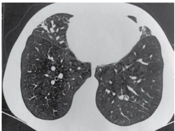
Because the productive cough persisted, the patient underwent chest CT, which revealed opacities in the middle lobe, the inferior segment of the lingula, and the anterior segment of the right upper lobe, as well as thickening of the peribronchial sheaths and signs of mucoid impaction (Figure 1). A second fiberoptic bronchoscopy revealed the persistence of a large quantity of diffuse yellowish mucus, and the microbiological analysis of the material revealed the growth of Staphylococcus aureus (50,000 CFU/mL). The patient underwent treatment with ciprofloxacin (500 mg, bid) for 21 days, after which she remained symptomatic.
Since the condition recurred, the patient underwent open lung biopsy. The histopathological study of the biopsy specimen revealed dense inflammatory lymphoplasmacytic peribronchiolar infiltration and lipid granuloma, leading to the diagnosis of lipoid pneumonia (Figure 2). The diagnosis was confirmed by the study of the BAL fluid, which revealed foamy macrophages surrounded by neutrophils and cell remnants (Figure 3).
The diagnosis of GERD was insufficient to explain the lipoid pneumonia. Upon further questioning, the patient reported that, for the last 12 years, she had been using an herbal remedy for the treatment of premenstrual tension syndrome and anxiety. She was asked to produce the medication, and we found that it was evening primrose oil.
The use of the evening primrose oil was discontinued, antireflux treatment was maintained, and there was progressive improvement.
Discussion
Exogenous lipoid pneumonia results from acute or chronic aspiration/inhalation of lipid particles into the lungs.(1) It was first described by Laughlen in 1925.(2) Exogenous lipoid pneumonia consists of a chronic inflammatory reaction of the lung parenchyma with interstitial
Figure 1 - Chest CT scan showing opacities in the middle lobe and in the inferior segment of the lingula.
Figure 2 - Histopathological study of the open lung
the lipid material present in the tracheobronchial tree is aspirated.(5)
The pathophysiology of lipoid pneumonia caused by aspiration of mineral or vegetable oil is basically that of a foreign body reaction in the lung. The aspirated oil is emulsified and phagocytosed by alveolar macrophages, which, filled with oil, reach the interlobular septum through the lymphatic channels. Therefore, there is thickening of the alveolar walls and some alveoli are destroyed. At a later stage, fibrotic proliferation can result in decreased lung volume.(4) Most of the oil coalesces, forming large fat drops surrounded by fibrous tissue and giant cells, creating a tumor mass known as paraffinoma.(10) Repeated massive aspiration results in diffuse parenchymal consolidation, similar to that found in lobar pneumonia. Severe destruction of the lung architecture can lead to terminal lung disease or cor pulmonale.(4)
The clinical profile of lipoid pneumonia is nonspecific and depends on the length of exposure and the quantity of oil that has been aspirated or inhaled. Patients can be asymptomatic or can present with dyspnea, with or without chronic productive cough. Less commonly, lipoid pneumonia patients have chest pain, hemoptysis, weight loss, and intermittent fever.(7) The clinical profile can be similar to that of bacterial pneumonia, with episodes of fever and cough.(5)
In general, what happens is a discrepancy between the clinical findings and the radiological findings. Patients for whom the clinical findings are unfavorable present with various radiological findings, usually including multiple lobe involvement and opacities (in the lung bases or throughout the lungs).(11) Because the examination lacks specificity, lipoid pneumonia can be confused with lung cancer, especially when oil ingestion has not been reported.(5)
Among imaging tests, HRCT is the best suited for diagnosing lipoid pneumonia, because it reveals alveolar consolidations, ground-glass opacities, nodular lesions, interlobular septal thickening, and intralobular interstitial thickening.(5) In the clinical case in question, the chest X-ray abnormalities, even taken together with the clinical abnormalities, were not sufficient to make the diagnosis. Further investigation (microbiological analysis of the BAL fluid) revealed S. aureus even after the 21-day treatment of premenstrual tension syndrome.
This herbal remedy is also used for treating other diseases, such as atopic dermatitis, rheumatoid arthritis, diabetic neuropathy, multiple sclerosis, menopausal symptoms (hot flashes), and mastalgia. However, its efficacy has yet to be established.(6)
Lipoid pneumonia is often triggered by risk factors such as gastrointestinal diseases, psychiatric diseases, and loss of consciousness or neurological diseases that affect the swallowing process or the cough reflex.(7) The patient in the case reported here was diagnosed with GERD, and, clinically, her only complaint was chronic cough. In patients with normal chest X-rays, GERD is the third leading cause of chronic cough, with a prevalence ranging from 21% to 41%.(8)
The pathophysiology of the respiratory manifestations of GERD is clarified by two major theories that are not mutually exclusive: the reflux theory, according to which direct contact between aspirated gastric contents and the airways damages the mucous membranes; and the reflex theory, according to which contact between gastric acid and the mucosa of the esophagus triggers an esophagobronchial reflex, which is mediated by the vagus nerve, causing the symptoms.(9)
When lipid herbal remedies are ingested, they remain supernatant within the stomach.(7) According to the reflux theory, the lipid particles would ascend and be preferably directed toward the lower airways. Since mineral oil and similar substances inhibit the cough reflex, as well as the mucociliary transport function of the epithelium,
Figure 3 - Photomicrograph of the BAL fluid showing
one of those substances is the etiologic agent of the disease.(14)
References
1. Laurent F, Philippe JC, Vergier B, Granger-Veron B, Darpeix B, Vergeret J, et al. Exogenous lipoid pneumonia: HRCT, MR, and pathologic findings. Eur Radiol. 1999;9(6):1190-6.
2. Laughlen GF. Studies on Pneumonia Following Naso-Pharyngeal Injections of Oil. Am J Pathol. 1925;1(4):407-14.
3. Sias SM, Ferreira AS, Daltro PA, Caetano RL, Moreira Jda S, Quirico-Santos T. Evolution of exogenous lipoid pneumonia in children: clinical aspects, radiological aspects and the role of bronchoalveolar lavage. J Bras Pneumol. 2009;35(9):839-45.
4. Kennedy JD, Costello P, Balikian JP, Herman PG. Exogenous lipoid pneumonia. AJR Am J Roentgenol. 1981;136(6):1145-9.
5. Marchiori E, Zanetti G, Escuissato DL, Souza Jr. AS, Neto CA, Nobre LF, et al. Pneumonia lipoídica em adultos: aspectos na tomografia computadorizada de alta resolução. Radiol Bras. 2007;40(5):315-9.
6. Kleijnen J. Evening Primrose Oil: Currently used in many conditions with little justification. BMJ. 1994;309:824-5.
7. Spickard A 3rd, Hirschmann JV. Exogenous lipoid pneumonia. Arch Intern Med. 1994;154(6):686-92. 8. Sociedade Brasileira de Pneumologia e Tisiologia. II
Diretrizes Brasileiras no Manejo da Tosse Crónica. J Bras Pneumol. 2006;32(Suppl 6):S403-S446.
9. Gastal OL, Palombini BC, DeMeester TR, Gastal CP, Corrêa da Silva MM, Macedo S. Influência dos níveis de refluxo gastroesofágico (RGE) na escolha do tratamento de pacientes com tosse crônica. J Pneumol. 1998;24(5):277-80.
10. Adkins D, Bensadoun ES. An 85-year-old man with a lung mass. Chest. 2004;125(3):1121-3.
11. Furuya ME, Martínez I, Zúñiga-Vásquez G, Hernández-Contreras I. Lipoid pneumonia in children: clinical and imagenological manifestations. Arch Med Res. 2000;31(1):42-7.
12. de Albuquerque Filho AP. Exogenous lipoid pneumonia: importance of clinical history to the diagnosis. J Bras Pneumol. 2006;32(6):596-8.
13. Tan JS. Nonresponses and treatment failures with conventional empiric regimens in patients with community-acquired pneumonia. Infect Dis Clin North Am. 2004;18(4):883-97.
14. Veiga Jr. VF. Estudo do consumo de plantas medicinais na Região Centro-Norte do Estado do Rio de Janeiro: aceitação pelos profissionais de saúde e modo de uso pela população. Rev Bras Farmacogn. 2008;18(2):308-13.
course of ciprofloxacin, and, as previously stated, the patient remained symptomatic. The presence of oil in the lungs can predispose to recurrent pneumonia and even to infections with atypical pathogens.(12)
Occasionally, lipoid pneumonia is diagnosed by lung biopsy performed for the investigation of community-acquired pneumonia that fails to respond to antibiotic therapy.(13) Since the case remained unresolved even after fiberoptic bronchoscopy and antimicrobial treatment had been carried out, an open lung biopsy was performed, and the anatomopathological study revealed lipoid pneumonia, a hypothesis that had not been raised during the investigation of the case.
The diagnosis of lipoid pneumonia, when suspected, can be made based on the clinical history and on suggestive complementary test results. The approved diagnostic method for lipoid pneumonia is BAL, through which it is possible to detect the presence of foamy alveolar macrophages with lipid vacuoles stained with Sudan.(3) In the present case, we used Papanicolaou staining in order to reveal such macrophages (Figure 3). Oil ingestion is rarely reported at the time of the first consultation, and the case reported here is a prime example of that. In most cases, except when focused history taking is employed, patients are questioned regarding oil ingestion only after complementary test results indicative of lipoid pneumonia are obtained.
About the authors
Marcelo Fouad Rabahi
Adjunct Professor. Department of Clinical Medicine, Federal University of Goiás School of Medicine, Goiânia, Brazil.
Andreia Alves Ferreira
Medical Student. Federal University of Goiás School of Medicine, Goiânia, Brazil.
João Gabriel Piccirilli Madeira
Medical Student. Federal University of Goiás School of Medicine, Goiânia, Brazil.
Paulo Menzel Galvao
Thoracic Surgeon. São Salvador Hospital, Goiânia, Brazil.
Sebastião Alves Pinto