Special Article
High-resolution computed tomography patterns of diffuse interstitial
lung disease with clinical and pathological correlation*
,**
Padrões tomográficos das doenças intersticiais pulmonares difusas com correlação clínica e patológica
Brett Elicker1, Carlos Alberto de Castro Pereira2, Richard Webb3, Kevin O. Leslie4
Abstract
High-resolution computed tomography (HRCT) is the radiological imaging technique best suited to revealing changes in lung structure. Various HRCT findings, taken together, can represent typical patterns. These patterns, in conjunction with the anatomical distribution of findings and with clinical data, can narrow the differential diagnosis of diffuse interstitial lung disease and, in many cases, indicate the correct diagnosis with a high degree of accuracy. The most common HRCT patterns seen in cases of diffuse interstitial lung diseases are the nodular pattern, linear/reticular opacities, cystic lesions, ground-glass opacities and consolidations. This article reviews the correlations between HRCT patterns and pathologic findings, summarizing the most common causes, as well as detailing the methods of investigation employed in order to diagnose the most common types of chronic diffuse lung disease.
Keywords: Lung diseases, interstitial/pathology; Tomography, X-Ray computed; Diagnostic techniques, respiratory system.
Resumo
A tomografia de alta resolução (TCAR) é a técnica de imagem radiológica que reflete mais de perto as alterações da estrutura pulmonar. Os vários achados tomográficos podem ser combinados para formar padrões típicos. Estes, conjuntamente com a distribuição anatômica dos achados, e com os dados clínicos, podem estreitar o diagnóstico das doenças intersticiais pulmonares difusas, e em vários casos sugerir o diagnóstico correto com alto grau de acurácia. Os padrões mais comuns das doenças intersticiais pulmonares difusas na TCAR são o nodular, linear e reticular, lesões císticas, opacidades em vidro fosco e consolidações. Este artigo revisa as correlações entre os padrões tomográficos na TCAR e os achados patológicos e resume as causas mais comuns e os métodos de investigação para se atingir um diagnóstico nas doenças pulmonares crônicas difusas mais comuns.
Descritores: Doenças pulmonares intersticiais/patologia; Tomografia computadorizada por raios X; Técnicas de diagnóstico do sistema respiratório.
* Study carried out in the Department of Radiology, University of California at San Francisco, San Francisco, CA, USA; the Department of Pulmonary Medicine, Hospital do Servidor Público Estadual de São Paulo/Universidade Federal de São Paulo, São Paulo, Brazil; and the Department of Laboratory Medicine and Pathology, Mayo Clinic Arizona, Scottsdale, AZ, USA.
1. Assistant Clinical Professor of Radiology. University of California at San Francisco, San Francisco, CA, USA.
2. Head of the Pulmonology Department. Hospital do Servidor Público Estadual de São Paulo – HSPE-SP, São Paulo Hospital for State Civil Servants – São Paulo, Brazil.
3. Professor of Radiology. University of California at San Francisco, San Francisco, CA, USA. 4. Professor of Pathology. Mayo Clinic College of Medicine, Scottsdale, AZ, USA.
Correspondence to: Carlos Alberto de Castro Pereira. Av. Iraí, 393, conjs 33/34, Moema, CEP 04083-001, São Paulo, SP, Brasil. Tel 55 11 5543-8070. E-mail: [email protected]
Financial support: None.
Submitted: 3 March 2008. Accepted, after review: 5 March 2008.
**A versão completa em português deste artigo está disponível em www.jornaldepneumologia.com.br
Introduction
High-resolution computed tomography (HRCT) is a crit-ical tool for the evaluation of lung disease. Because HRCT provides a global anatomic assessment of the lung, this imaging technique improves significantly the sensitivity and specificity of clinical and histopathological diagnosis. HRCT is particularly helpful in the evaluation of diffuse interstitial lung disease (DILD), as clinical presentation and histopatho-logic patterns can show significant overlap and there can be
significant heterogeneity of disease throughout the lung. These modalities together provide a clinical-radiological-histopathological approach to the patient with DILD that allows for accurate diagnosis and optimal management.
resolution and is a low radiation-dose technique. Volumetric imaging allows assessment of the entire lung.
Images are usually obtained in the supine and prone position. In normal patients, dependent lung opacity is often seen in the posterior, subpleural regions of the lung. In certain diffuse lung diseases, such as nonspecific interstitial pneumonia (NSIP), images can be identical to those seen in normal patients. Prone images will differentiate between these two possibilities, since normal dependent density in the posterior lung will disappear on prone images, whereas true lung disease-related density will persist. Dynamic expiratory images are also obtained to screen for air trapping.
A pattern-based approach to diagnosis
There are four general patterns of HRCT abnor-mality that are fundamental to the interpretation of HRCT scans: 1) reticular opacities; 2) nodules; 3) increased lung opacity; and 4) decreased lung opacity. Within each of these patterns, other features of the images can help narrow the differential diag-nosis, such as the distribution of abnormalities in the axial and coronal planes, as well as the co-occur-rence/overlap of patterns. Our purpose in this article is to describe these basic HRCT patterns, drawing important clinical and histopathological correla-tions. For the interested reader, histopathological findings are presented in greater detail elsewhere.(1)
Pattern 1. Reticular opacities
Thickening of the interstitial connective tissue network of the lung will result in reticular opaci-ties of varying morphology. This thickening can result from fluid/cellular infiltration or deposi-tion of fibrous tissue. Reticular opacities can be divided into interlobular septal thickening, reticu-lation associated with traction bronchiectasis, and honeycombing.
Interlobular septal thickening
Interlobular septal thickening (Figure 1) is defined as thickening of the interstitium that surrounds and delineates the secondary pulmo-nary lobule. Septal lines tend to be straight and 1-2 cm in length. The centrilobular artery, which lies in the center of the pulmonary lobule, is often (e.g., the typical lowest magnification microscope
objective has a field diameter of 1 cm), HRCT does allow for visualizing abnormalities at the level of the secondary pulmonary lobule. In this review, we present the key HRCT patterns in DILD, providing the clinical context and histopathological correla-tions for each.
Technique
There are multiple protocols in current use for obtaining HRCT scans. Examples of two such protocols—spaced axial images and multidetector volumetric HRCT—are given in Chart 1. Axial imaging has the advantage of having slightly better
Chart 1 - High-resolution computed tomography techniques.
Inspiration
Spaced axial imaging
Supine and prone positions 1-mm collimation
Axial images acquired at 1-cm intervals Pitch 1, standard gantry rotation speed, fixed or auto mA
Reconstruct with a high spatial frequency or edge enhancing algorithm
or
Multidetector HRCT Supine position Volumetric helical CT
0.625-mm to 1.25-mm detectors
Pitch 1, standard gantry rotation speed, fixed or auto mA
Reconstruct with a high spatial frequency or edge enhancing algorithm
Spaced axial prone scans as indicated above, if desired
Dynamic expiration
6 consecutive images during forced expiration at 1-s intervals
Obtain at three levels Aortic arch Tracheal carina Above diaphragm 1-mm collimation
Pitch 1, standard gantry rotation speed, low dose (e.g. 40 mA)
Reconstruct with edge enhancing algorithm
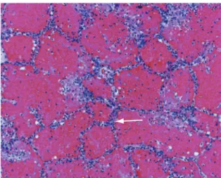
echocardiography can reveal diastolic dysfunction, such as mitral reflux, left atrial enlargement, and elevated pulmonary artery pressure. Brain natriu-retic peptide levels can also be elevated.(2) To rule out myocardial infarction, which can be asympto-matic, cardiac enzyme levels should be determined and an electrocardiogram should be performed. Elevated creatinine levels can indicate underlying renal failure, another cause of pulmonary edema. An example of pulmonary edema in a lung biopsy is presented in Figure 2.
Lymphangitic spread of tumor
Lymphangitic lung metastases can result from pulmonary and extrapulmonary tumors alike. Common extrathoracic origins include breast, stomach, pancreas, and prostate.(3) When lymphangitic metastases originate from a primary lung tumor, the metastases are commonly unilateral and a nodule or mass is seen.(4) In the presence of a visible. A few interlobular septa are often seen in
normal patients. In abnormal patients, however, many will be seen, outlining the polygonal lobules. It is important to note that septal thickening can be seen in a wide variety of diseases. This finding is most useful when it is the predominant abnor-mality, in which case the differential diagnosis is limited and depends upon whether the thickening is smooth, nodular, or irregular (Chart 2). The clinical context, especially information regarding the dura-tion of clinical symptoms and the tempo of disease progression, is extremely helpful in the interpreta-tion of interlobular septal thickening.
Predominant reticular opacities
Clinical and histopathological
correlations
Pulmonary edema
Although HRCT is not usually required for the diagnosis of pulmonary edema, it can be performed when there is a discrepancy between the clin-ical history and findings seen on the chest X-ray. In the absence of an enlarged heart silhouette,
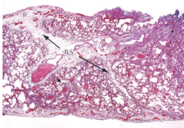
Figure 1 - Interlobular septal thickening in a patient with pulmonary edema. Note the thin, interconnecting lines forming polygonal shaped structures. The pulmonary arteries can be seen at the center of the lobules.
Chart 2 - Differential diagnosis of interlobular septal thickening as the predominant abnormality.
Smooth Nodular Irregular
Pulmonary edema Sarcoid Fibrosis (IPF, HP, sarcoid, etc.)
Lymphangitic spread of tumor Lymphangitic spread of tumor Erdheim-Chester disease
(Non-Langerhans cell histiocytosis)
Lymphoproliferative disease
IPF: idiopathic pulmonary fibrosis; and HP: hypersensitivity pneumonitis.
ILS ILS p
p
p p
the disease, fibrosis can be present, as manifested by irregular reticulation, traction bronchiectasis, and confluent masses of fibrotic tissue.
Erdheim-Chester disease
Erdheim-Chester disease is a rare systemic histi-ocytosis that typically affects the long bones, with lung involvement occurring in 15% of cases.(8) The disease is now recognized as a condition distinct from the systemic forms of Langerhans cell histi-ocytosis (LCH). A diagnosis of Erdheim-Chester disease should be considered in males over the age of 40 who present with diffuse bone pain and DILD. In such cases, metastatic carcinoma is the diagnosis to be ruled out. In patients with Erdheim-Chester disease, pulmonary involvement is suggested by the presence of symmetrical reticular shadows on chest X-rays, interlobular septal thickening (on chest X-rays and CT scans), centrilobular nodular opacities, ground-glass opacities, and fissural thick-ening.(9) The pathology is distinctive, showing bland fibrosis in the pleura and along the lymphatics of the interlobular septa. In contrast to what is seen in known primary tumor, typical HRCT findings can be
considered diagnostic. In the absence of a known primary tumor, the diagnosis requires cellular or tissue confirmation (bronchoalveolar lavage [BAL], pleural fluid collection, transbronchial biopsy, or surgical lung biopsy).(5) Because such tumors are typically distributed among the lymphatics of the bronchovascular bundles, bronchoscopic biopsy is a highly effective diagnostic method (Figure 3).
Sarcoidosis
The diagnosis of sarcoidosis requires a biopsy finding of nonnecrotizing granulomas, together with clinical-radiological findings that are consistent with the disease. Ruling out other causes of granulomatous disease (especially tuberculosis) is of paramount importance.(6) Biopsies should be performed at the most easily accessible sites, such as the skin or superficial lymph nodes. In some cases, fiberoptic bronchoscopy with bronchial and transbronchial biopsies is required, and, as a last resort, mediastinoscopy or surgical lung biopsy can be performed. Nodules are the hallmark of pulmo-nary sarcoidosis, being seen in ≈90% of all cases. Such nodules are widely distributed but tend to be concentrated around bronchovascular structures, the pleura, and the interlobular septa.(7) Transbronchial biopsy is highly effective in confirming the diagnosis (Figure 4). Hilar adenopathy is an expected finding, and there can be a confluence of nodules within larger parenchymal opacities. In the late stages of
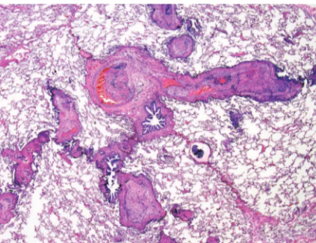
Figure 3 - Hematogenous and lymphangitic carcinoma. Irregular tumor nodules are randomly distributed along vascular and lymphatic routes. (Hematoxylin and eosin; magnification, ×40).
disease, bibasilar, peripheral, traction bronchiectasis accompanied by ground-glass attenuation can be considered diagnostic of NSIP. When the circum-stances are less diagnostic, a surgical biopsy might be required.
Honeycombing
Honeycomb lung remodeling (honeycombing) reflects the end stage of a number of diseases that cause parenchymal destruction. It presents a charac-teristic HRCT pattern, with subpleural, thick-walled cysts that share walls and, when advanced, are often stacked in multiple layers (Figure 6). It is typically accompanied by other signs of fibrosis (traction bronchiectasis and reticulation). Honeycombing is highly suggestive of a pathologic diagnosis of usual interstitial pneumonia (UIP), although it can be attributable to other diseases (Chart 3). Honeycombing seen on HRCT scans is often consid-ered diagnostic of UIP in patients presenting the appropriate clinical profile, and the majority of such patients will not be subjected to surgical lung biopsy. Because bilateral honeycombing on HRCT scans is considered diagnostic under these condi-tions, it is vitally important for the radiologist to be confident that honeycombing is truly present before describing it.
cases of LCH, histiocytes of patients with Erdheim-Chester disease are immunohistochemically negative for S100 protein and CD1a but positive for CD68.
Lymphoid pulmonary lesions
Lymphoid pulmonary lesions constitute a general category of disease that includes follic-ular bronchiolitis, nodfollic-ular lymphoid hyperplasia, lymphoid interstitial pneumonia, and low grade lymphoma.(9) In rare cases, lymphoid hyperplasia can simulate sarcoidosis, since the involvement is concentrated along the septa, in subpleural areas, and around airways (all of the locations of lymphatic channels in the lung).
Traction bronchiectasis
Bronchial dilatation occurring as a consequence of interstitial fibrosis is referred to as traction bron-chiectasis (Figure 5). The bronchi often appear irregular (corkscrewed) and are not associated with radiologic evidence of bronchial inflammation (gross bronchial wall thickening or mucous impac-tion). Traction bronchiectasis is often accompanied by other signs of lung fibrosis (honeycombing or irregular reticulation). While traction bronchiectasis is quite specific for fibrosis, the differential diagnosis is broader than that of honeycombing. Idiopathic pulmonary fibrosis (IPF) is commonly associated with traction bronchiectasis. However, in the absence of honeycombing, other diseases are more likely (Chart 3). In patients with known collagen vascular
Figure 5 - Traction bronchiectasis in a patient with nonspecific interstitial pneumonia. Dilated corkscrew-shaped bronchi are present in the posterior lungs. Note there is no bronchial wall thickening.
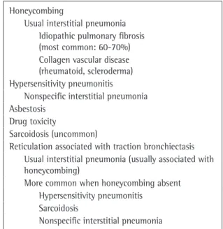
Chart 3 - Honeycombing and reticulation with traction bronchiectasis.
Honeycombing
Usual interstitial pneumonia Idiopathic pulmonary fibrosis (most common: 60-70%) Collagen vascular disease (rheumatoid, scleroderma) Hypersensitivity pneumonitis
Nonspecific interstitial pneumonia Asbestosis
Drug toxicity
Sarcoidosis (uncommon)
Reticulation associated with traction bronchiectasis Usual interstitial pneumonia (usually associated with honeycombing)
More common when honeycombing absent Hypersensitivity pneumonitis
Sarcoidosis
environmental and occupational exposures, use of fibrogenic drugs, and collagen vascular diseases
• abnormal pulmonary function test results
including evidence of restriction, impaired gas exchange (at rest or upon exertion), or
Clinical and histopathological
correlations
Idiopathic pulmonary fibrosis
A surgical lung biopsy finding of UIP is char-acteristic of IPF, which is a chronic idiopathic form of lung fibrosis. The histopathology of UIP is one of destructive fibrosis alternating with normal lung in the surgical biopsy specimen (Figures 7 and 8). A characteristic advancing edge of focal injury is always present and is referred to as “fibroblastic foci”. Clinical diagnostic criteria for UIP were proposed by a consensus committee of the American Thoracic Society/European Respiratory Society in 2000.(10) Since then, the following aspects have become apparent:
• Honeycombing is required for a reliable
HRCT-based diagnosis (i.e., reticular abnor-malities with minimal ground-glass are not sufficient).(11)
• Transbronchial biopsy is rarely indicated in the
diagnostic workup of suspected cases.(12) • The disease is uncommon in patients below
the age of 50.(13)
• The disease can be found in asymptomatic
patients.
Based on these observations, the criteria for the clinical diagnosis of IPF should include all of the following:
• age > 50 years
• exclusion of other potential causes of inter -stitial lung disease (ILD), such as relevant
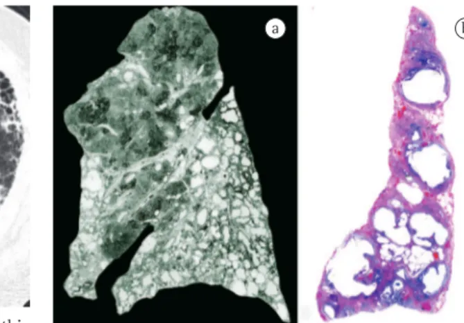
Figure 6 - Honeycombing in a patient with idiopathic pulmonary fibrosis. Subpleural cysts share walls, and some are stacked upon each other.
a b
Figure 7 - Honeycomb cystic remodeling. A. This paper-thin Gough lung section nicely demonstrates the aggregated thick-walled cysts of usual interstitial pneumonia (UIP). Note the lower lobe predominance and tendency for cysts to be present in the subpleural regions. Characteristically the upper lobes are relatively spared. B. At scanning magnification, the characteristic 3-5 mm honeycomb cysts of UIP can be identified in this peripheral lung biopsy. (Hematoxylin and eosin; magnification, ×12.5).
Figure 8 - Usual interstitial pneumonia. Irregular septal fibrosis, with relative centrilobular sparing, can be seen in this photomicrograph. Traction emphysema is present within the lobules, causing dilatation of alveolar spaces. A few fibroblast foci can be seen at the edge of dense fibrosis, where this interfaces with underlying lung (ff). (Hematoxylin and eosin; magnification, ×12.5).
other studies.(17,18) In patients with fibrosing lung disease and having been exposed to HP-provoking antigens, a clinical diagnosis can be accepted, provided that the CT context is appropriate and the BAL cytology reveals lymphocytosis.(19)
Fibrosing sarcoidosis (stage IV)
Advanced (stage IV) fibrosis in sarcoidosis
(fibro-sing sarcoidosis) is found in fewer than 10% of cases. Fibrosing sarcoidosis occurs predominately in the upper lung regions, in a central/dorsal distribu-tion, with bronchial/fissure distortion. Conglomerate hilar-perihilar masses are common. Septal lines, bronchovascular thickening, micronodules, and adenopathy (sometimes calcified) can be observed. When honeycombing is present, it is peripheral and involves the upper lung zones.(20) A pattern simu-lating UIP is quite rare, and, in this setting, there can be fewer granulomas than in the earlier stages of the disease. Histopathologic confirmation of sarcoidosis can be obtained through bronchial or transbronchial biopsies (positive in 60-80%),(20,21) biopsies from other sites, or, rarely, surgical lung biopsy.
Nonspecific interstitial pneumonia
Originally described in 1994 by Katzenstein and Fiorelli,(22) NSIP was introduced as a new form of idiopathic ILD separate and distinguishable from those proposed in Liebow’s original classification. (23) An inflammatory DILD characterized by temporal
uniformity of the disease process, NSIP presents varying degrees of interstitial inflammation or fibrosis. Pure inflammatory disease is rare. The prognosis is better in cases of NSIP than in cases of UIP. Patients with NSIP tend to be younger than do patients with IPF (mean age, 53 vs. 67 years). Most cases occur in the context of an underlying disorder such as connective tissue disease, drug-induced lung disease, or chronic HP.(24) The NSIP histopatholog-ical pattern is the predominant pattern seen in most rheumatic diseases, especially in systemic sclerosis, rheumatoid arthritis, dermatomyositis/polymyositis, and undifferentiated connective tissue disease, the last being a newly described distinct entity.(25) The NSIP pattern is a common presentation of HP.(26) However, in such cases, the distribution on HRCT scans is quite different from that seen in NSIP associ-ated with collagen vascular disease or drug reaction. decreased diffusing capacity of the lung for
carbon monoxide (DLCO)
• bibasilar inspiratory crackles
• HRCT findings of bibasilar reticular abnor -malities with honeycombing and absence of findings suggestive of other diseases (e.g., air trapping, centrilobular nodules, and extensive ground-glass opacities)
The specificity of these findings for IPF is approximately 90%.(14)
Collagen vascular diseases
All of the named rheumatic diseases can produce lung fibrosis. Rheumatoid arthritis and scleroderma are predominately implicated in cases where a UIP HRCT pattern is seen, and with similar functional abnormalities.
Hypersensitivity pneumonitis
mide, 1,3-bis(2-chloroethyl)-1-nitrosourea, and 1-(2-chloroethyl)-3-cyclohexyl-1-nitrosourea), statins, amiodarone, nitrofurantoin, methotrexate, and chest irradiation. Bronchoscopy with transbron-chial biopsy is often required in order to rule out infection. The surgical lung biopsy is not specific for a particular drug in the vast majority of cases.(37)
Asbestosis
Asbestosis is a pneumoconiosis caused by the inhalation of asbestos fibers and is characterized by slowly progressive pulmonary fibrosis. In the early stage of the disease, an irregular reticular pattern is a typical HRCT finding, whereas the cystic pattern is characteristic of the advanced stage. Asbestosis affects workers involved in the extraction of minerals, as well as those engaged in the manufacture and installation of products containing asbestos (indus-trial textiles, insulation, and manufactured cement goods). Asbestosis-related interstitial fibrosis varies in appearance. In some cases, the fibrosis is histopathologically indistinguishable from UIP,(38) although, in most instances, asbestosis is an airway-associated fibrotic lung disease and lacks the typical peripheral lobular accentuation of UIP. Parenchymal bands are more commonly a result of asbestosis. Asbestosis can be diagnosed without lung biopsy in the presence of three clinical signs (a restric-tive pattern of lung impairment, DLCO below the lower limit of the normal range, and bilateral fine crackles at the posterior lung base), together with irregular opacities (on chest X-rays or HRCT scans) and a history of relevant exposure.(39) A diagnosis of asbestosis can also be made based on the co-oc-currence of ILD with typical pleural plaques on CT scans. A finding of asbestos bodies in the BAL fluid is highly specific.(40)
Pattern 2. Nodules
There are several ways to classify nodules: well-defined vs. poorly-defined; upper vs. lower lobe distribution; and relationship to the secondary pulmonary lobule. The last is the most useful char-acteristic, since it provides a focused differential diagnosis and is reflective of the underlying disease pathophysiology. There are three possible HRCT distributions of nodules: perilymphatic, random, and centrilobular (Charts 4, 5, and 6).
This HRCT differentiation is especially helpful because typical histologic findings of HP, such as granulomas, giant cells, and interstitial bronchiolo-centric pneumonia, are absent by definition.
Several HRCT features are suggestive of a diag-nosis of NSIP.(27) Although UIP presents the same subpleural and basilar predominance, ground-glass opacities, which are rarely seen in UIP, are found in more than 75% of cases of NSIP. Reticular abnor-malities, with or without traction bronchiectasis, are common and appear to correlate with the amount of fibrosis observed histopathologically. In the axial plane, subpleural sparing (a thin rim of unaf-fected lung at the pleuroparenchymal interface) and tracking of opacities along lower-zone bron-chovascular bundles are two findings that often correlate with histopathologic findings of NSIP. Honeycombing is rare in NSIP, and there is debate as to whether this should be an exclusionary finding. In the original sample of 64 patients described by Katzenstein and Fiorelli, overall mortality was low and microscopic honeycombing was not present in any of the patients.(22) In a later study conducted by Travis et al.,(28) patients with microscopic honey-combing were included, and overall survival fell significantly. Since those early clinical studies, the reported occurrence of honeycombing on CT imaging in NSIP has been variable, ranging from 0% to 30% (mean, 20%).(29-34) In contrast, extensive honeycombing is much more commonly a manifes-tation of UIP.(35)
The findings described above for NSIP are not specific. Therefore, in the absence of a definable collagen vascular disease or exposure to fibrogenic drugs, surgical lung biopsy is necessary. Some cases of desquamative interstitial pneumonia (DIP), HP (with or without classical histological findings), and several less common diseases can also produce this pattern.
Drug-induced lung disease
cyclophospha-Perilymphatic nodules
Clinical and histopathological
correlations
Perilymphatic nodules are characterized by their distribution in the bronchovascular sheath, pleura, and interlobular septa, corresponding to the lymphatic routes in the lung (Figure 9). All of the conditions in this category necessarily have an affinity for the lymphatic channels.
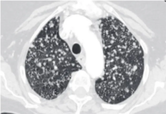
Sarcoidosis
Sarcoidosis is a noninfectious granulomatous disease of likely immune origin. The distribution of nodular granulomas along lymphatic routes is nearly diagnostic of sarcoidosis on HRCT scans. Lung biopsy performed using bronchoscopy might be required for diagnosis in cases in which there are no lesions at sites that are more accessible, such as the skin (except erythema nodosum, in which biopsy shows nonspecific findings) or superficial lymph nodes. A high degree of diagnostic accuracy is achieved if more than four bronchoscopic samples are taken. This is possible because lymphatic chan-nels traverse the bronchovascular bundles in great numbers and are therefore copiously sampled in the transbronchial biopsy specimen.(41) In addition, for a diagnosis of sarcoidosis, the sensitivity and
specifi-city of a CD4/CD8 ratio > 3.5 in the BAL fluid are
52-59% and 94-96%, respectively.(42) A surgical lung biopsy from a patient with sarcoidosis is presented in
Chart 4 - Perilymphatic nodules.
Primary lymphatic diseases or diseases involving lymphatics
Well-defined nodules Patchy, clustered abnormalities Effected structures
Bronchovascular interstitium Centrilobular region Interlobular septa Subpleural region Differential diagnoses
Sarcoidosis
Lymphangitic spread of tumor Silicosis (uncommon) Amyloid (rare)
Lymphoid interstitial pneumonia (rare)
Chart 5 - Differential diagnosis of random nodules.
Hematogenously spread diseases
Rarely lymphatic disease can appear random Uniform, symmetric distribution
Differential diagnoses Miliary tuberculosis
Miliary fungal infection (e.g. histoplasmosis, coccidiodomycosis)
Hematogenous metastases Sarcoid (rare)
Chart 6 - Centrilobular nodules.
Small airways or vascular disease
Most peripheral nodules spaced 5-10 mm from pleura Evenly distributed
Diffuse or patchy Differential diagnoses
Well-defined nodules Endobronchial infection (e.g., bronchopneumonia)
Endobronchial tumor (e.g., bronchioloalveolar cell carcinoma)
Aspiration
Ill-defined ground-glass nodules Hypersensitivity pneumonitis Respiratory bronchiolitis Follicular bronchiolitis Langerhans cell histiocytosis
Vascular causes (e.g., edema and hemorrhage)
noglobulin deficiency, Sjögren’s syndrome, or mixed autoimmune disease with Sjögren’s syndrome.(44,45)
Amyloidosis
Amyloidosis is a disorder of immunoglobulin protein folding in which normally soluble plasma proteins aggregate as an insoluble abnormal fibrillar form causing progressive disruption to tissue struc-ture and organ function. Diffuse amyloid deposition within the lung parenchyma is usually associated with involvement of other organs systems. Perilymphatic nodules are a rare manifestation of amyloidosis.(46)
Random nodules
Random nodules are defined by their seem-ingly haphazard occurrence in peribronchovascular regions, interlobular septa, and pleura, without a consistent perilymphatic pattern and absence of a consistent relationship with the secondary pulmo-nary lobule. An HRCT image from a patient with hematogenous metastases from thyroid cancer is shown in Figure 10.
Clinical and histopathological
correlations
Hematogenous metastasis
Hematogenous metastasis is the most common cause of multiple randomly distributed pulmonary nodules. Basilar predominance is typical, due to preferential blood flow to the lung bases. Since the Figure 4. The lymphatic distribution can be striking
and nicely recapitulates the HRCT findings.
Lymphangitic spread of tumor
For information regarding HRCT findings in cases of lymphangitic spread of tumor, see “Interlobular septal thickening” above.
Silicosis/silicatosis
Inhalation of significant quantities of pure silica dust, and/or mixed dusts with aluminum and magnesium silicates, is responsible for the clinical pneumoconiosis known as silicatosis, or silicosis. Pulmonary nodules visible on chest imaging occur mainly as the manifestation of an occupational disease, with nodules occurring in the centrilobular and subpleural regions. Nodules are distributed in the upper and middle lung regions, with a posterior predominance. Because the disease can persist or progress, even in cases of removal from exposure to silica particles, a detailed patient history should be taken. Given the long latency of the disease, particular attention should be paid to all previous occupations, even those from the remote past. The prevalence of silicosis remains high in certain areas of professional activity in Brazil, such as mining in general, metallurgy, and the manufacture of ceramic/ porcelain floor tiles, as well as sandblasting.(43)
The diagnosis of silicosis is based on the inter-pretation and analysis of radiographic imaging, as well as on the occupational history of the worker/ patient. Histopathological examination is limited to cases in which a discrepancy exists between these two analyses, thereby raising the significant possi-bility of another diagnosis.(43) Transbronchial biopsy is diagnostic in the majority of cases.
Nodular lymphoid hyperplasia
There is considerable ongoing debate regarding diffuse nodular lymphoid hyperplasia in the lung. The lesions are located around the airways and lymphatic routes in the lung. The histopathology reveals numerous apparently reactive lymphoid follicles with germinal centers. Low grade malignant lymphoma of the B-cell marginal zone type is the diagnosis to be ruled out. Diffuse pulmonary nodular lymphoid hyperplasia might be a preneoplastic condition, and some cases are related to
and, adjacent to them, supporting connective tissue with lymph vessels. Therefore, centrilobular nodular opacities can result from bronchiolar and peribronchiolar diseases, as well as from vascular and perivascular diseases. Mosaic attenuation asso-ciated with air trapping on expiratory HRCT, or functional evidence of airflow obstruction indi-cates diseases involving the peripheral airways. Centrilobular nodules are sometimes accompanied by the so-called “tree-in-bud” opacities, in which the abnormality resembles a budding tree. In the majority of cases, the tree-in-bud pattern occurs as a result of infectious diseases. Tree-in-bud is a subtype of a centrilobular pattern. Pathologically, this abnormality represents bronchiolar impaction and is almost always due to infection. The differen-tial diagnosis is detailed in Chart 7.
Clinical and histopathological
correlations
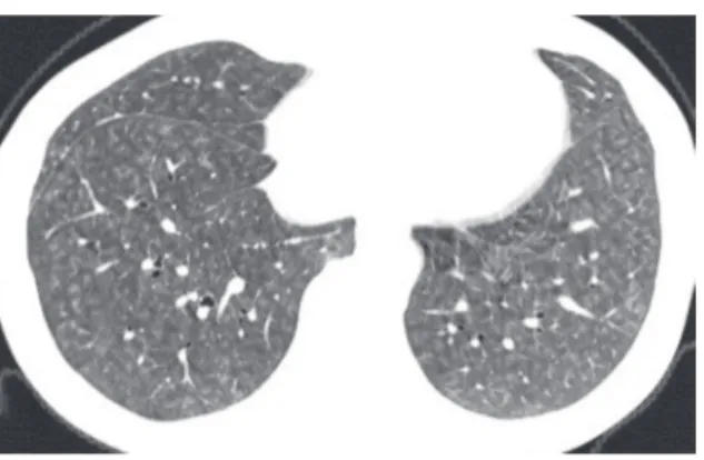
Subacute hypersensitivity pneumonitis
Innumerable ill-defined centrilobular ground-glass nodules with uniform distribution are characteristic of subacute HP (Figure 11).(50) This diagnosis can be confirmed by a history of exposure, clinical symptoms of a flu-like illness, lymphocytosis of the BAL fluid, and clinical improvement when the patient is removed from the offending envi-ronmental agent. In the subacute phase, surgical lung biopsy shows characteristic findings (chronic bronchiolitis, patchy cellular interstitial pneumonia, malignant cells enter the pulmonary lymphatics,
features of lymphangitic carcinomatosis with irregularly thickened interlobular septa and pleural effusion are common. In surgical lung biopsies, irregular nodules of endovascular and endolym-phatic tumor are present (Figure 3).
Although the appearance of miliary nodules has many causes, the most common are metastasis, tuber-culosis, fungal infections, and sarcoidosis.(47) Miliary metastasis are frequently due to thyroid cancer, renal cancer, melanoma, or other malignancies, whereas larger and less profuse metastases tend to be adeno-carcinomas in adults, typically originating from the lung, breast, or gastrointestinal tract.
Miliary tuberculosis
Miliary tuberculosis is often insidious. Reports of the yield of lung tissue and secretion assays in studies of miliary tuberculosis vary widely, prob-ably in part because of the great diversity in cases included in such studies. Overall, roughly 50% of sputum samples collected from suspected cases test positive in cultures. In a study conducted in South Africa,(48) granulomata was found in 30 (63%) of 48 transbronchial biopsies, 20 of which presented necrosis. Of the 30 presenting granulomata, 13 were smear-positive for acid-fast bacilli (AFB). Two sites likely to be involved and accessible for study are the liver and bone marrow. Among liver biopsies, gran-ulomata are found in 88%, necrotizing granulomas in 45%, and AFB in 40%. Among bone marrow biopsies, granulomata are found in 67%, necro-tizing granulomas in 42%, and AFB in 25%.(49)
Miliary fungal infections
Acute disseminated histoplasmosis is an uncommon presentation of miliary fungal infec-tion in adults. In patients with a miliary pattern and granulomas on transbronchial biopsies, without necrosis or infectious agents, an open lung biopsy with tissue culture can be necessary in order to rule out infectious disease and to establish more conclu-sively a diagnosis of sarcoidosis.
Centrilobular nodules
The central part of the secondary pulmonary lobule contains the branches of the terminal bron-chioles, their accompanying pulmonary arteries,
Chart 7 - Differential diagnosis of the tree-in-bud pattern.
Infection
Bronchopneumonia
Tuberculous and nontuberculous mycobacteria Fungal infection
Viral, parasitic (rare) Infectious variants
Cystic fibrosis
Allergic bronchopulmonary fungal disease Noninfectious causes (rare)
co-existing with LCH. Typically, RB-ILD produces poorly-defined centrilobular nodules in small numbers and predominantly in the upper lobes. In cases of RB-ILD, the HRCT findings can simu-late subacute HP, with widespread poorly-formed nodular abnormalities and areas of hypoattenua-tion (mosaic pattern). However, the history is often helpful (RB-ILD occurs only in smokers, whereas HP is rare in smokers) and the BAL profile is usually definitive in distinguishing between RB-ILD and HP. In RB-ILD and respiratory bronchiolitis alike, a characteristic brown pigmentation of macrophages is found. The histopathological difference resides in the extent of peribronchiolar macrophage reac-tion (much greater in RB-ILD). The BAL findings, in conjunction with the history and HRCT findings, usually allow the diagnosis of RB-ILD to be made without the need for thoracoscopic lung biopsy.(51)
Langerhans cell histiocytosis
In smokers, poorly defined centrilobular nodules can also be seen early in LCH, without the presence of cysts. Since LCH is an airway-centered disease, a transbronchial biopsy finding of LCH lesion is not unexpected. Langerhans cells can be found in the BAL fluid of patients with any one of a number of inflammatory conditions, potentially producing a false-positive result if this modality is used as a diagnostic test for LCH.(41) Knowledge of the CT findings is essential for accurate diagnosis. The exact incidence of LCH in the smoking population remains unknown.(52)
Follicular bronchiolitis
Reactive lymphoid hyperplasia, present in a peri-bronchiolar distribution, is referred to as follicular bronchiolitis. In the radiological and pathology liter-ature, follicular bronchiolitis and lymphoid interstitial pneumonia are thought to represent two ends of a spectrum, one being localized to the peribronchiolar regions (the former), and one being more diffuse (the latter). Underlying conditions associated with follicular bronchiolitis includes rheumatoid arthritis, mixed collagen vascular disease, Sjögren’s syndrome, other autoimmune disorders, and inherited or acquired immunodeficiency syndromes.(53,54) The disease can occur in an idiopathic form. Follicular bronchiolitis is a rare cause of a tree-in-bud pattern, and even less commonly occurs in a diffuse form. and small, scattered poorly-formed nonnecrotizing
granulomas in the alveolar walls in peribronchiolar regions; Figure 12), especially when avian antigens are responsible. Plasma cells are typically prominent in the interstitium in HP.
Respiratory bronchiolitis-associated interstitial lung disease
Respiratory bronchiolitis-associated intersti-tial lung disease (RB-ILD) appears to exist within a spectrum of smoking-related DILDs, sometime
Figure 11 - High-resolution computed tomography scan showing centrilobular nodules of ground-glass attenuation. The nodules are evenly spaced and spare the subpleural regions of the lung. This is a typical appearance of hypersensitivity pneumonitis.
Endobronchial spread of Mycobacterium
tuberculosis
Infection with Mycobacterium tuberculosis typically produces a tree-in-bud pattern, which indicates active disease. Associated cavitation is highly suggestive but can be absent.(58)
Infection with M. avium-intracellulare complex
When the tree-in-bud pattern is found in a thin elderly Caucasian woman, infection with M. avium-intracellulare complex should always be a consideration. The radiological manifestations consist of bronchiectasis and multiple centrilobular nodules. Disease is most severe in the lingula and middle lobe.(59) Sputum examination and cultures are essential to establish the diagnosis of
mycobac-Infectious pneumonia
In the immunocompromised host, bacterial (Staphylococcus aureus and Haemophilus influ-enzae), fungal (more commonly Aspergillus spp.) and, quite rarely, viral infection can all result in a tree-in-bud pattern (Figure 13) accompanied by variable consolidation (including cytomegalovirus and respiratory syncytial virus).(55)
Infectious bronchiolitis
In the normal host, acute diffuse bronchiolitis without associated consolidation (Figure 14) can occasionally result from viral or mycoplasma infec-tion.(56,57) Residual bronchiectasis can result. HP, which also can present with diffuse centrilobular nodules, virtually never demonstrates tree-in-bud lesions.
Figure 13 - Tree-in-bud opacities in the right upper lobe reflect bronchiolar impaction. This patient with allergic bronchopulmonary aspergillosis also had regions of cystic bronchiectasis elsewhere.
are the methods of choice for diagnosing infections (tuberculosis, fungi, viruses, and lobular bacte-rial pneumonia), neoplasms (bronchoalveolar cell carcinoma and lymphangitic carcinomatosis), and cryptogenic inflammatory lung disorders (HP, LCH, and sarcoidosis).(66) If the transbronchial biopsy and BAL are negative, surgical lung biopsy can be required.
Pattern 3. Increased lung opacity
Increased lung opacity can be described as ground-glass opacity or consolidation. Ground-glass opacity (Figure 15) is increased lung opacity that does not obscure the associated vessels and represents abnormalities below the resolution of HRCT. Consolidation (Figure 16) is increased lung opacity in which the vessels are obscured and repre-terial infections. Bronchoscopy might be needed to
obtain secretions and biopsies for culture.
Bronchiectasis
Diseases that result in bronchiectasis are commonly accompanied by tree-in-bud lesions.(60) In cystic fibrosis, the tree-in-bud pattern can be an early sign of disease.
Diffuse panbronchiolitis
Diffuse panbronchiolitis is a histopathologically characteristic disease represented by an inflam-matory condition with extensive involvement of the peripheral airways, producing a tree-in-bud pattern with or without associated bronchiectasis. Although the term diffuse panbronchiolitis implies a generic inflammatory disease of the bronchioles, the histopathology is sufficiently distinctive that, once seen, is rarely forgotten and is not easily confused with other inflammatory disorders. The disease is reported primarily in Asians. However, some cases have been described in non-Asian Brazilians.(61,62) The disease is predominantly seen between the second and fifth decade of life. Chronic sinusitis is common. In Western countries, where the disease is rare, a lung biopsy is usually necessary for diag-nosis.(63) Transbronchial or surgical lung biopsy shows a distinctive accumulation of foamy histio-cytes in bronchiolar walls and in the immediate peribronchiolar regions.(64)
Diffuse aspiration bronchiolitis
Chronic inflammatory reaction to repeated aspi-ration of foreign material results in diffuse aspiaspi-ration bronchiolitis.(65) Conditions favoring aspiration, such as esophageal disorders and neurological defects, are typically found.
Tumor emboli
In rare cases, tumor emboli can expand small vessels and produce a tree-in-bud pattern.(55)
Clinical approach to diagnosis in patients
with centrilobular opacities
In cases of the tree-in-bud pattern and centrilob-ular nodules (with or without alveolar/ground-glass attenuation), transbronchial lung biopsy and BAL
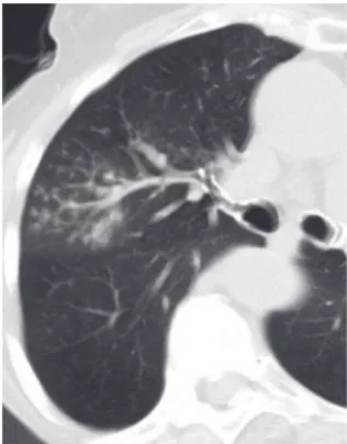
Figure 15 - Symmetric perihilar ground-glass opacity, representing pulmonary hemorrhage in a patient with Wegener’s granulomatosis.
diseases, environmental inhalants, and drug use also should be considered when increased diffuse lung opacity is present.
Acute increased opacity
Clinical and histopathological
correlations
Pulmonary edema
The most common cause of acute diffuse pulmonary disorders with consolidation/ground-glass opacities is pulmonary edema. In hydrostatic edema, there is generally a combination of septal thickening and ground-glass opacities. Heart size can be normal or enlarged. Thickening of the peri-hilar peribronchovascular interstitial (peribronchial cuffing) and fissures are common. The vascular distribution is balanced or inverted. Hazy, poorly-sents confluent disease. These findings are quite
nonspecific and can reflect diseases that are prima-rily alveolar, interstitial, or mixed. The differential diagnosis of ground-glass opacity and consolidation overlaps greatly and is predominantly based upon symptom duration: acute or chronic (Chart 8). The distribution of findings (focal, patchy, or diffuse/ symmetric) can be helpful in further narrowing the differential diagnosis (Chart 9).
Parenchymal consolidation and ground-glass opacity are HRCT findings that have been associ-ated with active or reversible lung disease. However, ground-glass opacity can also be seen in cases in which fibrosis is the predominant abnormality. Ground-glass attenuation can only be considered as reflecting the presence of potentially revers-ible disease if there are no associated findings of fibrosis in the same area. The differential diagnosis of ground-glass and consolidation opacities should be based upon the host immune status and dura-tion of symptoms. The presence of connective tissue
Chart 8 - Differential diagnosis of ground-glass opacity and consolidation based upon duration of symptoms.
Acute Chronic
• Edema • Hypersensitivity pneumonitis
• DAD/ARDS/AIP • Smoking related interstitial lung disease (RB-ILD,
DIP)
• Infections (bacterial, viral, Pneumocystis jiroveci,
Mycoplasma pneumoniae)
• Interstitial pneumonia (NSIP, rarely UIP)
• Hemorrhage • Bronchioloalveolar carcinoma • Hypersensitivity pneumonitis • Organizing pneumonia • Eosinophilic pneumonia (acute) • Lymphoid interstitial pneumonia • Radiation pneumonitis (acute) • Eosinophilic pneumonia (chronic)
• Exogenous lipoid pneumonia • Alveolar proteinosis • Sarcoidosis
DAD: diffuse alveolar damage; ARDS: acute respiratory distress syndrome; AIP: acute interstitial pneumonia; RB-ILD: respiratory bronchiolitis-associated interstitial lung disease; DIP: desquamative interstitial pneumonia; NSIP: nonspecific interstitial pneu-monia; and UIP: usual interstitial pneumonia.
Chart 9 - Typical distribution of diseases that produce ground-glass opacity and consolidation.
Focal Diffuse/symmetric Patchy
Infection Edema Infection
Aspiration DAD/ARDS/AIP Sarcoid
Hemorrhage Infections (viral, atypical) Hypersensitivity pneumonitis
Bronchoalveolar cell carcinoma Interstitial pneumonias Organizing pneumonia
Infarct Hemorrhage Bronchoalveolar cell carcinoma
Bronchoalveolar cell carcinoma Hemorrhage
Alveolar proteinosis Eosinophilic pneumonia
lungs are characteristic of HP. In other cases, areas of ground-glass attenuation, centrilobular nodules, and patchy air-space opacifications with micronodules are seen. In the acute stage air-space consolidation can also be found.(50)
Diffuse alveolar hemorrhage (DAH)
Diffuse alveolar hemorrhage (DAH), defined as active hemorrhage into the alveolar parenchyma, produces increased lung opacity. The causes of DAH are many. Renal involvement should always suggest an etiology of vasculitis or connective tissue disease. When DAH occurs as an immunologic phenomenon, it is frequently accompanied by systemic vasculitis, and, in such cases, serologic studies are essential to the diagnosis and management.(71) Hemoptysis can be absent. A surgical lung biopsy sample from a patient with Wegener’s granulomatosis who presented with hemoptysis and perihilar ground-glass opacity is shown in Figure 17. To avoid confusion with traumatic hemorrhage caused by the biopsy procedure, DAH should never be diag-nosed in the absence of siderophores and fibrin, signs that lung hemorrhage is a true manifestation of immunologic injury (the cause of the majority of DAH cases). Although confusion can occur in a patient with airway-associated acute hemorrhage (such as that seen in bronchiectasis), this is nearly always a segmental or lobar, rather than a diffuse, phenomenon.
defined, centrilobular opacities can also be seen. There is a tendency for hydrostatic edema to have a perihilar and gravitational distribution. In noncar-diogenic edema, the heart size is normal, the vascular distribution is normal or balanced, and the distribu-tion of edema is patchy or peripheral. Peribronchial cuffing and septal lines are generally absent.(67)
Acute respiratory distress syndrome
Acute respiratory distress syndrome (ARDS) can result from a wide variety of lung injuries, including trauma, aspiration, sepsis, and infectious pneu-monia.(68) The distribution of CT abnormalities is characteristically bilateral, gravity-dependent, and accentuated at the lung bases. When ARDS is caused by pulmonary disease, it tends to be asymmetric with a mix of consolidation and ground-glass opacity, whereas ARDS caused by extrapulmonary disease presents predominantly symmetrical ground-glass opacity. In hydrostatic edema and ARDS alike, pleural effusion and air bronchograms are common. Traction bronchiectasis in diffuse alveolar damage (DAD) suggests that the disease is in the prolifera-tive or fibrotic phase.(69)
Infections
Bacterial pneumonia. Bacterial pneu-monia, especially when caused by Streptococcus pneumoniae, Legionella, or other agents including mycoplasma, can result in fulminant pneumonia, with ARDS.
Pneumocystis jiroveci. Pneumocystis jiroveci should always be considered. Cystic changes accom-panied by diffuse ground-glass opacities are highly suggestive but are found in less than one third of all cases.
Cytomegalovirus pneumonia. Cytomegalovirus pneumonia can lead to interstitial pneumonia, and, in severe cases, DAD. The most helpful finding in distinguishing infectious from noninfectious causes of acute diffuse lung disease in the normal host is that of centrilobular nodules. When these are present in a patchy distribution, they suggest infec-tious disease.(70)
Hypersensitivity pneumonitis
Centrilobular nodules of ground-glass opacity distributed diffusely and profusely through the
among which are chemotherapeutic drugs such as bleomycin.
Connective tissue disease-related interstitial lung disease
Like drugs, systemic autoimmune diseases can produce a wide variety of histopathological patterns.(76) In patients previously diagnosed with connective tissue disease (CTD), DAD or acute OP can occur—or the pneumonitis can represent the initial manifestation of disease, especially in systemic lupus erythematosus, polymyositis, and adult-onset Still’s disease.(77,78) Serum creatine phosphokinase and ferritin should be measured in order to evaluate the last two.
Acute eosinophilic pneumonia
Acute eosinophilic pneumonia can occur in asth-matic patients but can also result from the use of medications or illicit drugs, or even from heavy cigarette smoking. An idiopathic form has been described. Only one third of patients have an elevated peripheral
eosi-nophil count. The BAL shows eosieosi-nophils > 25%.(79) Treatment of the patient with even a single dose of corticosteroids prior to biopsy can markedly reduce the number of eosinophils in the tissue and thereby complicate the diagnostic evaluation.
Organizing pneumonia
Organizing lung injury from any cause can be clinically acute, resulting in a presentation with respiratory failure.(80) The acute noninfectious form is either idiopathic, associated with a CTD, or related to drug toxicity. An intermediate form between DAD and OP, designated acute fibrinous and organizing pneumonia, is characterized by rich fibrinous alve-olar exudates, although without hyaline membranes. By definition, infection is absent. Acute fibrinous and organizing pneumonia can either be idiopathic or be associated with an underlying or concomitant condition, such as collagen vascular disease, drug reaction, and occupational exposure.
Clinical management
Acute noninfectious DILDs often present symp-toms that are consistent with pneumonia.(81) Patients thought to have ARDS on the basis of pneumonia, and those considered to have ARDS but without
Acute interstitial pneumonia
The idiopathic form of DAD is referred to clini-cally as acute interstitial pneumonia (AIP), which is the same idiopathic clinico-pathologic entity originally described by Hamman and Rich in 4 patients.(72) Patients with AIP present progressive respiratory symptoms and respiratory insufficiency occurring over the course of days to weeks. The disease is distinguished from other forms of DAD by the absence of an identifiable cause or predisposing disease. Infection is the diagnosis to be ruled out, and specific staining for organisms (at the minimum, Grocott silver staining for fungi and pneumocystis) should always be performed, as in all cases of DAD. The biopsy shows hyaline membranes lining alveolar spaces, typically with variable interstitial and airspace organization by the time biopsy is performed.(73)
The CT findings are typically indistinguishable from those of ARDS and include extensive bilateral air-space consolidation and patchy or diffuse bilateral areas of ground-glass attenuation. Traction bron-chiectasis is often seen as a delayed manifestation in the areas of air-space consolidation or ground-glass attenuation. Some patients with IPF (or other interstitial pneumonias) can experience a precipi-tous course, with periods of acute deterioration in respiratory status, together with DAD and other manifestations of acute injury.(74) Digital clubbing is limited to patients with acute exacerbation of under-lying fibrotic lung disease and serves as a helpful clue to separate such patients from those with AIP.
Radiation pneumonitis
Acute lung manifestations can occur approxi-mately 8 weeks after completion of radiation therapy involving doses of 40 Gy or more. Thoracic irradiation is a relatively uncommon cause of acute increased lung opacity.(75)
Drug-induced interstitial lung disease
A history of smoking might be an important additional factor in this population. Patients with DIP or RB-ILD are almost exclusively smokers. The BAL findings in this group of diseases can be highly specific and can directly confirm a particular diag-nosis or condition, effectively supplanting lung biopsy. Diffuse diseases presenting chronic increased lung opacity and often diagnosable through BAL include alveolar proteinosis, BAC, and CEP. In addition, supportive BAL cytology combined with clinical and HRCT features is frequently sufficient for the diagnosis of HP (lymphocytes, plasma cells, and foamy macrophages) or OP (mixed cellularity and low CD4/CD8 ratio).(86)
Clinical and histopathological
correlations
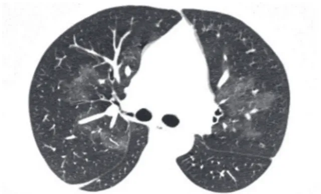
Hypersensitivity pneumonitis
In a normal host who is a nonsmoker and does not have cardiac failure, the most common cause of ground-glass opacity is HP.(84) In HP, ground-glass opacity indicates subacute disease. With relevant exposure, a ground-glass opacity pattern associated with centrilobular nodules and lobular air trapping (the so called “head-cheese pattern” or “terrine sign”, Figure 18) is highly suggestive of the diagnosis.
Nonspecific interstitial pneumonia
It has been shown that CTD, HP, drug-in-duced lung disease, and resolved acute respiratory distress syndrome can all produce an NSIP histologic a defined predisposing condition, should undergo
BAL and, depending on the findings, a lung biopsy, to rule out one of the acute noninfectious DILDs.
Lymphocytosis (>25%) in BAL fluid is the rule in
HP. A predominance of neutrophils in BAL fluid suggests DAD or infection; blood in sequential BAL samples indicates DAH; eosinophilia, or acute eosi-nophilic pneumonia.
In the setting of acute onset lung disease, infec-tion always heads the list of differential diagnoses. Transbronchial biopsies, bronchial washings, and BAL play a crucial role in the clinical evaluation. When fibrin, hyaline membranes, or organization in alveolar spaces is identified, the differential diag-nosis includes infection, drug toxicity, CTD, and AIP.(1) Specific staining for bacterial, fungal, and mycobacterial infections should be performed. In the absence of these agents, a careful search for viral cytopathic changes is warranted, mainly if the patient is known to be immunocompromised, and especially if necrosis is present. In cases of acute HP, biopsy can reveal DAD with typical HP findings (granulomas and cellular pneumonitis).
The causes of DAD diagnosed through surgical lung biopsy have been described in some studies.(82,83) Infections and AIP are the most common causes. Other major causes are CTD, exacerbation of IPF, and drug use. Infections are more common in immuno-compromised hosts. In selected patients with clinical ARDS, surgical lung biopsy can be performed safely, often reveals an unsuspected diagnosis, and leads to alterations in therapy.(82)
Chronic consolidation/ground-glass
opacities
The previously healthy patient who presents with mild chronic dyspnea and ground-glass opacity (or diffuse consolidation) should be investigated for HP, DIP, RB-ILD, NSIP, AIP, OP, bronchiolitis obliterans organizing pneumonia, chronic eosi-nophilic pneumonia (CEP), and sarcoidosis.(84) Rare patients will present some of the atypical causes of chronic consolidation/ground-glass opacity, such as pulmonary alveolar proteinosis (PAP), bronchoal-veolar carcinoma (BAC), and lymphoma. Although ground-glass opacity and consolidation can denote interstitial or alveolar disease,(85) ground-glass opacity is more often seen in HP, DIP, RB-ILD, and NSIP. Consolidation is mainly seen in OP, CEP, and BAC.
sharply defined than those encountered in thick-ened interlobular septa, and with an arcade-like or polygonal appearance) was observed in more than half of the patients.(94)
The diagnosis of OP is based on a combination of clinical, imaging, and histological findings.(95) In cases of CTD, cases of exposure to drugs or environ-mental antigens, and cases of aspiration, localized areas of OP can be a secondary pathologic finding. Therefore, a diagnosis of OP should be made only in the presence of typical HRCT findings, absence of findings indicative of fibrosis on HRCT, and a good response to corticosteroids. If these criteria are not met, surgical lung biopsy should be performed.
Acute interstitial pneumonia
Acute interstitial pneumonia is typically char-acterized by the rapid development of progressive dyspnea and cough; in rare cases, lower respiratory symptoms can persist for up to 60 days.(96)
Lymphocytic interstitial pneumonia
Lymphocytic interstitial pneumonia is usually associated with Sjögren’s syndrome in adults and with HIV infection in children.(97) Diffuse ground-glass opacity and consolidation are the most common CT findings, and thin-walled cysts can also be present, presumably due to follicular bron-chiolitis. Lymphoid interstitial pneumonia, first described by Liebow, is overwhelmingly represented by low-grade B-cell mucosa-associated lymphoid tissue lymphoma (the so-called “MALToma”) of the lung. It should be borne in mind that the accrual of dense lymphoid tissue in the lung is always consid-ered lymphoma until proven otherwise. Patients with Sjögren’s syndrome who present with obstruc-tive physiology, as well as with cysts or centrilobular nodules on HRCT scans, but do not present with ground-glass opacity can be diagnosed with follic-ular bronchiolitis without open lung biopsy. In other cases, a surgical biopsy should be performed in order to rule out lymphoma.(98)
Pulmonary lymphomas
Primary lymphomas of the lung are rare. One of the most common is MALToma, which is also referred to as “extranodal marginal zone B-cell lymphoma”. These pulmonary lymphomas are characteristically pattern.(87) However, there is still considerable debate
as to whether NSIP is a condition distinct from UIP. In addition, including HRCT honeycombing in the diagnostic algorithm further obscures this distinction. Although response to therapy and survival are better in NSIP than in UIP, recent studies have shown that the two diseases can have minor differences in gene expression.(88) In other cases, NSIP can exhibit a gene profile indistinguishable from that of HP.(89) In NSIP, ground-glass opacity is common and, when accompanied by traction bronchiectasis or irregular reticulation, reflects the fibrotic form of disease (cellular NSIP is quite rare). At this juncture, given the inherent diversity of conditions known to produce the NSIP pattern on HRCT scans and histopathologically, a degree of caution is advisable when attempting to predict prognosis for a given patient.
Chronic forms of organizing pneumonia
Chronic smoldering forms of noninfectious OP can produce clinical findings of progressive dyspnea, low fever, constitutional symptoms, and lung consolidations that are unresponsive to the standard treatment for infectious pneumonia. Many conditions can result in OP. In a study conducted in the city of São Paulo, Brazil and involving 95 patients, OP was idiopathic in one third of the cases and secondary to an identifiable cause in of the remaining cases.(90) The most common causes were: drugs (especially amiodarone and MTX), environ-mental exposure (such as that seen in HP), chronic aspiration, and CTD. Consolidations, central or peripheral, were seen in 64% of cases, ground-glass opacity in 53%, and nodules in 26%. Transbronchial biopsy was diagnostic in 58% of cases.
eosinophilia: a careful history and examination to exclude systemic diseases (Churg-Strauss syndrome, sarcoidosis, etc.) as well as a careful review of concomitant drug intake to rule out drug-induced pulmonary eosinophilia is necessary. Examination of the stool for ova and parasites is important.(105) It is not uncommon for CEP to be misdiagnosed as bacterial pneumonia. The hallmark of CEP is a rapid, dramatic response to oral corticosteroids.
Exogenous lipoid pneumonia
Prolonged microaspiration of lipid emulsions can produce lung disease with a distinctive HRCT pattern sometimes referred to as a “crazy-paving” pattern: consolidation with low attenuation and ground-glass opacities. The most common chronic form of the disease is caused by the prolonged inges-tion of mineral oil-based laxatives for the treatment of obstipation.(107) The diagnosis is suggested by the finding of free lipid or lipids in the alveolar cell vacuoles in the BAL fluid. If this is not confirmed, transbronchial or surgical lung biopsy becomes necessary. In many cases, the cause is determined in retrospect, after the diagnosis has been established through surgical lung biopsy.
Bronchioloalveolar carcinoma
The definition of BAC is adenocarcinoma showing growth of neoplastic cells among alve-olar structures (lepidic growth) without evidence of stromal, vascular, or pleural invasion.(108) There are three subtypes of BAC: nonmucinous, muci-nous, and mixed. The pneumonia pattern is more common in patients with the mucinous type, and such patients are often mistakenly diagnosed with infectious pneumonia. Radiographic findings such as ground-glass opacities, nonresolving consoli-dation, and centrilobular satellite nodules due to bronchogenic dissemination should raise the suspi-cion of BAC.(109) Classically, BAC demonstrates a relatively slow growth pattern and an indolent clin-ical course. However, in a subset of patients, rapid growth and death from bilateral diffuse consoli-dative disease occurs within months of diagnosis. Patients with advanced diffuse BAC can present with severe bronchorrhea and refractory hypoxemia from intrapulmonary shunting.(110) The BAL often reveals the presence of well-differentiated neoplastic alveolar cells, although this finding is not sufficient of indolent, low-grade morphology and can present
in otherwise healthy individuals or in patients with Sjögren’s syndrome. High-grade lymphomas also occur in the lung, although these tend to be more localized on imaging and are much easier to diag-nose histopathologically. Low-grade MALTomas can be difficult or impossible to distinguish from benign lymphoid hyperplasia and lymphoid intersti-tial pneumonia. Fortunately, the progression of the low-grade form of the disease is quite slow.(99)
Sarcoidosis
In rare cases, sarcoidosis can present ground-glass opacity or consolidations. The reported frequency of ground-glass opacity varies widely, and this finding is occasionally the predominant abnormality. Ground-glass opacity is typically multi-focal rather than diffuse.(100) Pathologic correlation of ground-glass opacity in patients with sarcoidosis has shown that conglomerate granulomas can occur, as can delicate fibrosis below the limits of HRCT resolution.(101-103) Consolidations, mimicking OP, are uncommon in sarcoidosis.(104) The presenta-tion is acute and the prognosis is excellent. Other CT findings of sarcoidosis and miliary nodules are usually seen as well.
Chronic eosinophilic pneumonia
Significant accumulation of eosinophils in the lungs is characteristic of CEP. It has been suggested that a differential cell count of greater than 40% eosinophils in the BAL fluid is diagnostic of CEP.(105) The symptoms are similar to those found in COP (fever, weight loss, night sweats, cough, and dyspnea), evolving over weeks or months. Asthma antedates the diagnosis in 50% of the cases. Similar to what is seen in COP, consolidations can be migratory. In fact, the two diseases can be difficult or impossible to distinguish on HRCT scans,(106) in some cases also overlapping in lung biopsies. The diagnosis of CEP is based on a history of insidious clinical onset, characteristic chest X-ray appearance of peripheral infiltrates with transient opacities, and peripheral eosinophilia. In this scenario, most authors do not recommend lung biopsy. BAL analysis can be helpful in cases without peripheral eosinophilia.