* Study carried out in the Pulmonology Sector of the Department of Clinical Medicine of the Universidade Estadual de Campinas – Unicamp, State University at Campinas – School of Medical Sciences, Campinas, Brazil.
1. Graduate Student in the Surgery Department. Universidade Estadual de Campinas – Unicamp, State University at Campinas – School of Medical Sciences, Campinas, Brazil
2. Full Professor in the Surgery Department. Universidade Estadual de Campinas – Unicamp, State University at Campinas – School of Medical Sciences, Campinas, Brazil.
3. Attending Physician in the Pulmonary Sector of the Department of Clinical Medicine of the Universidade Estadual de Campinas – Unicamp, State University at Campinas – School of Medical Sciences, Campinas, Brazil.
4. Pulmonologist at the Hospital das Clínicas of the Universidade Estadual de Campinas– Unicamp – State University at Campinas – Campinas, Brasil.
5. Associate Professor in the Department of Clinical Medicine of the Universidade Estadual de Campinas – Unicamp, State University at Campinas – School of Medical Sciences, Campinas, Brazil.
Correspondence to: Marcos Mello Moreira. Rua Celso Egídio de Souza Santos, 181, Jardim Chapadão, CEP 13070-570, Campinas, SP, Brasil. Tel 55 -19-3521-7830. E-mail: [email protected]
Submitted: 17 May 2007. Accepted, after review: 18 July 2007.
Capnografia volumétrica como auxílio diagnóstico não-invasivo no tromboembolismo pulmonar agudo
Marcos Mello Moreira1, Renato Giuseppe Giovanni Terzi2, Mônica Corso Pereira3, Tiago de Araújo Guerra Grangeia4, Ilma Aparecida Paschoal5
Abstract
Pulmonary thromboembolism is a common condition. Its diagnosis usually requires pulmonary scintigraphy, computed angiotomography, pulmonary arteriography and, in order to rule out other diagnoses, the measurement of D-dimer levels. Due to the fact that these diagnostic methods are not available in most Brazilian hospitals, the validation of other diagnostic techniques is of fundamental importance. We describe a case of a woman with chronic pulmonary hypertension who experienced a pulmonary thromboembolism event. Pulmonary scintigraphy, computed angiotomography and pulmonary arteriography were used in the diagnosis. The D-dimer test result was positive. Volumetric capnography was performed at admission and after treatment. The values obtained were compared with the imaging test results.
Keywords: Thromboembolism; Hypertension, pulmonary; Capnography; Schistosomiasis.
Resumo
O tromboembolismo pulmonar é uma situação freqüente que pode ser diagnosticada pela cintilografia pulmonar, angiotomografia computa-dorizada, arteriografia pulmonar e, como método de exclusão, dosagem do dímero-D. Como estes exames nem sempre estão disponíveis, a validação de outros métodos diagnósticos é fundamental. Relata-se o caso de uma paciente com hipertensão pulmonar crônica, agudizada por tromboembolismo pulmonar. Confirmou-se o diagnóstico por cintilografia, angiotomografia computadorizada, arteriografia pulmonar; a dosagem do dímero-D resultou positiva. A capnografia volumétrica associada à gasometria arterial foi realizada na admissão e após o tratamento. As variáveis obtidas foram comparadas com os resultados dos exames de imagem.
Descritores: Tromboembolismo; Hipertensão pulmonar; Capnografia; Esquistossomose.
Introduction
Pulmonary thromboembolism (PTE) is a disease frequently observed and difficult to diagnose.(1) Pulmonary
arteriogram, considered the gold standard for the diag-nosis, is an invasive test and is not without risk. Pulmonary ventilation/perfusion scintigraphy, computed angiography and D-Dimer tests are not always available, especially in secondary hospitals. Volumetric capnography (VCap) is a noninvasive test that can facilitate the diagnosis of PTE.
Here we report the case of a patient with chronic pulmonary arterial hypertension (PAH) who had
exacer-bation of the clinical profile due to PTE. Traditional tests were compared to VCap combined with arterial blood gas analysis, all of which were performed at admission, during treatment and at hospital discharge.
Case Report
estimated at 157 mmHg; and Doppler echocardio-gram of the lower limbs with no thrombi.
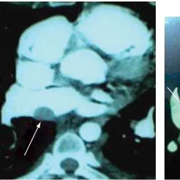
The patient received treatment for schisto-somiasis and was transferred to a referral center. Pulmonary scintigraphy was performed, showing high probability of PTE, with hypoperfusion in the right upper lobe and hypoperfusion in the left lung, without ventilatory alterations. Computed angiography (Figures 1 and 2) revealed thrombi in the pulmonary artery and in the right atrium. The D-Dimer testing, with a cut-off point of 500 ng/mL, showed > 8000 ng/mL using the qualitative method (Diagnostica Stago, Asnières-Sur-Seine, France), compared with 5154.02 ng/mL using an enzyme-linked immunosorbent assay.
Respiratory dead space and functional space were determined through VCap (CO2SMO PLUS 8100; Dixtal/Novametrix, Manaus, Brazil) associ-ated with arterial blood gas analysis (Radiometer ABL® 700 Series; Radiometer Medical ApS, Bronshoj,
Denmark). The indices derived from these values were the end-tidal alveolar dead space fraction (AVDSf) and the late dead space fraction (fDlate) The initial values obtained were an AVDSf of 0.52 and an fDlate of 0.56. Arterial blood gas analysis (O2 at 4 L/min) showed pH de 7.44; PaO2 of 90 mmHg; PaCO2 of 21 mmHg; HCO3 of 14 mmol/L; BE of
−8 mmol/L; SpO2 at 98%; and a respiratory rate of
20 breaths/min. dyspnea during the four final weeks of pregnancy,
and that the dyspnea intensified two weeks before going to the emergency room. She reported no fever or cough. In the physical examination, she presented a respiratory rate of 25 breaths/min, orthopnea, tachycardia, hypotension, edema from the knees down and no tightening of the skin on the calves. Pulmonary auscultation was normal. In cardiac auscultation, there was hyperphonesis of the second heart sound in pulmonary focus. The gyne-cological examination was normal
The personal history of the patient included three pregnancies, all resulting in natural childbirth, and no miscarriages or abortions. During pregnancy, infection with Schistosoma mansoni was detected but left untreated. She denied being a smoker.
The initial tests included an electrocardiogram, involving measurement of the sinus rhythm and right heart overload and arterial blood gas analysis (O2 at 4 L/min): pH of 7.19, arterial oxygen tension (PaO2) of 106 mmHg; arterial carbon dioxide tension (PaCO2) of 9.5 mmHg; bicarbonate (HCO3)
of 3.5 mmol/L; base excess (BE) of −24 mmol/L;
and peripheral oxygen saturation (SpO2) of 97.3%. Orotracheal intubation was not necessary. In the echocardiogram, we observed the following: dilated right heart chambers; preserved left ventricular function; pulmonary artery systolic pressure (PASP)
A week after this scintigraphy, the patient was hospitalized with chest pain and episode of presyn-cope. Another scintigraphy showed low probability of acute PTE. The following values were obtained in the VCap: an AVDSf of 0.17 and an fDlate of 0.07. Another echocardiogram showed pronounced dilation of the right heart chambers, paradoxical interventricular septal motion and moderate PAH (PASP of 67 mmHg and PADP of 42 mmHg), together with severe tricuspid and pulmonary insufficiency.
Discussion
In PAH, there is a significant and persistent increase in the mPAP. Initially, the mPAP is elevated only during physical activities, although, in more severe cases, it is elevated even at rest. When the adaptive mechanisms (dilatation and hypertrophy of the right ventricle) are insufficient to compensate for the afterload increase imposed by the PAH, there is right ventricular failure.
A diagnosis of PAH is made based on the mPAP (higher than 25 mmHg at rest or higher than 30 mmHg during physical activity), which is obtained using a catheter inserted into the pulmo-nary artery.
Doppler echocardiogram allows noninvasive eval-uation of mPAP by estimating the systolic pressure. Systolic pressures between 30 and 50 mmHg are Treated with oxygen therapy, low molecular
weight heparin and sildenafil (75 mg/day), the patient presented favorable evolution. After one week, a transesophageal Doppler echocardiogram revealed PASP of 138 mmHg and pulmonary artery diastolic pressure (PADP) of 58 mmHg, dilated right heart chambers and dilated pulmonary artery, thrombus in the right atrium and pericardial effu-sion. Twelve days after this echocardiogram was performed, pulmonary arteriography revealed a reduction in the vascularization of the upper, right middle and upper left lobes (consistent with acute or chronic PTE), PASP of 109 mmHg, mean pulmonary arterial pressure (mPAP) of 70 mmHg and absence of a response to sodium nitroprusside infusion.
There was progressive clinical improvement and improvement in the blood gas analysis results (room air: pH of 7.54; PaO2 of 102 mmHg; PaCO2 of 30.2 mmHg; HCO3 of 25.4 mmol/L; BE of
3.9 mmol/L; and SpO2 of 99.2%), which allowed
hospital discharge after 37 days. The VCap results prior to hospital discharge were an AVDSf of 0.24 and an fDlate of 0.23. The patient was discharged using oral anticoagulants, and home oxygen therapy was prescribed. Maintenance of sildenafil was not possible.
Pulmonary scintigraphy performed 45 days after discharge showed significant improvement in both lungs.
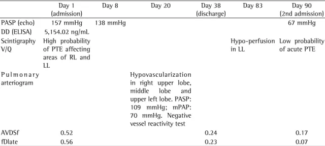
Table 1 - Results of the tests performed on the patient. Day 1
(admission)
Day 8 Day 20 Day 38
(discharge)
Day 83 Day 90 (2nd admission)
PASP (echo) 157 mmHg 138 mmHg 67 mmHg
DD (ELISA) 5,154.02 ng/mL Scintigraphy
V/Q
High probability of PTE affecting areas of RL and LL
Hypo-perfusion in LL
Low probability of acute PTE
P u l m o n a r y arteriogram
Hypovascularization in right upper lobe, middle lobe and upper left lobe. PASP: 109 mmHg; mPAP: 70 mmHg. Negative vessel reactivity test
AVDSf 0.52 0.24 0.17
fDlate 0.56 0.23 0.07
observe the following, higher, cut-off values for the studied variables: 0.15 for AVDSf (4) and 0.12 for
fDlate.(5) Considering the fact that a higher calculated
value indicates greater obstruction of the vascular system (and a larger alveolar dead space), we infer that there is correlation between the extent of the area without perfusion and the value obtained.(3,7,8)
Improvement in pulmonary perfusion by the lytic process of the thrombus resulted in a significant improvement in the patient, as well as a signifi-cant improvement in the respiratory variables, with parallel reduction of the VCap variables. The AVDSf dropped from 0.52 to 0.24 (46%), and the fDtale dropped from 0.56 to 0.23 (50%) The decrease in the AVDSf and fDlate after treatment and in the period before hospital discharge corroborate the findings of many studies evaluating patients after chemical thrombolysis due to massive PTE(for AVDSf)(7,8) and
surgical removal of pulmonary emboli (for fDlate).(3)
Despite the nearly 50% decrease in these variables, there was no normalization, since the alveolar dead space was still increasing in relation to the normal values, and the presence of thrombi in the pulmo-nary arteriogram was detected two weeks prior to hospital discharge. During the second hospitalization (52 days after scintigraphy and computed angiog-raphy were performed) the VCap values obtained were practically normal: an AVDSf of 0.17 and an fDlate of 0.07.
It is known that in the PTE, anticoagulation aims at reducing the possibility of a new embolic phenomenon, as well as reducing the risk of death. Therefore, imaging tests are requested whenever there is clinical suspicion of PTE. Noninvasive methods that rule out the possibility of PTE would reduce the number of patients unnecessarily submitted to imaging tests.
In summary, we have presented the case of a patient with a diagnosis of PTE, which was confirmed through imaging tests and with altered functional variables obtained through VCap. These variables decreased through clinical intervention, which indi-cates the potential worth of VCap as a noninvasive diagnostic tool when used in conjunction with the analysis of D-dimer levels and clinical history.
References
1. Owings JT, Kraut E, Battistella F, Cornelius JT, O’Malley R. Timing of the occurrence of pulmonary embolism in trauma patients. Arch Surg. 1997;132(8):862-6; discussion 866-7.
considered normal. However, the mPAP estimated by the Doppler effect during echocardiography is critically dependent upon age, body mass index and right atrial pressure.(2)
The PASP obtained through Doppler echocar-diogram when the patient was first hospitalized was extremely high, which indicates the pre-existence of a chronic condition.
Considering the presence of S. mansoni in the stool of the patient, it is possible that she had pulmonary schistosomiasis with chronic PAH, and that this condition was aggravated by an embolic event.
The D-dimer levels were elevated (Table 1), a finding compatible with the possibility of a recent embolic episode superimposed on chronic PAH.
The VCap estimates the functional dead space. Combining the VCap results with those of the arte-rial blood gas analysis allows the calculation of various indices, whose variations enable us to infer occlusion or recanalization of the vessels of the pulmonary arterial system.(3)
The following are the patterns typically observed:
• AVDSf, which is calculated using the equation
PaCO2 − PetCO2, in which PetCo2 is end-tidal
CO2, obtained based on the VCap(4)
• fDlate; which is obtained through the extrap -olation of the expiratory tidal volume at 15% of the estimated total lung capacity (TLC); the equation used in order to calculate fDlate is fDlate = PaCO2 − Pet (15% CPT)CO2/PaCO2
(5)
The calculation of fDlate attempts to avoid differences introduced in the functional dead space value by height, gender and age variations. The use of an estimated tidal volume also eliminates the effect of the respiratory rate on tidal volume. In addition, the mathematical extrapolation of phase 3 of the CO2 spirogram at 15% of the TLC aims at bringing PetCO2 and PaCO2 into closer proximity. The TLC was obtained using the method devised by Grimby et al.,(6) with reference values for women
calculated using the equation ([6.71 × height] − [0.015 × age] − 5.77).
For this patient, two parameters were deter-mined: AVDSf and fDlate.
6. Grimby G, Söderholm B. Spirometric studies in normal subjects. III. Static lung volumes and maximum voluntary ventilation in adults with a note on physical fitness. Acta Med Scand. 1963;173(2):199-206.
7. Thys F, Elamly A, Marion E, Roeseler J, Janssens P, El Gariani A, et al. PaCO(2)/ETCO(2) gradient: early indicator of thrombolysis efficacy in a massive pulmonary embolism. Resuscitation. 2001;49(1):105-8.
8. Verschuren F, Heinonen E, Clause D, Roeseler J, Thys F, Meert P, et al. Volumetric capnography as a bedside monitoring of thrombolysis in major pulmonary embolism. Intensive Care Med. 2004;30(11):2129-32.
2. Chemla D, Castelain V, Hervé P, Lecarpentier Y, Brimioulle S. Haemodynamic evaluation of pulmonary hypertension. Eur Respir J. 2002;20(5):1314-31.
3. Moreira MM, Terzi RGG, Vieira RW, Petrucci Jr O. Fração tardia do espaço morto (fDlate) antes e após embolectomia pulmonar. Rev Bras Cir Cardiovasc. 2005;20(1):81-4. 4. Rodger MA, Jones G, Rasuli P, Raymond F, Djunaedi H,
Bredeson CN, et al. Steady-state end-tidal alveolar dead space fraction and D-dimer: bedside tests to exclude pulmonary embolism. Chest. 2001;120(1):115-9.