Natriuria and calciuria levels in preeclampsia: a
cross-sectional study
Valores de natriúria e calciúria na pré-eclâmpsia: estudo transversal
Rose Gasnier
I, Edimárlei Gonsales Valério
I, Janete Vettorazzi
I, Sérgio Hofmeister Martins-Costa
II, Elvino Guardão Barros
III,
José Geraldo Lopes Ramos
IIPostgraduate Program in Medicine: Medical Sciences, Department of Obstetrics and Gynecology, School of Medicine, Universidade
Federal do Rio Grande do Sul (UFRGS), and Division of Obstetrics and Gynecology and Division of Nephrology, Hospital de Clínicas de
Porto Alegre (HCPA), Brazil
ABSTRACT
CONTEXT AND OBJECTIVE: Sodium excretion abnormalities in preeclampsia have been studied in rela-tion to several factors. The objective of this study was to compare natriuria (mEq/24 h) and calciuria levels (mg/24 h) in preeclamptic patients.
DESIGN AND SETTING: An analytical cross-sectional study with a control group was conducted in the ob-stetric center and the high-risk pregnancy outpatient clinic at a university hospital in southern Brazil, and in a primary healthcare unit in the same city, including pregnant women with mild preeclampsia, severe preeclampsia or chronic hypertension, and women with normal pregnancies (14 patients in each group). METHOD: Natriuria was measured using an ion-selective electrode in an automated clinical chemistry an-alyzer (Hitache 917, Roche). All the patients collected 24-hour urine, at home or at the hospital, for analysis of proteins, creatinine, calcium, sodium and uric acid. Quantitative variables with asymmetrical distribution were described using the median, minimum and maximum, and were compared using the Kruskal-Wallis test. The results were logarithmically transformed, with one-way analysis of variance (ANOVA) by ranks and then the post-hoc Tukey test, and were analyzed by means of the Spearman correlation and receiver operating characteristic (ROC) curve. The signiicance level used was 0.05.
RESULTS: There were signiicant diferences between the groups in comparing severe preeclampsia with chronic hypertension and severe preeclampsia with controls (P < 0.0001 for both measurements). CONCLUSION: Natriuria levels may be lower in preeclampsia when associated with calciuria. Natriuria as-sessment is an additional test for diferential diagnosis of hypertensive diseases in pregnancy, but is a poor predictor when used alone.
RESUMO
CONTEXTO E OBJETIVO: Alterações na excreção de sódio têm sido estudadas na pré-eclâmpsia relacio-nadas a vários fatores. O objetivo deste estudo foi comparar natriúria (mEq/24 h) com os níveis de calciúria (mg/24 h) em pacientes pré-eclâmpticas.
ESTUDO E LOCAL: Estudo transversal analítico com grupo controle foi realizado no Centro Obstétrico e no Ambulatório de Gestação de Alto Risco em um hospital universitário no sul do Brasil, e na Unidade Bá-sica de Saúde na mesma cidade, incluindo gestantes com pré-eclâmpsia leve e grave, hipertensão crônica e gestações normais, com 14 pacientes em cada grupo.
MÉTODO: A natriuria foi dosada através da medida de íon eletrodo seletivo, utilizando analizadores au-tomáticos de química clínica (Hitache 917 Roche). Todas as pacientes coletaram urina de 24 h, em casa ou no hospital, para análise de proteínas, creatinina, cálcio, ácido úrico e sódio. As variáveis quantitativas com distribuição assimétrica foram descritas por mediana, mínimo e máximo, e comparadas por teste Kruskal-Wallis. Os resultados foram transformados logaritmicamente, com ANOVA one-way por ranking e, posteriormente, teste post-hoc de Tukey, e foram analisados por médias de correlações de Spearman e curva ROC (receiver operating characteristic). O nível de signiicância adotado foi de 0.05.
RESULTADOS: Foram encontradas diferenças signiicativas entre os grupos quando comparados pré-eclâmpsia grave com hipertensão crônica e pré-eclâmpsia grave com controles (P < 0.0001 para am-bas as medidas).
CONCLUSÃO: Natriúria pode estar reduzida na pré-eclâmpsia quando associada com calciúria. Avaliação da natriúria é um teste adicional para diagnóstico diferencial de doenças hipertensivas na gestação, mas é um pobre preditor quando utilizado isolado.
IMD. Gynecologist and Obstetrician, Hospital de Clínicas de Porto Alegre (HCPA), Department of Obstetrics and Gynecology, Universidade Federal do Rio Grande do Sul (UFRGS), Porto Alegre, Rio Grande do Sul, Brazil.
IIMD. Gynecologist and Obstetrician and Professor, Hospital de Clínicas de Porto Alegre (HCPA), Department of Obstetrics and Gynecology, Universidade Federal do Rio Grande do Sul (UFRGS), Porto Alegre, Rio Grande do Sul, Brazil.
IIIMD, PhD. Nephrologist and Professor, Hospital de Clínicas de Porto Alegre (HCPA), Department of Obstetrics and Gynecology, Universidade Federal do Rio Grande do Sul (UFRGS), Porto Alegre, Rio Grande do Sul, Brazil.
KEY WORDS:
Pregnancy, high-risk. Pre-eclampsia.
Hypertension, pregnancy-induced. Natriuresis.
Diagnosis, diferential.
PALAVRAS-CHAVE:
Gravidez de alto risco. Pré-eclâmpsia.
Hipertensão induzida pela gravidez. Natriurese.
INTRODUCTION
Preeclampsia is one of the major causes of maternal morbidity, preterm birth, intrauterine growth restriction and perinatal mor-tality.1,2 Its pathophysiology has been extensively studied, and its
etiology is probably multifactorial. he disease is characterized by volume contraction, intravascular coagulation and vasocon-striction. It was previously thought to be triggered by an over-active renin-angiotensin-aldosterone (RAA) system, but studies have shown that the system is more complex than this. here is an inverse relationship between the plasma-active renin to pro-renin ratio and the clinical severity of preeclampsia.3
Natriuretic factors also appear to be altered in preeclampsia. Several studies have reported increased atrial natriuretic peptide (ANP) in preeclampsia, but this is not a uniform inding.4 his
event can precede the clinical emergence of the disease. In addi-tion, changes in cell sodium transport are likely to accompany hypertension-induced pregnancy.5 In a case-control
cross-sec-tional study, Reis et al. demonstrated that aggravation of hyper-tension in preeclampsia correlates with serum atrial natriuretic peptide (ANP) and brain naturiuretic peptide (BNP) concentra-tions, although BNP values may be inluenced by the existence of a prior hypertensive state.6
Some studies have found reduced natriuria in preeclamp-sia, probably related to the hypocalciuria process. Because of renal involvement, reabsorption of sodium linked to calcium in the ascending loop of Henle has been described.7 McGrowder
demonstrated a signiicant diference in natriuria levels between preeclampsia and normal pregnancies: 100.43 ± 16.61 for pre-eclampsia, 106.46 ± 14.98 for chronic hypertension, and 144.42 ±
16.37 mEq/24-hour for normotensive patients.8
OBJECTIVE
he aim of our study was to evaluate the relationship between hyponatriuria and preeclampsia, and the possibility of its use, in combination with calciuria measurement, for preeclampsia diagnosis and to diferentiate the forms of hypertension in preg-nancy, by evaluating the natriuria and calciuria levels in pregnant women with chronic hypertension or preeclampsia and in nor-mal controls.
METHODS
An analytical cross-sectional study with a control group was performed, in which the factors evaluated were calciuria and natriuria in relation to preeclampsia. he patients selected were women in their 20th to 37th week of pregnancy, between
March 2008 and November 2009. hey were divided into four groups: severe preeclampsia, mild preeclampsia, chronic hyper-tension and normal pregnancy. he preeclamptic subjects were recruited at the obstetric emergency clinic of a university
hospital located in the south of Brazil, upon hospitalization. he group of hypertensive women was recruited at the high-risk pregnancy outpatient clinic at the same hospital. he con-trol group was normotensive, with no history of preeclampsia or hypertension in previous pregnancies, and was recruited at a primary healthcare unit.
The group with chronic hypertension and the normoten-sive controls collected a 24-hour urine pool at home, unlike the patients in the preeclampsia group, who were hospitalized. The patients were instructed to collect urine for 24 hours, starting with the second morning urine, until the first urine of the next day, thus completing 24 hours. The patients were asked to store the urine in plastic bottles in the refrigerator and deliver it to the laboratory at the end of the collection period. The collections from hospitalized patients were made in the same way and then sent for analysis.
he exclusion criteria for cases and controls were malnutri-tion, previous or gestational diabetes, renal diseases, previous signiicant proteinuria, superimposed preeclampsia, continuous use of calcium supplements or calcium channel blockers, drugs that alter sodium levels, major fetal malformations, intrauterine fetal death and multiple pregnancies.
he criteria used to diagnose diabetes followed the recom-mendations of the Fourth International Workshop Conference on Gestational Diabetes Mellitus.9
he criteria adopted for diagnosing preeclampsia (systolic arterial blood pressure ≥ 140 mmHg and/or diastolic arterial blood pressure ≥ 90 mmHg in two measurements, separated by 6 hours, with ≥ 300 mg/24 hours proteinuria). For classifying preeclampsia as severe, the criteria were those presented by the United States National High Blood Pressure Education Program
Working Group (NHBPEP) in 2000.10
he patients’ 24-hour urine pools were needed in order to analyze their protein (mg/24 hours), creatinine (mg/24 hours), calcium (mg/24 hours), uric acid (mg/24 hours) and sodium levels (mEq/24 hours). In order to guarantee adequate 24-hour urine pooling, creatinine was assayed in the pool (24-hour urine pools with less than 600 mg of creatinine were discarded).11 Ater
preeclampsia had been diagnosed, laboratory tests were carried out to assess its severity.
his study was approved by the Ethics Committee (project 07-563), and written informed consent was obtained from each subject before she joined the study protocol.
he laboratory tests were done at the clinical pathology lab-oratories at the same hospital. Natriuria was measured using an ion-selective electrode in an automated clinical chemistry ana-lyzer (Hitache 917, Roche), and was expressed as mEq/l.
calciuria was used. In that study, the preeclampsia group showed calciuria of 82 ± 15.1 mg/24 h and the control group showed calci-uria of 317 ± 86 mg/24 h,12 with 0.01 alpha and 0.90 beta risks. he
number of patients calculated as necessary in each group was 14. he quantitative variables with asymmetrical distribution were described in terms of the median, minimum and maximum, and were compared by means of the Kruskal-Wallis test. Next, the results were logarithmically transformed, with one-way anal-ysis of variance (ANOVA) by ranks and then a post-hoc Tukey test. he results were analyzed by means of the Spearman corre-lation and the receiver operating characteristic (ROC) curve. he signiicance level used was 0.05. he database was built in Excel, and the analyses were done using the Statistical Package for the Social Sciences (SPSS) 16.0 sotware.
RESULTS
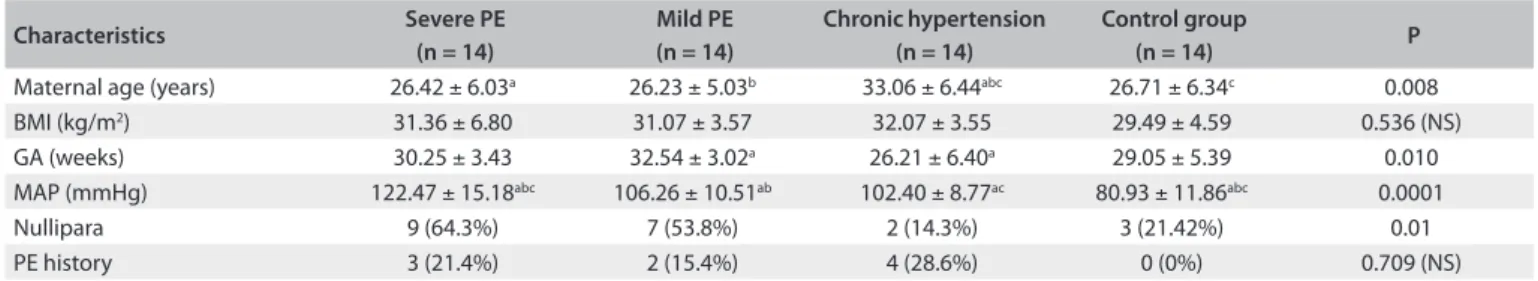
he characteristics of the patient population at the time of inclusion in the study are presented in Table 1. Maternal age was statistically greater in the group with chronic hypertension than in the other groups. Fetal gestational age was signiicantly greater in patients with mild preeclampsia than in those with chronic hypertension. Mean arterial pressure was signiicantly higher in the group with severe preeclampsia than in the other groups. here were more primigravi-dae in the preeclampsia groups than in the other groups.
he calciuria and natriuria levels can be seen in Table 2, showing progressive hypocalciuria and hyponatriuria from nor-mal pregnancy to severe preeclampsia. he diferences in these
levels between patients with severe preeclampsia and chronic hypertension and between patients with severe preeclampsia and normal pregnancy were statistically signiicant (P < 0.0001).
he correlation between natriuria/proteinuria and natri-uria/calciuria can be seen in Figures 1 and 2, respectively. he Spearman correlation was 0.734 between calciuria and natriuria.
he ROC curve demonstrated concordance between the sen-sitivity and speciicity of 24-hour natriuria and the preeclampsia diagnosis, with an area under the curve of 0.841 (P = 0.0001), as seen in Figure 3. he best cutof point was 177 mEq, showing sen-sitivity and speciicity of 67%.
DISCUSSION
Prediction of preeclampsia has been based on detection of risk factors and measurement of arterial pressure, proteinuria and edema. However, some pregnant women without risk factors will develop preeclampsia, thus demonstrating the necessity for bio-chemical markers that could predict this condition. he impor-tance of predicting which women will develop preeclampsia lies in the need for special medical care and preventive measures that might prolong the pregnancy and reduce the maternal and fetal risks.13 he purpose of our study was to assess the correlation
between natriuria and preeclampsia, and to evaluate the possi-bility of using this in the diferential diagnosis of hypertension in pregnancy, in combination with calciuria levels. he classical methods of diferential diagnosis between chronic arterial hyper-tension and preeclampsia work well, but biochemical analysis on
Characteristics Severe PE
(n = 14)
Mild PE (n = 14)
Chronic hypertension (n = 14)
Control group
(n = 14) P
Maternal age (years) 26.42 ± 6.03a 26.23 ± 5.03b 33.06 ± 6.44abc 26.71 ± 6.34c 0.008
BMI (kg/m2) 31.36 ± 6.80 31.07 ± 3.57 32.07 ± 3.55 29.49 ± 4.59 0.536 (NS)
GA (weeks) 30.25 ± 3.43 32.54 ± 3.02a 26.21 ± 6.40a 29.05 ± 5.39 0.010
MAP (mmHg) 122.47 ± 15.18abc 106.26 ± 10.51ab 102.40 ± 8.77ac 80.93 ± 11.86abc 0.0001
Nullipara 9 (64.3%) 7 (53.8%) 2 (14.3%) 3 (21.42%) 0.01
PE history 3 (21.4%) 2 (15.4%) 4 (28.6%) 0 (0%) 0.709 (NS)
Quantitative variables for symmetrical distribution are expressed as mean ± standard deviation, compared using one-way ANOVA and, subsequently, Tukey’s test. Categorical variables are expressed as n (%) and were compared using the chi-square test.
NS = not signiicant; PE = preeclampsia; BMI = body mass index; GA = gestational age; MAP = mean arterial pressure. abc = is an indication of which group is
diferent in relation to the others.
Table 1. Characteristics of the study groups
Severe PE (n 14)
Mild PE (n 14)
Chronic hypertension (n 14)
Control group
(n 14) P
24-hour urinary sodium (mEq) 109
ab 166 203a 206.5b
0.043
(198-932) (15-343) (91-346) (88-568)
24-hour urinary calcium (mg) 81.5
ab 118 226a 272b
< 0.0001
(3-164) (8-564) (50-162) (60-489)
Table 2. Natriuria and calciuria levels
Quantitative variables for asymmetrical distribution are expressed as median and minimum-maximum, calculated using the Kruskal-Wallis test, with subsequent logarithmic transformation ranking for one-way ANOVA and then the Tukey post-hoc test. PE = preeclampsia; abc = indication of which group is diferent in
urine could make a contribution in diicult cases, towards diagnoses that are more complex and accurate, thereby adding to knowledge of the renal pathophysiology of pregnancy-induced hypertension.
Our observation that preeclamptic patients present hypo-calciuria is in agreement with reports from other investiga-tors.14-18 hese authors suggested that urinary calcium excretion
levels may serve as a diagnostic tool for diferentiating between the various forms of hypertension in pregnancy. his might be explained by an increase in tubular calcium reabsorption, rather than a decrease in glomerular iltration, since similar creatinine clearance values were found among the groups.
Frenkel et al. compared natriuria among preeclampsia, chronic hypertensive and normotensive patients and found val-ues of 142 ± 42, 130 ± 35, and 122 ± 38 mEq/l, respectively, with no signiicant diferences among the groups.17 Halhali et al.
stud-ied preeclampsia and normotensive groups, and showed natriuria levels of 87 ± 31 and 90 ± 29 mEq/l, which were non-signiicant values.18 Likewise, according to Selly et al., urine sodium
excre-tion did not difer between groups (preeclampsia group with 124 ± 13 and normotensive group with 100 ± 8 mmol/24 hours).19
here are few studies on renal management in preeclamp-sia other than in relation to proteinuria. Ultrastructural glomer-ular alterations were demonstrated in one study: subendothelial deposits and fusion of podocytes were the most common fea-tures, and fusion of podocytes was correlated with the level of proteinuria.20 One important point has been the diferential
diag-nosis with other forms of hypertension. Natriuria measurement, just like hypocalciuria and uric acid measurement, can help in this diferentiation. Recent studies have demonstrated that the glomerular lesions present in preeclampsia could be related to placental antiangiogenic protein factors, but tubular function is not altered, as shown in the present study.
he strong point of this study is that it may lead the way towards a new kind of research on preeclampsia, especially with regard to analysis on medications for treating gestational hyper-tension. On the other hand, the small number of patients and the need for studies in other places with the same efect could repre-sent weak points of this investigation. Other authors did not ind any signiicant diference, but they also did not control for renal function or collect data on proteinuria and creatinuria levels.
One of the early events in preeclampsia may be excessive expansion of the extracellular luid volume, which causes cir-culation of factors that modify the remodeling of the decidual vasculature, thus preventing normal placentation.21 Many
stud-ies have suggested that the plasma volume is lower in preeclamp-sia than in normal pregnancies. However, serum sodium con-centration does not difer.22 A reduced glomerular iltration rate
would contribute towards the reduced rate of sodium excre-tion in preeclamptic patients. Recent studies have shown that
Figure 1. Proteinuria and natriuria correlation.
24 h proteinuria (mg)
24 h na
tr
iur
ia (mE
q)
5000.00
0.00 10000.00 15000.00 20000.00
0.00 200.00
100.00 300.00 400.00 500.00 600.00
Figure 2. Calciuria and natriuria correlation.
24 h calciuria (mg)
24 h na
tr
iur
ia (mE
q)
0.00 0.00
200.00 200.00
100.00 300.00 400.00 500.00 600.00
400.00 600.00
Figure 3. Natriuria and preeclampsia diagnosis.
1 - Specificity ROC Curve
S
ensitivit
y
0.0 0.0
0.2 0.2
0.4 0.4
0.6 0.6
0.8 0.8
1.0 1.0
autoantibodies against the angiotensin II type 1 (AT1) receptor are present in the serum of preeclamptic patients. In cases of pre-eclampsia, compared with normal pregnancies, it was found that the AT1 receptor gene was upregulated ivefold in the decidua.23
It has been suggested that a maternal autoantibody with the abil-ity to activate AT1 receptors might be implicated in the renal damage in preeclampsia.24
he kidneys have an important role in regulating blood pres-sure, extracellular luid volume, sodium balance and water excre-tion. Glomerular iltration rate and renal plasma low increase by 40% to 65% and 50 to 85%, respectively, during normal preg-nancy. Increased renal perfusion pressure (RPP) causes a potent natriuretic efect (pressure natriuresis) that is greater in situations of hypertension and during pregnancy.25 Our results showed
hyponatriuria in preeclampsia, rather than the natriuretic efects of progesterone, arginine vasopressin, atrial natriuretic factor, prostaglandins and other factors that may lead to excessive loss of iltered sodium.
Digitalis-like cardiotonic steroids may be involved in the patho-physiology of preeclampsia, as shown by observations that Digibind (a digoxin antibody) lowers blood pressure in patients with the dis-ease.26 Adair et al. showed that there was signiicantly lower
eryth-rocyte sodium-pump activity in severe preeclampsia than in normotensive pregnancy, and suggested that the plasma levels of bio-logically active endogenous digitalis-like factors (EDLF) are elevated in patients with severe preeclampsia.25
Current research is more strongly in favor of cell sodium alterations, perhaps mediated by circulating sodium-pump inhibitors, oten leading to increased cell sodium. Increased cell sodium in vascular tissue has been shown to enhance vascular sensitivity to vasoconstrictor agents or lead directly to increased vasoconstriction, which causes elevated pressure levels. he low sodium content in extracellular tissues may have caused the hyponatriuria seen in our study.
he pathophysiology of natriuria and the levels to be consid-ered require further studies before this criterion can be used in clinical practice. Such studies may help towards developing new medications for preeclampsia management, since there are insui-cient reports in the literature to deine this. he home collection of urine by some patients may have afected our results, even though the collections were controlled for creatinine levels, as described above, because of evidence that sodium, uric acid and water renal management may be abnormal in the inal stages of pregnancy, depending on patient rest.
CONCLUSIONS
In conclusion, natriuria levels may be lower in preeclampsia when associated with calciuria. his forms an additional test for the difer-ential diagnosis of hypertensive diseases in pregnancy, but it is a poor predictor when used alone.
REFERENCES
1. Lindheimer MD, Umans JG. Explaining and predicting preeclampsia. N Engl J Med. 2006;355(10):1056-8.
2. Sibai B, Dekker G, Kupferminc M. Pre-eclampsia. Lancet. 2005;365(9461):785-99.
3. Brown MA, Wang J, Whitworth JA. The renin-angiotensin-aldosterone system in pre-eclampsia. Clin Exp Hypertens. 1997;19(5-6):713-26. 4. Tihtonen KM, Kööbi T, Vuolteenaho O, Huhtala HS, Uotila JT. Natriuretic
peptides and hemodynamics in preeclampsia. Am J Obstet Gynecol. 2007;196(4):328.e1-7.
5. Graves SW. Sodium regulation, sodium pump function and sodium pump inhibitors in uncomplicated pregnancy and preeclampsia. Front Biosci. 2007;12:2438-46.
6. Reis ZSN, Cabral ACV, Barra JS, et al. Pressão arterial e concentração plasmática do peptídeo atrial natriurético e do peptídeo natriurético tipo B, em gestações complicadas pela pré-eclâmpsia [Inluence of atrial natriuretic peptide and type B natriuretic peptide plasma levels on arterial pressure in pregnancies complicated by preeclampsia]. Rev Bras Gynecol Obstet. 2003;25(6):413-8.
7. Cunningham FG, Gant NF, Leveno KJ, et al. Hypertensive disorders in pregnancy. In: Cunningham FG, Gant NF, Leveno KJ, et al (eds.). Williams Obstetrics. 21st ed. United States of America: McGraw-Hill Companies;
2001. p. 567-618.
8. McGrowder DA, Williams A, Gordon L, et al. Hypocalciuria in pre-eclampsia and gestational hypertension due to decreased fractional excretion of calcium. Archives of Medical Science. 2009;5(1):80-5. Available from: http://www.termedia.pl/Hypocalciuria-in-pre-eclampsia- and-gestational-hypertension-due-to-decreased-fractional-excretion-of-calcium,19,12290,0,1.html. Accessed in 2012 (Aug 7).
9. Metzger BE, Coustan DR. Summary and recommendations of the Fourth International Workshop-Conference on Gestational Diabetes Mellitus. The Organizing Committee. Diabetes Care. 1998;21 Suppl 2:B161-7. 10. Report of the National High Blood Pressure Education Program Working
Group on High Blood Pressure in Pregnancy. Am J Obstet Gynecol. 2000;183(1):S1-S22.
11. Kaisiske BL, Keane WF. Laboratory assessment of renal disease: clearance, urinalysis, and renal biopsy. In: Brenner B, Rector M, editors. The kidney. Philadelphia: WB Saunders: 1996. p. 1137-74.
12. Ramos JG, Martins-Costa SH, Kessler JB, Costa CA, Barros E. Calciuria and preeclampsia. Braz J Med Biol Res. 1998;31(4):519-22.
13. Cavalli RC, Sandrim VC, Santos JET, Duarte G. Predição de pré-eclâmpsia [Preeclampsia prediction}. Rev Bras Ginecol Obstet. 2009;31(1):1-4. 14. Szmidt-Adjidé V, Vendittelli F, David S, Brédent-Bangou J, Janky E. Calciuria
and preeclampsia: a case-control study. Eur J Obstet Gynecol Reprod Biol. 2006;125(2):193-8.
16. Ingec M, Nazik H, Kadanali S. Urinary calcium excretion in severe preeclampsia and eclampsia. Clin Chem Lab Med. 2006;44(1):51-3. 17. Frenkel Y, Barkai G, Mashiach S, et al. Hypocalciuria of preeclampsia
is independent of parathyroid hormone level. Obstet Gynecol. 1991;77(5):689-91.
18. Halhali A, Díaz L, Avila E, et al. Decreased fractional urinary calcium excretion and serum 1,25-dihydroxyvitamin D and IGF-I levels in preeclampsia. J Steroid Biochem Mol Biol. 2007;103(3-5):803-6.
19. Seely EW, Wood RJ, Brown EM, Graves SW. Lower serum ionized calcium and abnormal calciotropic hormone levels in preeclampsia. J Clin Endocrinol Metab. 1992;74(6):1436-40.
20. Rio SMP, Melo VH, Godoy P, Yokota M. Alterações ultra-estruturais do glomérulo na pré-eclâmpsia [Ultrastructural glomerular alterations in preeclampsia]. Rev Bras Ginecol Obstet. 2004;26(3):185-92.
21. Puschett JB. The role of excessive volume expansion in the pathogenesis of preeclampsia. Med Hypotheses. 2006;67(5):1125-32.
22. Rizk DE. A study of alpha-human atrial natriuretic peptide in normal pregnancy and in pre-eclampsia. J Obstet Gynaecol. 1997;17(3):234-8. 23. Khraibi AA. Renal interstitial hydrostatic pressure and sodium excretion in
hypertension and pregnancy. J Hypertens Suppl. 2002;20(3):S21-7. 24. Averina IV, Tapilskaya NI, Reznik VA, et al. Endogenous Na/K-ATPase
inhibitors in patients with preeclampsia. Cell Mol Biol (Noisy-le-grand). 2006;52(8):19-23.
25. Adair CD, Haupert GT Jr, Koh HP, et al. Erythrocyte sodium/potassium ATPase activity in severe preeclampsia. J Perinatol. 2009;29(4):280-3. 26. Stoupakis G, Klapholz M. Natriuretic peptides: biochemistry, physiology,
and therapeutic role in heart failure. Heart Dis. 2003;5(3):215-23.
The abstract was presented at the 17th World Congress of the
International Society for the Study of Hypertension in Pregnancy, in
Melbourne, Australia, in 2010
Sources of funding: This project was supported by the Fundo de Incentivo à Pesquisa do Hospital de Clínicas de Porto Alegre (FIP-HCPA); Grant number 07-563
Conlict of interest: None
Date of irst submission: July 31, 2011 Last received: August 9, 2012 Accepted: September 4, 2012
Address for correspondence:
Rose Gasnier
Rua Ramiro Barcelos, 1691/11 Porto Alegre (RS) — Brasil CEP 90035-006