ABSTRACT
BACKGROUND AND OBJECTIVES: Diabetic neuropathy is a major cause of neuropathy worldwide and may lead to amputations and incapacity. his study aimed at a detailed and updated review on diabetic neuropathy, focusing on its classiication, diagnostic investigation and treatment. CONTENTS: It is estimated that 371 million people aged from 20 to 79 years, worldwide, have diabetes mellitus and that at least half of them are unware of the diagnosis. Its prevalence in Central and South America was estimated in 26.4 million people, corresponding to approximately 6.5% of the population. Among microvascular complications, diabetic neuropathy is the most prevalent, leading to the highest rates of hospitalization, atrau-matic amputations and incapacity. Diabetic neuropathy may have diferent clinical presentations, being distal symmetric polyneuropathy its most fre-quent presentation and major mechanism to the development of diabetic foot. Predominantly it presents with positive (burning, tingling) and negative (numbness, loss of sensitivity) sensory symptoms. In general it is associated to autonomic signs and symptoms and seldom there is motor manifestation. Approximately 20% of patients with distal symmetric polyneuropathy have neuropathic pain, which sometimes becomes chronic and disabling. CONCLUSION: Early and accurate diagnosis allows for adequate treat-ment, preventing progression of neuropathy and severe complications. For such, it is necessary to obtain an acurate clinical history, in addition to thor-ough neurological tests and additional tests, to identify signs of nervous i-bers involvement. Its treatment depends on adequate glycemic control and neuropathic pain treatment, when present.
Keywords: Diabetes mellitus, Diabetic neuropathy, Neuropathic pain, Pe-ripheral neuropathy.
RESUMO
JUSTIFICATIVA E OBJETIVOS: A neuropatia diabética constitui uma das principais causas de neuropatia no mundo, podendo levar a amputacoes e incapacidade. O objetivo deste estudo foi fazer uma revisão detalhada e atu-alizada sobre neuropatia diabética, focando em sua classiicação, investigação diagnóstica e tratamento.
CONTEÚDO: Estima-se que 371 milhões de pessoas, entre 20 e 79 anos, em todo o mundo apresentem diabetes mellitus e que pelo menos metade destas
desconheça o diagnóstico. Sua prevalência na América Central e do Sul foi estimada em 26,4 milhões de pessoas e projetada para 40 milhões, em 2030. O Brasil ocupa a 4ª posição mundial com maior prevalência de diabetes mel-litus com 13.4 milhões de pessoas com a doença, correspondendo a
aproxi-madamente 6,5% da população. Dentre as complicações microvasculares, a neuropatia diabética apresenta maior prevalência, levando a maiores taxas de internações hospitalares, amputações não traumáticas e incapacidade. A neuropatia diabética pode se manifestar de diferentes formas clínicas, sendo a polineuropatia simétrica distal sua apresentação mais frequente e princi-pal mecanismo de desenvolvimento do pé diabético. Predominantemente, apresenta-se com sintomas sensitivos positivos (queimação, formigamento) e negativos (dormência, perda de sensibilidade); porém, pode se desenvolver de
Diabetic neuropathy
Neuropatia diabética
Osvaldo José Moreira do Nascimento1, Camila Castelo Branco Pupe1, Eduardo Boiteux Uchôa Cavalcanti2
1. Universidade Federal Fluminense, Disciplina de Neurologia, Rio de Janeiro, RJ, Brasil. 2. Rede Sarah, Departamento de Neurologia, Brasília, DF, Brasil.
Conlict of interests: none – Sponsoring sources: none.
Correspondence to:
Rua Siqueira Campos, 53 – Sala 1204 – Copacabana 22031-071 Rio de Janeiro, RJ, Brasil.
E-mail: [email protected]
© Sociedade Brasileira para o Estudo da Dor
maneira assintomática. Geralmente associa-se a sinais e sintomas autonômi-cos e raramente há manifestação motora. Aproximadamente, 20% dos paci-entes com polineuropatia simétrica distal apresentam dor neuropática que, por vezes, torna-se crônica e incapacitante.
CONCLUSAO: O diagnóstico realizado precoce e corretamente possibilita o adequado tratamento, evitando-se a progressão da neuropatia e complica-ções graves. Para isso, é necessária a obtenção de cuidadosa história clínica, além de minucioso exame neurológico e exames complementares, a im de identiicar sinais de comprometimento de ibras nervosas. Seu tratamento depende do adequado controle glicêmico e quando presente, tratamento da dor neuropática.
Descritores: Diabetes mellitus, Dor neuropática, Neuropatia diabética, Neu-ropatia periférica.
INTRODUCTION
Diabetic neuropathy (DN) is a heterogeneous set of clinical or subclinical manifestations afecting the peripheral nervous system (PNS) as complica-tion of diabetes mellitus (DM). It may have diferent clinical manifestations,
pathophysiologic mechanisms, onset and evolution1,2.
It was only in 1864 that DM was recognized as cause of peripheral neu-ropathy (PN). Some years later, the involvement of cranial nerves of diabetic patients has been observed3. he loss of tendinous relexes in lower limbs
(LLll) was described by Bouchard in 18844 and the presence of spontaneous
symptoms such as pain and hyperesthesia was described by Pavy in 18855.
Motor manifestations were documented by Buzzard in 18906.
he irst DN classiication was suggested by Leyden (1893)7 who subdivided
it in sensory and motor manifestations. Jordon and Crabtree (1935)8 in turn,
were the irst to mention pathophysiologic DN mechanisms.
After the discovery of insulin in the 1930s to treat DM, the prevalence of DN has signiicantly increased since diabetic patients started to have longer life expectation.
Studies by Fagerberg9, Mulder et al.10 and Pirart, Lauvaux and Rey11, have
proven the correlation of DN with other microvascular complications such as diabetic nephropathy and retinopathy12.
In face of an alarming number of DM patients, the prevalence of DN is following this growth and is already appearing as major cause of NP in devel-oped countries. One should stress that for being the most prevalent micro-vascular complication, it is estimated that at least half the diabetic patients shall develop this neuropathy in some moment of their clinical evolution13.
Distal symmetrical polyneuropathy is its most frequent clinical presentation, being in general asymptomatic14. Less than half the patients have some type
of neuropathic symptom, being mostly sensory symptoms15. Among DN
pa-tients, approximately 20% have neuropathic pain, implying signiicant de-crease in quality of life and functional capacity16.
In addition, DN is a major risk factor for LLII ulcers, deformities and am-putations and for the development of other microvascular complications. In addition, it increases hospitalization and cardiovascular mortality rates in diabetic patients due to autonomic involvement.
In 2003, it has been estimated in the USA an annual cost directly related to DN and its complications of US$ 10.9 billion, being even higher in patients with painful presentation17.
CONTENTS
Since the irst classiication suggested by Leyden in 18937, many other
siications were proposed and currently the most widely accepted are the clas-siication based on clinical presentations published by homas18 and the
clas-siication according to its pathophysiologic mechanism proposed by Dyck and Giannini19 (Figure 1; Table 1).
It is important to highlight that, although didactically subdivided in difer-ent clinical presdifer-entations, these presdifer-entations may coexist in a same patidifer-ent during disease evolution.
ASYMMETRICAL OR FOCAL AND MULTIFOCAL PRESENTATIONS
Acute mononeuropathies
hey refer to acute onset of the afection of one or more nerves, in general associated to sensory (pain and paresthesia) and motor symptoms in the ter-ritory supplied by such nerve. It is more prevalent in older patients, having as major cause vascular obstruction with consequent nervous ibers ischemia. In general it presents with self-limited course and good clinical evolution, with recovery within six to eight weeks. It is more frequent in cranial nerves such as oculomotor, trochlear and facial, and in peripheral nerves, such as ulnar and ibular23.
Chronic compressive mononeuropathies
hey have insidious onset with sensory symptoms and in their most severe form with motor involvement in speciic compression sites such as wrist me-dian nerve (Carpal Tunnel syndrome – CTS), elbow ulnar, common ibular in ibular head and lateral and medial plantar nerves in tarsal tunnel syn-drome. heir prevalence is three times higher than in general population, having as pathogenesis the participation of micro traumas associated to perineural edema secondary to DM metabolic changes, which peak in nerve compression. Its course is in general progressive, and may have severe motor presentations which often need surgical interventions23.
Radiculoplexus neuropathies (RPNP)
RPNP are asymmetrical sensory-motor presentations of acute onset, involv-ing proximal and distal segments. In general they evolve with severe and disabling painful symptoms and may present autonomic symptoms in up to 50% of cases24. hey may afect cervico-brachial, thoracic, abdominal or
lum-bosacral segments in isolation or even concomitantly25. heir
pathophysiol-ogy seems to be closely related to immunopathic mechanisms, and signs of microvasculitis and consequent ischemic injury were shown by Said et al.26
and Dyck, Norell and Dyck25 in LLll peripheral nerve biopsies.
Is spite of the severity of nervous fibers involvement, prognosis is in gen-eral favorable, even without therapeutic intervention. However, it is still not clear in the literature whether treatment with immunomodulators such as steroids, intravenous human immunoglobulin (IgIV) or plasmapheresis might be effective24.
SYMMETRICAL OR DIFFUSE PRESENTATIONS
Insulinic neuritis
his was irstly described by Carvati27, who has observed patients with distal
sensory symptoms in LLII after starting insulin therapy. Its pathophysiologic mechanism is unknown and its course is in general benign.
Hypoglycemic neuropathy
Uncommon condition associated to prolonged and repeated hypoglycemic states, in general secondary to insulinomas (insulin-producing pancreatic tumor). It is presented with sensory-motor pattern, with predominance of upper limbs (UULL), with presence of atrophy, and may be reversible after treating the hypoglycemic condition28,29.
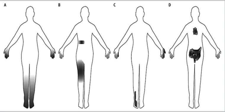
Figure 1. Schematic drawing – different clinical patterns of diabetic neuropathy. Modiied20
A) Distal symmetrical polyneuropathy, B) Radiculoplexus neuropathies, C) compressive focal neuropathies, D) Autonomic neuropathy.
Table 1. Clinical classiication of diabetic neuropathies. Modiied21,22
I- Symmetrical polyneuropathies: Relatively stable conditions:
Symmetrical distal sensory polyneuropathy (SDSP)
Variants: acute, severe SDSP in beginning of diabetes, pseudosyrin-gomyelia neuropathy, pseudodiabetic neuropathy, autonomic neuropa-thies.
Episodic (transient) symptoms:
Diabetic cachexia neuropathy Hyperglycemic neuropathy
Treatment-induced diabetic neuropathy or insulinic neuritis Chronic inflammatory demyelinating polyneuropathy (CIDP-plus) Hypoglycemic neuropathy
Symmetrical distal sensory polyneuropathy (SDSP)
Variants: acute, severe SDSP in beginning of diabetes, pseudosyrin-gomyelia neuropathy, pseudodiabetic neuropathy, autonomic neuropa-thies.
II - Asymmetrical/focal and multifocal neuropathies:
Diabetic lumbosacral radiculoplexus neuropathy (DLSRN; Bruns-Gar-land syndrome, diabetic amyotrophy, proximal diabetic neuropathy). Cervicobrachial radiculoplexus neuropathy
Trunk neuropathies (thoracic/abdominal radiculopathy) Cranial neuropathies
Post-ketoacidosis polyneuropathy
CNS manifestations secondary to the state of ketoacidosis, acute complica-tion of glycemic decompensacomplica-tion in general experienced by DM type 1 pa-tients, are widely known. However, PNS involvement, in addition to being uncommon, is not totally understood. Case reports of these conditions show predominantly motor polyneuropathy with rapid and spontaneous recovery after reversion of this basic condition24.
Acute painful sensory neuropathy
Also known as diabetic cachexia neuropathy, so called because it is in general developed after major weight loss secondary to uncontrolled DM glycemia. It evolves in a single-phase manner with acute onset of symptoms on LLll, pre-doninantly painful, severe and disabling. Due to a strong correlation between glycemic uncontrol and development of this neuropathy, it is especulated the participation of metabolic changes in its pathophysiology, however these mechanisms have not been totally explained. Its treatment is based on glyce-mia and pain control. It has good prognosis evolving with pain improvement and weight gain after glycemic control30.
Glucose intolerance-associated neuropathy
his has remained as a questionable clinical entity for a long time until Lu et al.31 have shown in a broad population study glucose intolerance as
inde-pendent risk factor for PN. It is manifested by predominantly sensory and autonomic symptoms with further involvement of small ibers. It shares the same pathophysiologic mechanisms of DSP, suggesting that this is an early type of this clinical presentation with already established DM.
Autonomic neuropathy (AN)
Disorder afecting autonomic nervous system involving small PNS unmyelin-ated ibers (C ibers), resulting from chronic hyperglycemia metabolic changes. In few occasions, DM and pre-diabetes autonomic neuropathy is present in isolation. In its vast majority, it develops simultaneously with other DN types, more frequently with DSP and it is thought to be part of the same spectrum of chronic afection of DM32.
In general asymptomatic and underdiagnosed, it is estimated that approxi-mately 50% of patients with DM type 1 and 70% of patients with DM type2 have some autonomic involvement, although only 14% have moderate to severe disease32.
AN may result in involvement of cardiovascular, gastrointestinal and uro-genital system, sudomotor function and pupilary motility.
Autonomic cardiovascular dysfunction has been broadly recognized as an in-dependent risk factor for mortality secondary to cardiovascular disease33,34,
increasing the risk of post-surgical complications and mortality35,36. Its major
symptoms include postural hypotension, arrhythmias, silent myocardial isch-emia, pressure lability and intolerance to exercise33.
Sensory, motor and secretory functions of the gastrointestinal system may be involved in diabetic AN, producing symptoms such as nausea, early saciety, vomiting, alternance between diarrhea and constipation and, in more severe cases, postprandial hypotension and syncope37.
Among autonomic DN changes, erectile dysfunction may be the irst mani-festation of the disease, however it shares other pathogenic mechanisms, such as internal pundendal artery atherosclerosis. It has major psychosocial im-pact, leading to severe decrease in quality of life38.
Diabetic cytopathy involves urinary complications caused by changes in de-trusor smooth muscles and urothelial dysfunction, secondary to autonomic involvement of the urogenital system. Its major symptoms are dysuria, po-laciuria, nocturia, urinary urgency and incomplete bladder emptying. hese factors, added to DM-related immunosuppression, increase the prevalence of repetitive urinary tract infections, contributing to the development of renal failure among these patients39.
Sudomotor dysfunction in diabetic AN results in trophic changes in extremi-ties, being associated to Charcot arthropathy, LLll ulcers and amputations. In general it presents with change in LLll color and distal temperature, added to hair loss, intolerance to heat, skin dryness, decreased sweating and perforat-ing plantar disease40.
Not uncommonly, there are pupilary changes, such as the presence of Argyll Robertson pupil, characterized at exam for becoming smaller and present-ing dissociations between light and convergence reactions, that is, they react weakly or do not react to light, by react very well to proximity. his is due to the involvement of oculomotor nerve parasympathetic ibers41.
Distal symmetrical polyneuropathy (DSP)
Being the most common type of DN its prevalence is estimated in 50% of both type 1 and type 1 diabetec patients, being that it is already present in 20% of patients at the moment they are diagnosed with DM.
It remains subclinic in most cases, becoming symptomatic in less than half the patients with DSP15.
It develops slowly, progressively and symmetrically, primarily presenting sensory and autonomic symptoms with predominant involvement of small ibers, evolving with the involvement of sensory large ibers and inally mo-tor ibers in its more severe stages. Classically, it is distally distributed in LLll with length-dependent progression, then afecting upper limbs (UULL), central abdominal region and vortex, in a pattern known as “socks, gloves and apron”.
It may be associated to other types of DN, especially chronic compressive mononeuropathies such as CTS, however in some cases it may be associated to inlammatory neuropathies such as chronic inlammatory unmyelinating polyradiculopathy (CIUP). here is still no clear causality relation between both conditions, however chronic involvement of peripheral nerve by DM seems to be a risk factor for its development30
.
Pathogenesis
DSP pathogenesis is associated to multiple factors related to metabolic, vas-cular, inlammatory and neurodegenerative pathways.
Chronic hyperglycemia plays critical role and is the major triggering factor of DSP pathogenic pathways42.
Metabolic pathway
Glucose penetrates in high levels in peripheral nerves and generates diferent pathologic metabolic reactions. An example is polyol pathway which trans-forms glucose into sorbitol by means of the aldose reductase enzyme. Build up of intracellular sorbitol and fructose decreases active transport of several metabolytes, among them myo-inositol
his process changes intracellular regulatory mechanisms decreasing Na/K pump activity with consequent build up of intracellular sodium. With this, intracellular osmolarity is increased, which generates oxidative stress. hese abnormalities decrease nervous conduction velocity and produce the irst and reversible structural changes in Ranvier nodes.
Another pathologic metabolic mechanism caused by chronic hyperglycemia comes from the formation of inal advanced glycosylation products (FAGP), which are obtained by non-enzymatic reaction of groups of amino acids and glucose reduction products.
FAGP act changing intracellular function of several proteins, changing ex-tracellular components such as laminin and ibronectin, which are essential for axonal regeneration and, inally, promoting irreversible binding in recep-tors of macrophages and endothelial cells. hese changes result in oxidative stress, cytokines secretion and extracellular matrix degradation, peaking in cell apoptosis.
In addition, high glucose levels promote excessive protein C kinase activa-tion, determining the production of nitric oxyde which leads to ischemic peripheral nerve injury43.
Recently, due to high association rate between DM and dyslipidemia (DLD)44 it has been observed the participation of excessive lipids as cofactor
in DN pathogenesis45. It has been proven in vitro direct injury of free fatty
acids in Schwann cells46. In addition, systemic DLP efects promote increased
pro-inlammatory substances and oxidative stress.
Associated to all these metabolic pathways, there is the activation of hexos-amine pathway which, induced by hyperglycemia, results in changes in the expression of some genes and on the functioning of intracellular proteins47.
Oxidative stress leads to increased formation of free radicals, both by the polyol pathway and by FAGP and protein kinase C. his mechanism gener-ates mytochondrial dysfunction, which when critically afected, activgener-ates cell apoptosis cascade43.
Vascular pathway
Neurodegenerative pathway
Another mechanism possibly involved with DN pathophysiology is loss of cell neurotrophism. In DM, quantitative and qualitative decline of insulin also leads to partial reduction of activity of the insulin-like growth factor I and of the neuronal growth factor, with consequent decrease in production of proteins essential for the formation of neuroilaments and maintenance of axonal transport, indispensable for their growth and regeneration. his way, there is axonal degeneration and neuronal body apoptosis, making neuropa-thy to be gradually installed43.
Inflammatory pathway
here are substantial evidences pointing to an immunopathic mechanism in the development of DN. he presence of pro-inlammatory agents has been proven in diabetic patients with neuropathy, promoting inlammatory cells recruitment, cytokines production and decreased blood low48. In summary,
these mechanisms increase peripheral nerve hypoxia and ischemia, making diicult its regeneration49.
Histopathologic changes
Electronic microscopy has observed poorly oriented ilaments in the subax-olemal region, relecting the slowing down of axonal transport. hese neuro-ilaments are later scavenged by Schwann cells which, added to decrease pro-duction capacity of cytoskeleton proteins, decrease axonoplasmatic volume determining axonal atrophy in a way that it peaks with Wallerian degenera-tion. Most common afection pattern is, then, compatible with dying-back axonal degeneration, which preferably afects longer ibers, determining the length-dependent clinical pattern.
hese factors lead to most important DN histopathologic change: multifocal loss of nervous ibers, with axonal degeneration in activity and depending on its chronicity some level of regeneration, characterized by the presence of sproutings. In addition, there are obliterated blood vessels, with endothe-lial basal thickening and neoangiogenesis, revealing the participation of the ischemic component. It is also possible to observe segmentar demyelination and remyelination, which relects the mixed neurophysiologic involvement pattern (axonal and unmyelinating) of this condition50.
Risk factors
here are several risk factors associated to the development and progression of DSP, among them advanced age, male gender, non-hispanic blacks, longer DM duration, glycosilated hemoglobin (HbA1c) higher than 7%, insulin therapy and history of systemic hypertension (SH), DLP and albuminuria51.
CLINICAL MANIFESTATIONS
Sensory signs and symptoms
Most symptomatic patients have positive sensory symptoms (excessive re-sponse to a stimulus or spontaneously), such as paresthesia and pain, and in some cases may present proprioceptive ataxia. hese are referred as sensations of numbness, tingling, imbalance and falls, shocks, pricks and especially burning. hey are distributed in LLll extremities and may evolve to UULL and characteristically patients refer worsening at night. In general these are mild symptoms, however they may be severe and disabling. Negative sensory symptoms (decreased response to a certain stimulus) are those referred as loss of sensitivity in involved segment.
At neurologic evaluation there is distal hypoesthesia/hyperesthesia in seg-ments, initially in thermoalgesic sensitivity modalities. In the presence of severe painful neuropathy, there may be hyperesthesia (exaggerated response to tactile stimuli), hyperalgesia (exaggerated sensitivity to painful stimuli), hyperpathia (persistence of pain even afer painful stimulus removal) or even allodynia (painful sensation caused by painless stimuli). It may evolve to deep sensitivity hypo/anesthesia such as tactile, vibratory and proprioceptive. In addition, when there is large ibers sensory impairment, there is deep hypo/ arrelexia, primarily in Achillean relex, and there might be global arrelexia in very severe cases30.
Autonomic signs and symptoms Already described.
Motor signs and symptoms
Patients with this clinical presentation of neuropathy seldom refer motor
symptoms. When present, they start in the most advanced phase of the dis-ease with mild distal LLll weakness, and mild muscle atrophy of LLll and UULL extremities may be observed
In the presence of major motor signs and symptoms, one should investigate over-lapping causes, such as PIDC or inlammatory radiculoplexus neuropathy30.
Differential diagnosis
DSP secondary to DM is similar to a series of conditions eliciting distal sensory-motor polyneuropathy with axonal predominance. Among them, most frequent are those of toxic-metabolic etiology such as ethyl deiciency, uremic, hypothyroidism, etc. One should also rule out infectious, inlamma-tory and paraneoplastic causes as well as hereditary neuropathies. It is not uncommon the association of DM with other peripheral neuropathies, very often delaying its diagnosis and possible speciic treatment.
Other clinical conditions, such as intermittent lameness, osteoarthritis and Morton’s neuroma share painful symptoms which may simulate DSP. For this reason it is necessary to obtain detailed clinical history, followed by careful neurological and physical evaluation, in addition to complementary investigation with neurophysiologic and laboratory tests52.
Diagnostic tests
Several clinical scales and additional tests have been proposed along decades to early detect DPS and follow its progression with regard to the level of PNS involvement. Among additional tests, one should stress neurophysiologic, autonomic and morphologic tests.
Clinical scales
Clinical scales are based on questionnaires answered by patients about their symptoms and on scores regarding patients’ neurological exams indings, illed by the examiner. Currently, most commonly used scales in population studies and clinical trials are the Michigan Neuropathy Screening Instru-ment53 and the Neuropathic Involvement Score (NIS), Neuropathy
Disabil-ity Scale (NDS) or Neuropathy Impairment Scale (NIS)54,55.
NEUROPHYSIOLOGIC TESTS
Electroneuromyography (ENMG)
For years, ENMG has remained as the golden standard for DSP diagnosis. Still today, it is the diagnostic method most commonly used and available in Brazil. Notwithstanding the inability of the test to identify the early involve-ment of small ibers in this condition, it remains with major importance not only to document the involvement of large ibers but also to evaluate symmetry, severity and progression of the disease, ruling out other coexistent conditions, such as myopathy, motor plate or inferior motor neuron diseases, in addition to primary demyelinating diseases such as PIDC or hereditary neuropathies. By means of needle test (electromyography – EMG) it is pos-sible to characterize both time of evolution (acute versus chronic) and the distribution of neurophysiologic changes.
he routine of neuroconduction study in diabetic patients with DSP involves motor evaluation of median, ulnar, tibial and ibular nerves and sensory eval-uation of median, ulnar, radial and sural nerves. EMG should be performed when diferential diagnosis with other etiologies is needed56.
In general it presents as pure sensory or sensory-motor polyneuropathy or mixed polyneuropathy with axonal and distal predominance, preferably af-fecting LLll. Firs observed changes in the neuroconduction of DSP patients are sensory changes in distal LLll nerves with decreased sensory action po-tential amplitude in plantar, supericial ibular and sural nerves. he dis-ease progresses with sensory UULL involvement and 10 to 30% decrdis-ease in conduction velocities (initially of LLll progressing to UULL) until, in its most advanced stages, present decreased compound muscle action potentials (CMAP) with predominance of LLll57,58.
here is often focal slowdown of conduction velocity with possible presence of conduction blockade (more than 50% decrease in CMAP amplitude from a proximal stimulation point to a distal point) in some nerves susceptible to compression such as wrist median, elbow ulnar and common ibular nerves of ibular head30.
Quantitative sensitivity test (QST)
Method used to indentify and quantify sensory changes of polyneuropathy thermal, painful and vibratory modalities. It may be performed in diferent sites by applying thermal hot and cold stimuli and checking the temperature at the moment patients start to refer beginning of stimulus sensation and pain. It is also possible to check the level of vibration experienced by patients. It is a useful tool in the clinical practice for being a rapid, noninvasive and easy to perform test. However, this method has low repeatability rate because it depends on patients’ cooperation, attention and motivation, being results vulnerable to emotional status. In addition, this test captures changes in any point of the neuraxis and may lead to error in the analysis56.
Evoked potentials
Evoked potentials represent central nervous system electric responses to an external stimulus60. Of interest for the study of small ibers
polyneuropa-thy are laser evoked potential stimulation (LEPS) and contact heat evoked potential stimulation (CHEPS). Such methods allow examining peripheral and central conduction of Aδ and C ibers. However, LEPS may cause skin injuries in laser-stimulated areas, while CHEPS, in addition to being more sensitive and speciic, is noninvaise and able to generate reproducible evoked potentials. here is still no standardization for both methods for the clinical practice61.
Autonomic tests
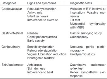
here are several autonomic tests to identify the involvement of C ibers. In clinical practice, most accessible tests for cardiac and sudomotor evaluation are, respectively: tilt test, Valsalva maneuver and R-R interval calculation at ECG and relex sympathetic skin response and sudomotor relex quantitative test (Table 2)41.
Table 2. Signs, symptoms and autonomic tests. Modiied41
Categories Signs and symptoms Diagnostic tests
Cardiovascular Postural hypotension Arrhythmia
Silent ischemia Intolerance to exercise
Variation of R-R interval at inspiration/ Valsalva ma-neuver
Tilt test
Myocardial cyntigraphy with MIBG
Gastrointestinal Nausea
Constipation/diarrhea Early saciety
Gastric emptying study Colonoscopy
Genitourinary Erectile dysfunction Retrograde ejaculation Vaginal lubrication reduction Neurogenic bladder
Nocturnal penile pletis-mography
Urodynamic study
Skin/sudomotor Anhidrosis Skin dryness Intolerance to heat
Quantitative sudomotor relex test
Relex sympathetic skin response
MORPHOLOGIC TESTS
Nerve biopsy
Peripheral nerve biopsy was used for a long time for morphologic and patho-physiologic study of nervous ibers involvement in DN62. Since this is an
invasive exam with possibility of generating complications and sequelae, cur-rently this method has been reserved for atypical situations of clinical pre-sentations when there is doubt about the overlapping with other etiologies, such as inlammatory/infectious neuropathies and amyloidosis. In addition, this exam requires highly specialized material and qualiied professionals to examine the blades63. In general, in research, fascicular biopsy of the
superi-cial sensory nerve is used for being less harmful.
Skin biopsy
With a fragment of approximately 3mm of hairless skin, obtained by punch biopsy, it is possible to identify small epidermal nervous ibers, being a use-ful tool to diagnose small ibers neuropathy. his method is performed as from immune-hystochemical marking of PGP 9.5 – protein gene product, which is present throughout nervous iber extension, allowing direct view of
epidermal ibers.
Currently, intraepidermal ibers density quantiication as from skin biopsy is suggested as diagnostic method for small ibers neuropathy, and its standard-ization for gender and age has been published. Its limitations include being an invasive process which does not add information about the etiology of the neuropathy64.
Confocal corneal microscopy
Recently, human cornea sub-basal plexus, made up of small ibers, has been mapped by confocal microscopy in vivo allowing its characterization and distribution pattern of nervous ibers in healthy individuals of both genders and of diferent ages65.
Malik et al.66 have shown for the irst time, in a series of 18 diabetic
pa-tients compared to controls, a signiicant decrease in sub-basal plexus ibers by means of corneal confocal microscopy (CCM) in vivo, highlighting this test as a rapid, noninvasive and reproducible diagnostic tool to identify DSP. Since then, this method has been pointed in diferent studies as able to iden-tify neuropathy, as well as disease progression and possible improvement after treatment.
Treatment
A strict glycemic control seems to be critical for stabilization and even to improve DN67. his way, all efort should be done to maintain patients
nor-moglycemic.
Many evidences indicate that oxidative stress is involved with DN genesis. So, antioxidant drugs would be an excellent therapeutic alternative. Intra-venous α-lipoic acid (thioctacideMR) (600mg/day for 3 weeks) is currently
the only treatment based on disease mechanism with proven eicacy and amenable to be used in the clinical practice68. he same drug orally (600mg/
day in fasting), the only presentation currently available in Brazil, still need further evidential studies, although evidences suggest its eicacy67. Other
treatment modalities were proposed, but still lack data conirming that they are efective67.
Among available drugs for symptomatic pain treatment, there is evidence level A supporting the use of tricyclic antidepressants, anticonvulsants gaba-pentin and pregabalin, and antidepressant duloxetine, selective dual inhibitor of serotonin and norepinephrine reuptake. here is also second line evidence for the use of opioids such as tramadol and oxicodone67. he combination of
irst line drugs should be considered before using opioids67.
Tricyclic antidepressants have proven eicacy but their adverse efects are ma-jor limiting factors because they might be associated to cardiac conduction changes (A/V blocks, arrhythmias), xerostomy, sweating, dizziness, sedation, urinary retention and glaucoma. Above 100mg/day, their use seems to be associated to sudden death risk, reason why they should be carefully used in cardiopathic patients. It is recommended to start with 10 to 25mg/day and gradually increase the dose with careful follow up of patients. Although doses of up to 150mg/day are indicated, it is hard to go beyond 75mg/day. he choice of the speciic drug should take into consideration patients’ manifesta-tions and drugs adverse efects67.
Among anticonvulsants, gabapentin and pregabalin, both inhibitors of cal-cium channel alpha-2-delta subunit, are currently the best options for this group of patients67. Among dual antidepressants, serotonin and
norepineph-rine reuptake blockers, duloxetine, as compared to venlafaxine, have the best cost-beneit and control of painful neuropathy69. Duloxetine may be
admin-istered in the initial dose of 30mg/day, titrating in one week to 60mg/day as maintenance. Some patients need 120mg/day to control NP.
Whenever there is clinical and/or electromyographic evidence of signiicant arrest, with major motor involvement, decompressive surgeries are theoreti-cally indicated, however the risk of no improvement or even of worsening is signiicant and should be explained to patients.
Maybe one of the most important functions of neurologists in managing DN is orienting prevention and treating diabetic foot, which basically results in insensitivity and autonomic dysfunction. Periodic exams, orientation for self-evaluation, and immediate rest at the onset of any injury, are simple but very important measures.
CONCLUSION
of knowing its primary clinical manifestations, available investigation meth-ods and proposed treatments to establish early diagnosis with possibility of preventing disease progression and its complications.
REFERENCES
1. Greene DA, Sima AA, Stevens MJ, Feldman EL, Lattimer SA. Complications: neuropathy, pathoge-netic considerations. Diabetes Care. 1992;15(12):1902-25.
2. Greene DA, Sima AF, Pfeifer MA, Albers JW. Diabetic neuropathy. Annu Rev Med. 1990;41:303-17.
3. Ogle JW. On disease of the brain as a result of diabetes mellitus. St George’s Hosp. Rep. 1866;1:157. 4. Bouchard CH. Sur la perte des relexes tendineux dans le diabète sucré. Progr Med (Paris).
1884;12:819-27.
5. Pavy FW. Introductory address to the discussion of the clinical aspect of glycosuria. Lancet. 1885;126(3250):1085-7.
6. Buzzard F. Illustrations of some less known forms of peripheral neuritis, especially alcoholic mono-plegia and diabetic neuritis. Br Med J. 1890;1:1419
7. Leyden E. Beitrag zur klinik des diabetes mellitus. Wien Med Wochenschrift. 1893;43:926. 8. Jordon WR, Crabtree HH. Paralysis of bladder in diabetic subjects. Arch Intern Med. 1935;55(1):17. 9. Fagerberg SE. Diabetic neuropathy: a clinical and histological study on the signiicance of vascular
afections. Acta Med Scand Suppl. 1959;345:1-97.
10. Mulder DW, Lambert EH, Bastron JA, Sprague RG. he neuropathies associated with diabetes mellitus. A clinical and electromyographic study of 103 unselected diabetic patients. Neurology. 1961;11(4)Pt1:275-84.
11. Pirart J, Lauvaux JP, Rey W. Blood sugar and diabetic complications. N Engl J Med. 1978;298(20):1149
12. Dyck PJ, homas PK. Peripheral neuropathy. 4th ed. Philadelphia: Elsevier Saunders; 2005. 1951p. 13. Abbott CA, Malik RA, van Ross ER, Kulkarni J, Boulton AJ. Prevalence and characteristics of pain-ful diabetic neuropathy in a large community-based diabetic population in the U.K. Diabetes Care. 2011;34(10):2220-4.
14. Partanen J, Niskanen L, Lehtinen J, Mervaala E, Siitonen O, Uusitupa M. Natural history of peripheral neuropathy in patients with non-insulin dependent diabetes mellitus. N Engl J Med. 1995;333(2):89-94.
15. Dyck PJ, Kratz KM, Karnes JL, Litchy WJ, Klein R, Pach JM, et al. he prevalence by staged severity of various types of diabetic neuropathy, retinopathy, and nephropathy in a population-based cohort: the Rochester Diabetic Neuropathy Study. Neurology. 1993;43(4):817-24
16. Dibonaventura MD, Cappelleri JC, Joshi AV. Association between pain severity and health care resource use, health status, productivity and related costs in painful diabetic peripheral neuropathy patients. Pain Med. 2011;12(5):799-807
17. Gordois A, Scufham P, Shearer A, Oglesby A, Tobian JA. he health care costs of diabetic peripheral neuropathy in the US. Diabetes Care. 2003;26(6):1790-5
18. homas PK. Classiication, diferential diagnosis and staging of diabetic peripheral neuropathy. Dia-betes. 1997;46(Suppl 2):S54-7
19. Dyck PJ, Giannini C. Pathologic alterations in the diabetic neuropathies of humans: a review. J Neuropathol Exp Neurol. 1996;55(12):1181-93
20. Callaghan BC, Cheng HT, Stables CL, Smith AL, Feldman EL. Diabetic neuropathy: clinical mani-festations and current treatments. Lancet Neurol. 2012;11(6):521-34.
21. Nascimento OJM. Neuropatia diabética: diagnóstico e tratamento. In: Oliveira JEP, Milech A. (editores) Diabetes mellitus: clínica, diagnóstico e tratamento interdisciplinar. São Paulo: Atheneu; 2004; 183-97p
22. Marques W Jr, Nascimento O. Neuropatias diabéticas. In: Melo-Souza SE. (editores) Tratamento das doenças Neurológicas. 3a ed. Rio de Janeiro: Guanabara Koogan Ltda; 2013. 582-6p. 23. Vinik AI, Mehrabyan A. Diabetic neuropathies. Med Clin North Am. 2004;88(4):947-99 24. Dyck PJ, Windebank AJ. Diabetic and nondiabetic lumbosacral radiculoplexus neuropathies: new
insights into pathophysiology and treatment. Muscle Nerve. 2002;25(4):477-91
25. Dyck PJ, Norell JE, Dyck PJ. Microvasculitis and ischemia in diabetic lumbosacral radiculoplexus neuropathy. Neurology. 1999;53(9):2113-21.
26. Said G, Goulon-Goeau C, Lacroix C, Moulonguet A. Nerve biopsy indings in diferent patterns of proximal diabetic neuropathy. Ann Neurol. 1994;35(5):559-69
27. Carvati C. Insulin neuritis: a case report. Va Med Mon. 1933;59:745-6.
28. Jaspan JB, Wollman RL, Bernstein L, Rebenstein AH. Hypoglycemic peripheral neuropathy in as-sociation with insulinoma: Implication of glucopenia rather than hyperinsulinism. Case report and literatura review. Medicine. 1982;61(1):33-44.
29. de Freitas MR, Chimelli L, Nascimento OJ, Barbosa GM [Hypoglycemic polyneuropathy: report of a case with insulinoma]. Arq Neuropsiquiatr. 1989;47(2):235-40. Portuguese.
30. Sinnreich M, Taylor BV, Dyck PJ. Diabetic neuropathies. Classiication, clinical features and patho-physiological basis. Neurologist. 2005;11(2):63-79.
31. Lu B, Hu J, Wen J, Zhang Z, Zhou L, Li T, Hu R. Determination of peripheral neuropathy preva-lence and associated factors in Chinese subjects with diabetes and pre-diabetes - ShangHai Diabetic Neuropathy Epidemiology and Molecular Genetics Study (SH-DREAMS). PLoS One. 2013;8(4) e61053.
32. Low PA, Benrud-Larson LM, Sletten DM, Opfer-Gehrking TL, Weigand SD, O’Brien PC, et al. Autonomic symptoms and diabetic neuropathy: a population-based study. Diabetes Care. 2004;27(12):2942-7.
33. Vinik AI. Diabetic autonomic neuropathy. Diabetes Care. 2003;26:1553-79.
34. Suarez GA, Clark VM, Norell JE, Kottke TE, Callahan MJ, O’Brien PC, et al. Sudden cardiac death in diabetes mellitus: risk factors in the Rochester diabetic neuropathy study. J Neurol Neurosurg Psychiatry. 2005;76(2):240-5.
35. Burgos LG, Ebert TJ, Asiddao C, Turner LA, Pattison CS, Wang-Cheng R, et al. Increased
in-traoperative cardiovascular morbidity in diabetics with autonomic neuropathy. Anesthesiology. 1989;70(4):591-7, 1989.
36. Maser RE, Mitchell BD, Vinik AI, Freeman R. he association between cardiovascular autono-mic neuropathy and mortality in individuals with diabetes: a meta-analysis. Diabetes Care. 2003;26(6):1895-901.
37. Rayner CK, Horowitz M. Gastrointestinal motility and glycemic control in diabetes: the chicken and the egg revisited? J Clin Invest. 2006;116(2):299–302.
38. Malavige LS, Levy JC. Erectile dysfunction in diabetes mellitus. J Sex Med. 2009;6(5):1232-47. 39. Hill SR, Fayyad AM, Jones GR. Diabetes mellitus and female lower urinary tract symptoms: a
review. Neurourol Urodyn. 2008;27(5):362-7.
40. Tentolouris N, Marinou K, Kokotis P, Karanti A, Diakoumopoulou E, Katsilambros N. Sudomotor dysfunction is associated with foot ulceration in diabetes. Diabet Med. 2009;26(3):302-5. 41. Smith AG, Singleton JR. Diabetic neuropathy. Continuum. 2012;18(1):60-84.
42. Zochodne DW. Diabetes mellitus and the peripheral nervous system: manifestations and mechanis-ms. Muscle Nerve. 2007;36(2):144-66.
43. Edwards JL, Vincent AM, Cheng HT, Feldman EL. Diabetic neuropathy: mechanisms to manage-ment. Pharmacol her. 2008;120(1):1-34.
44. Clemens A, Siegel E, Gallwitz B. Global risk management in type 2 diabetes: blood glucose, blood pressure, and lipids--update on the background of the current guidelines. Exp Clin Endocrinol Diabetes. 2004;112(9):493-503.
45. Wiggin TD, Sullivan KA, Pop-Busui R, Amato A, Sima AA, Feldman EL. Elevated triglycerides correlate with progression of diabetic neuropathy. Diabetes. 2009;58(7):1634-40
46. Padilla A, Descorbeth M, Almeyda AL, Payne K, De Leon M. Hyperglycemia magniies Schwann cell dysfunction and cell death triggered by PA-induced lipotoxicity. Brain Res. 2011;1370:64-79. 47. Farmer KL, Li C, Dobrowsky RT. Diabetic peripheral neuropathy: should a chaperone accompany
our therapeutic approach? Pharmacol Rev. 2012;64(4):880-900.
48. Gruden G, Bruno G, Chaturvedi N, Burt D, Schalkwijk C, Pinach S, et al. Serum heat shock protein 27 and diabetes complications in the EURODIAB prospective complications study: a novel circulating marker for diabetic neuropathy. Diabetes. 2008;57(7):1966-70.
49. McDonald DS, Cheng C, Martinez JA, Zochodne DW. Regenerative arrest of inlamed peripheral nerves: role of nitric oxide. Neuroreport. 2007;18(16):1635-40.
50. Yasuda H, Dyck PJ. Abnormalities of endoneurial microvessels and sural nerve pathology in diabetic neuropathy. Neurology. 1987;37(1):20-8.
51. Pop-Busui R, Lu J, Lopes N, Jones TL. Prevalence of diabetic peripheral neuropathy and relation to glycemic control therapies at baseline in the BARI 2D cohort. J Peripher Nerv Syst. 2009;14(1):1-13.
52. Vinik AI. Diabetic neuropathy. Endocrinol Metab Clin North Am. 2013;42:747-87. 53. Herman WH, Pop-Busui R, Brafett BH, Martin CL, Cleary PA, Albers JW, et al. Use of the
Mi-chigan Neuropathy Screening Instrument as a measure of distal symmetrical peripheral neuropathy in type 1 diabetes: results from the Diabetes Control and Complications Trial/ Epidemiology of Diabetes Interventions and Complications. Diabet Med. 2012;29(7):937-44.
54. Dyck PJ, Karnes JL, Daube J, O’Brien P, Service FJ. Clinical and neuropathological criteria for the diagnosis and staging of the diabetic polyneuropathy. Brain. 1985;108(Pt4):861-80.
55. Young M, Boulton AJ, MacLeod AF, Williams DR, Sonksen PH. A multicentre study of the preva-lence of diabetic peripheral neuropathy in the United Kingdom hospital clinic population. Diabeto-logia. 1993;36(2):150-4.
56. Perkins BA, Bril V. Diabetic neuropathy: a review emphasizing diagnostic methods. Clin Neurophy-siol. 2003;114(7):1167-75.
57. Redmond JM, McKenna MJ, Feingold M, Ahmad BK. Sensory testing versus nerve conduction velocity in diabetic polyneuropathy. Muscle Nerve. 1992;15(12):1334-9.
58. Albers JW, Brown MB, Sima AA, Greene DA. Nerve conduction measures in mild diabetic neuropa-thy in the Early Diabetes Intervention trial: the efects of age, sex, type of diabetes, disease duration, and anthropometric factors. Neurology. 1996;46:85-91 1996;46(1):85-91.
59. Wooten K. Clinical features and eletrocdiagnosis of diabetic peripheral neuropathy in the dysvascu-lar patients. Phys Med Rehabil Clin N Am. 2009;20(4):657-76.
60. American EEG Society. Clinical evoked potentials guidelines: recommended standards for norma-tive studies of evoked potentials, statistical analyses of results and criteria for clinically signiicant abnormality. J Clin Neurophysiol. 1994;11(1):45-47.
61. Casanova-Molla J, Grau-Junyent JM, Morales M, Valls-Solé J. On the relationship between nocicep-tive evoked potentials and intraepidermal nerve iber density in painful sensory polyneuropathies. Pain. 2011;152(2):410-8.
62. Sima AA. Diabetic neuropathy--the utility of nerve biopsy. Eletroencephalogr Clin Neurophysiol Suppl. 1999;50:525-33.
63. homas PK. Nerve biopsy. Diabet Med. 1997;14(5):345-6.
64. Lauria G, Hsieh ST, Johansson O, Kennedy WR, Leger JM, Mellgren SI, et al. European Federation of Neurological Societies/Peripheral Nerve Society Guideline on the use of skin biopsy in the diagno-sis of small iber neuropathy. Report of a joint task force of the European Federation of Neurological Societies and the Peripheral Nerve Society. Eur J Neurol. 2010;17(7):903-12.
65. Oliveira-Soto L, Efron N. Morphology of corneal nerves using confocal microscopy. Cornea. 2001;20(4):374-84.
66. Malik RA, Kallinikos P, Abbott CA, van Schie CH, Morgan P, Efron N, et al. Corneal confocal microscopy: a non-invasive surrogate of nerve ibre damage and repair in diabetic patients. Diabeto-logia. 2003;46(5):683-8.
67. Tesfaye S, Boulton AJ, Dyck PJ, Freeman R, Horowitz M, Kempler P, et al. Diabetic neuropathies: update on deinitions, diagnostic criteria, estimation of severity, and treatments. Diabetes Care. 2010;33(10):2285-93.
68. Ziegler D. hioctic acid for patients with symptomatic diabetic polyneuropathy: a critical review. Treat Endocrinol. 2004;3(3):173-89.