27
ABSTRACT
BACKGROUND AND OBJECTIVES:Ideal models should reproduce just sensory deicits, such as alodynia, hyperalgesia and spontaneous pain for short periods. here are diferent types of animal models to evaluate diferent neuro-pathic pain etiologies and manifestations. Some models study neuroneuro-pathic pain peripheral mechanisms and other study its central mechanisms. his review fo-cuses on animal models most commonly used for neuropathic pain research. CONTENTS: Animal models based on peripheral nerves ligation which are more commonly used are described. From all models described in this review, spared nerve injury is that producing more reproducible behavioral abnormali-ties for a longer period, while chronic sciatic nerve compression produces be-havioral signs of less predictable painful neuropathies. Spinal hemisection and cytokines-induced spinal injury are the models of choice for the study of central pain mechanisms. Other speciic models are used for the study of the speciic etiology of pain.
CONCLUSION:Since neuropathic pain is multifactorial, diferent neuropathic pain animal models were developed throughout the years, which have been criti-cal for the study of neuropathic pain, since much of current knowledge comes from studies with rats and mice. Current animal models need to be further re-ined and more eforts should be made to determine which animal models may be more predictive, with less biases and more complex and objective analysis parameters.
Keywords: Experimental models of neuropathic pain, Neuropathic pain.
RESUMO
JUSTIFICATIVA E OBJETIVOS: Os modelos ideais deveriam reproduzir ape-nas déicits sensitivos, como alodínea, hiperalgesia e dor espontânea por curtos períodos de tempo. Existem diversos tipos de modelos animais, que avaliam as diversas etiologias e manifestações da dor neuropática. Alguns modelos estudam os mecanismos periféricos e outros estudam mecanismos centrais da dor neu-ropática. Esta revisão enfoca os modelos animais mais comumente utilizados para pesquisa em dor neuropática.
CONTEÚDO: São descritos modelos animais baseados em ligadura de nervos periféricos que são mais comumente empregados. De todos os modelos descritos nesta revisão, a lesão poupadora de nervo é aquela que produz anormalidades comportamentais mais reprodutíveis, por um período mais longo, ao passo que a constrição crônica do ciático produz sinais comportamentais de neuropatia dolorosas menos previsíveis. Hemisecção espinhal e lesão espinhal induzida por citocinas são os modelos de escolha para estudar mecanismos de dor central. Outros modelos especíicos são utilizados para estudo da etiologia especíica da condição dolorosa.
CONCLUSÃO:Como a dor neuropática é multifatorial, diferentes modelos animais de dor neuropática foram desenvolvidos ao longo dos anos que têm sido fundamentais para o estudo da dor neuropática, uma vez que muito do
conheci-Experimental models for the study of neuropathic pain
Modelos experimentais para o estudo da dor neuropática
Angela Maria Sousa1, Gustavo Veloso Lages2, Carla Leal Pereira3, Alexandre Slullitel4
1. Universidade de São Paulo, Faculdade de Medicina, Anestesiologista, Chefe de Equipe de Dor do Insti-tuto do Câncer do Estado de São Paulo, São Paulo, SP, Brasil.
2. Hospital Santa Casa, Hospital Dilson Godinho, Departamento de Neurocirurgia, Montes Claros, MG. 3. Hospital São Luís, Anestesiologista, Supervisor Responsável pelo serviço de dor - Unidade Brasil. São Paulo, SP, Brasil.
4. Hospital Santa Paula, Anestesiologista, Corresponsável pelo Centro de Ensino e Treinamento, Hospital Campo Limpo, São Paulo, SP, Brasil.
Conlict of interests: none – Sponsoring sources: none.
Correspondence to:
Av. Dr. Enéas de Carvalho Aguiar, 255, 8º andar 05403-000 São Paulo, SP, Brasil.
E-mail: angela.sousa@icesp.org.b
© Sociedade Brasileira para o Estudo da Dor
mento atual provém de estudos em ratos e camundongos. São necessários maio-res reinamentos nos modelos animais atualmente empregados e mais esforços para determinar quais modelos animais podem ser mais preditivos, com menos vieses e com parâmetros de análises mais complexos e objetivos.
Descritores: Dor neuropática, Modelos experimentais de dor neuropática.
INTRODUCTION
Neuropathic pain (NP) is characterized by sensory abnormalities such as abnormal unpleasant sensation (dysesthesia), increased intensity of response to painful stimuli (hyperalgesia) and pain in response to normally painless stimuli (allodynia)1.
Most experimental NP models were developed as from peripheral traumatic, metabolic or toxic injuries2. Such models are relevant for the study of
periph-eral disorders in humans (neuropathies, plexopathies and radiculopathies), which represent a considerable percentage of NP-inducing diseases. Diabetes and alcoholism, for example, promote sensory disorders which may induce pain by painful peripheral neuropathy2.
In cancer patients, in addition to tumor growth and compression of nervous structures causing tissue injury, treatment itself may induce NP3. Global
inci-dence of chemotherapy-induced neuropathy is high, between 20 and 100%, because more efective drugs to treat solid tumors, in high doses, induce pe-ripheral neuropathy as secondary efect4. As opposed to diabetes-related NP,
which starts on feet and then afects hands in a period of time varying from months to years, chemotherapy-induced NP may simultaneously appear on hands and feet4.
Experimental evaluation of NP in humans is complex, because most stimuli necessary to produce pain also produce neural injury. So, animal studies are needed to understand mechanisms involved with NP2.
NEUROPATHIC PAIN MODELS IN ANIMALS
Ideal models should produce just sensory deicits, such as allodynia, hyperal-gesia and spontaneous pain for short periods of time.
here are diferent animal models to evaluate diferent NP etiologies and manifestations (Table 1)2. Some models study peripheral NP mechanisms
and others study central mechanisms2. his review focuses on most
com-monly used animal models to study NP.
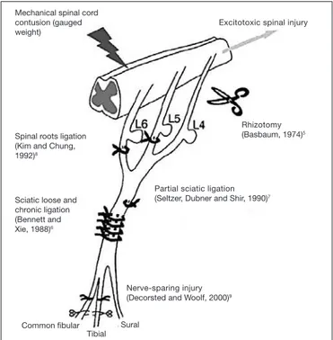
Peripheral nerves injury
Axotomy of a variable portion of nervous ibers has become a primordial technique to generate pain behaviors in animals, being performed in rhi-zotomy5, partial sciatic nerve ligation (PSL), L5 spinal nerve ligation (SNL)
and spared nerve injury (SNI). In all these models, injured animals develop self-protection behaviors and have exaggerated reactions to mechanical (al-lodynia) and thermal stimuli2.
Axotomy
These are extensive injuries coursing with complete sciatic nerve transec-tion in the medial region of the thigh. Animal is anesthetized, common sciatic nerve is exposed and approximately 5mm of the nerve are removed. Adjacent saphenous nerve is also injured, inducing complete limb denerva-tion. Animal develops painful anesthesia and autoctomy (self-mutilation) behavior. In this model, animals are monitored for the extension of the injury caused by autoctomy. However, the interpretation of the mutilation is ambiguous. Although being considered by some investigators as response to mutilated limb chronic pain, it is considered by others just as a reaction to complete anesthesia of deafferented limb or to local pain induced by the injury2.
Such model was replaced by milder injuries to such nerves, which are equally efective to induce NP behaviors in animals.
Rev Dor. São Paulo, 2016;17(Suppl 1):S27-30
REVIEW ARTICLE
28
Sousa AM, Lages GV, Pereira CL and Slullitel A
Rev Dor. São Paulo, 2016;17(Suppl 1):S27-30
Chronic sciatic nerve constriction (loose sciatic ligation)
Under anesthesia, an incision of approximately 3cm is made on the skin cover-ing the area between gluteus and femoral biceps muscles of the hind paw of rats. Common sciatic nerve is exposed and four loose knots with chrome plated Catgug 4-0 are tied around the nerve at a distance of 5mm between them6.
Animals develop behaviors resembling NP in humans by protecting the in-jured limb (animal tries to hide the paw) suggesting spontaneous pain. In addition, there is pain evoked by warmth and mechanical stimulation, char-acterizing thermal and mechanical hyperalgesia, chemical hyperactivity and allodynia to cold, which are detected one week after injury. Pain mechanism may be explained by immune reaction triggered by suture thread, causing nerve edema and compression, in addition to axotomy and Wallerian de-generation.
Electrophysiological studies in this model have revealed slower conduction velocity and histopathology shows major injury in myelinated ibers, A delta ibers and unmyelinated C ibers, which are responsible for pain6.
Peripheral mononeuropathy is similar to that of causalgia and complex regional pain syndrome in humans.
Partial sciatic nerve ligation (Seltzer’s model)7
he sciatic nerve is exposed in upper thigh. he dorsal third (or half ) of the sciatic nerve is tied by a silk thread 8-0 with a loose knot at the region distal to the point where posterior branches of semitendinous biceps emerge. After injury, animals develop protection behavior and licking of the afected limb, suggesting spontaneous pain. Other behavioral changes are also reported in this model one week after surgery and lasting for approximately six weeks.
Seltzer’s model7 represents pain not mediated by sympathetic in the irst
post-operative week. In further weeks, however, there is change in the sympathetic nervous system in this model.
Hyperalgesia duration depends on the type of suture material. his is a good model to study causalgia.
Spinal roots ligation (SNL)8
Animals are anesthetized and a 2cm incision is made at the height of the poste-rior iliac crest to allow access to rats’ lumbar spine roots. Spinal nerves L5 and L6 are identiied and carefully dissected to release the root of L4 (which is not involved in the ligation) and a silk thread 6.0 is used to promote loose ligation of mentioned roots.
Animals develop mechanical allodynia to cold, thermal hyperalgesia and spon-taneous pain 24 to 48 hours after surgery, which persist for 10 to 16 weeks8.
It may be considered an animal model of pain mediated by sympathetic, since sympathetic block relieves hyperalgesia.
As compared to CCI6 and PSL7, nervous ligation extension and site are more
consistent. In addition, spinal segments are preserved intact while others are being injured, thus closer to reality. Drawback is larger surgical extension.
Spared nerve injury (SNL)9
Animals are anesthetized and an incision is performed on femoral biceps of right hind paw of rats. Sciatic nerve and its three terminal branches are ex-posed: sural, common ibular and tibial. Tibial and common peroneal are tied with a silk thread 5-0 and then they are sectioned at 2mm of their end. Sural nerve is not manipulated, that is, it is spared. he other two nerves (tibial and
Table 1. Neuropathic pain models2
Model name Type of injury Animal
Axotomy Complete sciatic section Rat
Chronic sciatic constriction 4 ligatures around the nerve Rats and mice
Partial sciatic ligature Ligature of 1/3 to ½ the nerve Rats and mice
Spinal roots ligation 1. Ligation of L5/L6
2. Ligation of L7
Rats Monkeys
Nerve-sparing injury Tibial and peroneal axotomy Rats and mice
Tibial and sural nerve transection Tibial and sural axotomy Rats
Common peroneal ligation Common peroneal ligation Mice
Sciatic cryoneurolysis Nerve freezing Rats
Caudal trunk resection Caudal trunk resection Rats and mice
Sciatic inlammatory neuritis Zymozan injection, TNF around the nerve Rats and mice
Balloon-induced sciatic injury Implant of polyethylene balloon around the nerve Rats and mice
Laser-induced sciatic injury Decreased blood low to the nerve mediated by radiation Rats
Spinal injury by contusion A weight is dropped on exposed spinal cord Rats and mice
Excitotoxic spinal cord injury Spinal injection of aminoacids Rats and mice
Spinal hemisection Laminectomy of T11-T12 Rats
Drug-induced
1. antineoplastic drugs (vincristine, cisplatin, oxaliplatin, paclitaxel)
2. anti-HIV (2,3- dideoxycytidine, didanosine)
Direct drug injury to peripheral nerves 1.Rats, mice, Guinea pigs
2. Rabbits, rats
Diabetes-induced neuropathy 1. induced by streptozocin 2. genetic models
Persistent changes in nerves induced by hyperglycemia Rats and mice
Bone pain models Inoculation of cancer cells in bones Rats and mice
HIV-induced neuropathy Inoculation of HIV gp120 protein in sciatic nerve Rats
Postherpetic neuralgia Injection of viral cells Rats
Alcoholic neuropathy Ethanol administration for long periods Rats
Pyridoxine-induced neuropathy Administration of high doses of pyridoxine for long periods Dogs and rats
Trigeminal neuralgia Trigeminal compression; chronic infraorbitary nerve constriction Rats
Orofacial pain Formalin of carrageenin injection in temporomandibular joint and jaw Rats and mice
29
Experimental models for the study of neuropathic pain Rev Dor. São Paulo, 2016;17(Suppl 1):S27-30
common ibular) are sectioned and that is why the technique is called spared nerve injury9.
he response to nociceptive and non-nociceptive stimuli is increased in the territory of the ipsilateral sural nerve and at a lesser extent, in the territory of the saphenous nerve.
Pain induced by this model is independent from sympathetic nervous system and, diferently from other models, allows the comparison of thermal and me-chanical sensitivity of non-injured territories close to injured areas. his way, it is possible to evaluate the contribution of injured primary sensory aferent neurons to hypersensitivity2.
Mechanical and thermal hyperalgesia are present 4 days after surgery and per-sist for several weeks, up to 6 month4.
Excitotoxic spinal injury Mechanical spinal cord
contusion (gauged weight)
Spinal roots ligation (Kim and Chung, 1992)8
Sciatic loose and chronic ligation (Bennett and Xie, 1988)6
Common ibular Tibial
Sural
Nerve-sparing injury (Decorsted and Woolf, 2000)9
Partial sciatic ligation (Seltzer, Dubner and Shir, 1990)7
Rhizotomy (Basbaum, 1974)5
Figure 1. Experimental model of neuropathic pain. Roots ligation (SNL), partial nerve ligation (PNL), chronic nerve constriction (CNC), rhizotomy, spinal cord mechanical contusion, spared nerve injury. Adapted16
CENTRAL INJURIES
Contusion models
Most spinal injuries in humans are result of fracture/luxations induced by com-pressive contusions. So, models resulting in nervous tissue contusions seem to be relevant to evaluate clinical phenomena. In the model described by Al-len10, spinal cord is exposed by laminectomy in the thoracolumbar region and
a weight is left to fall over spinal cord of dogs. As a consequence, there is paraplegia and complete segmental necrosis. Several diferent contusion mod-els11-13 were developed since then, in the attempt to control injury level. New
“pendulums” were projected using the force of the impact rather than tissue displacement.
EXCITOTOXIC MODELS
Diabetic neuropathy
Streptozocin-induced neuropathy14
Streptozocin (2-desoxi-2 (3-methyl-3-nitro shureido)-D-glucopyranose) is an anti-cancer agent clinically classiied as antibiotic and chemically analog to ni-trosoureas chemotherapeutic drugs. Intravenous injection of 60mg/kg strep-tozocin in rats induces diabetes after three days, due to beta cells destruction. his model is used to study diabetes-induced neuropathy.
However, animals develop other metabolic disorders caused by hyperglycemia, such as ketoacidosis, lipid metabolism changes, widespread physical weakness (decreased growth and motor activity, lethargy, bladder distention, polyuria and diarrhea). Some of those symptoms complicate data interpretation in animal pain studies.
Chemotherapy-induced neuropathy2
Vincristine
Vincristine is a puriied alkaloid extracted from the Linn pervinca plant from the Apocynaceae family.
Diferent animal models of vincristine-induced neuropathy were developed to study mechanisms involved in the development of neurotoxicity. Intravenous injection of multiple vincristine doses (20, 75, 100 or 200mg/kg) in the tail of rats, triggers fast onset of painful neuropathy10.
Dose regimen of intravenous 75mg/kg (in days 1-5 and 8-11, in a total of nine consecutive doses) has been more commonly used for resulting in maximum hyperalgesia with relative absence of motor deicit in most rats. In the fourth day after vincristine administration, there is allodynia with peak of efect in the 11th day. Cumulative dose above 1000mg/kg leads to severe sensory and motor
changes and very often to death2.
Other model able to induce neuropathy was described with a single vincristine dose (50, 100 or 200mg/kg i.v.) and is characterized by allodynia and mechani-cal hyperalgesia 5 days after injection2.
Surgical implant of an osmotic mini-pump in right external jugular vein, with administration of vincristine sulfate (30mg/kg/day) is another proposed mod-el. Vincristine infusion has been associated to the development of mechanical allodynia and allodynia to cold the irst week after infusion, with no efect on mechanical and thermal nociception2.
Platinum
Platinum-derived products (oxaliplatin, cisplatin and carboplatin) are agents inhibiting DNA synthesis and the replication by means of cross-links estab-lished by the complex.
Cisplatin
Koning et al.15 have studied the development of cisplatin-induced neurotoxicity,
although this is a model diicult to reproduce because animals develop renal failure before neurotoxicity. In addition, it is diicult to determine whether neu-rotoxicity is induced by renal disease or by platinum. Animals’ hydration dur-ing the model development period and administration of escalatdur-ing doses of the chemotherapeutic drug decrease this complication. Another model uses 1mg/kg three times a week for ive weeks.
Another possibility is the administration of 1 to 2mg/kg cisplatin once a week for nine weeks, leading to the development of peripheral neuropathy2,16. Other models with
dif-ferent doses and administration regimens have been described in the literature. Mechanical and thermal to cold allodynia and mechanical and thermal hyper-algesia were documented with no signs of motor dysfunction.
Oxaliplatin
he toxicity proile of this drug is favorable as compared to its antecessors, that is, less nephrotoxicity, ototoxicity and hematotoxicity. However, it also induces peripheral sensory toxicity. Cavaletti et al.17 have observed electrophysiological
changes after two cumulative oxaliplatin doses (36 and 48 mg/kg) inducing slower sensory nerves conduction velocity due to injuries in dorsal root gan-glion neuronal cells2.
Other more recent model of oxaliplatin-induced neuropathy with single dose infu-sion was developed in the attempt to reproduce pain signals which are observed after administration of a single oxaliplatin dose in patients with metastatic colorec-tal cancer. A single oxaliplatin dose varying from 3 to 12mg/kg is administered2.
Mechanical and thermal to cold allodynia, and mechanical and thermal hyper-algesia were documented with no sign of motor dysfunction.
Diabetic rats
Normal rats 500
400
300
200
100
0
0 20 40 60 80 Days
30
Sousa AM, Lages GV, Pereira CL and Slullitel A
Rev Dor. São Paulo, 2016;17(Suppl 1):S27-30
Taxanes
Taxanes improve life expectancy of lung, head and neck, breast and ovary cancer patients. hey afect sensory neurons and especially nervous ibers conducting vibration and proprioception sensations.
Taxanes, especially paclitaxel, are agents promoting the union of microtubules as from tubulin dimers, stabilizing them and preventing their depolymerization. his interferes with the dynamic of physiologic reorganization of the network of microtubules, which is essential for vital cell functions2,16.
Paclitaxel
Intraperitoneal 1 to 2mg/kg in rats promotes signs of hyperalgesia without systemic toxicity signs. Paclitaxel administered in four alternate days, with cu-mulative dose of 4 or 8mg/kg induces peripheral neuropathy characterized by allodynia to cold, long lasting mechanical allodynia and sciatic nerve endoneu-ral edema. Changes in pain threshold are observed in the ifth day of paclitaxel administration and persist for 3 weeks after the last dose.
Animals treated with vincristin and paclitaxel have mechanical hypersensitivity and hypersensitivity to cold, but few, if any, hyperalgesia to warmth. Models using higher paclitaxel doses (cumulative dose of 80mg/kg) induce thermal hy-poalgesia, indicating loss of thermal sensitivity2,16.
Loss of pain sensation, morphological abnormalities, neurophysiologic disorders and motor function changes may be well studied with the model with higher doses, be-cause these changes are uncommon or absent in models with low doses of paclitaxel16.
However, severe autonomic deicits and neurophysiologic abnormalities are drawbacks of the high cumulative dose model.
Docetaxel
he model of docetaxel-induced neuropathy consists of injection of 5, 10 or 12.5mg/kg during four weeks. Animals present slower neural conduction veloc-ity, changes in thermal thresholds and degeneration of skin and paw nerves2.
Cancer-induced neuropathies18
An animal model of NP in cancer was developed in mice, where tumor growth is induced close to the sciatic nerve. Under anesthesia, right sciatic nerve is exposed in the gluteus and ascites luid containing 50 thousand of tumor cells is depos-ited on the nerve. Tumor growth leads to nerve compression and development of thermal and mechanical hyperalgia in the ipsilateral paw. Signs of spontane-ous pain and paw licking are observed. However, as the tumor grows, there is mechanical hyposensitivity of the afected limb, while thermal hyperalgesia and spontaneous pain signs persist.
Gradual compression by tumor cells results in injury to both myelinated and unmyelinated ibers.
Alcohol-induced neuropathy
Animals (rats) receive 6.5% ethanol every day for 12 weeks. Ethanol doses as from initial concentration of 2.5% maintained from the irst to the third day are progressively and consecutively increased to 4% from the fourth to the sixth day; to 4.5% as from the seventh to the 16th day and inally to 5% from the 17th to the
70th consecutive day. After this 70-day period, alcohol-containing diet is
progres-sively decreased in three days. Mechanical hyperalgesia is present after 12 weeks of alcohol consumption and persists after alcohol withdrawal from the diet2.
Other neuropathic pain models2
Human immunodefficiency virus-induced neuropathy
Sciatic nerve is surgically exposed and impregnated with a cellulose solution con-taining HIV gp120 proteins. Allodynia and hyperalgesia are developed within three days.
Postherpetic neuralgia
Cells infected with varicella-zoster virus are injected in paws of rats. Animals develop hyperalgesia behavior three days after infection.
Pyridoxine-induced neuropathy
Oral administration of 50 to 300mg/kg/day pyridoxine for 112 days induces sensory neuropathic abnormalities.
Trigeminal neuralgia
Anesthetized rats are submitted to the implant of a 21G cannula in left trigemi-nal nerve ganglion, through which 10µL of 4% agar solution is injected to pro-mote nerve constriction without injury.
Orofacial pain
Formalin is injected in the temporomandibular joint of rats, leading to stereo-typed behaviors such as shaking the head, letting the head fall to the injured side, and scratching the afected area of the face.
Acrylamide-induced neuropathy: administration of intraperitoneal acrylamide in doses varying from 20 to 40mg/kg, 3 days a week for 8 weeks induces neuro-pathic symptoms.
CONCLUSION
Since NP is multifactorial, diferent NP animal models were developed along the years. Models based on peripheral nerves ligation are the most commonly used. From all models described in this review, spared nerve injury is that producing more reproducible behavioral abnormalities for a longer period (>6 months), while chronic sciatic constriction produces less predictable behavioral signs of painful neuropathy.
Hemisection and citokines-induced spinal injury are models of choice to study central pain mechanisms. Other speciic models are used to study the speciic etiology of the painful condition.
Animal models are critical for the study of NP and much of current knowledge has come from studies with rats and mice. However, few discoveries in basic sci-ences were efectively translated from rodent models to efective pain therapies. So, behavioral tests with animals should be carefully evaluated19.
Further reinements of currently used animal models are needed. Further eforts are needed to determine which animal models may be more predictive, with less investigator biases and with the introduction of more complex and objective evaluation parameters.
REFERENCES
1. Woolf CJ, Mannion RJ. Neuropathic pain: aetiology, symptoms, mechanisms, and management. Lancet, 1999;353(9168):1959-64.
2. Jaggi AS, Jain V, Singh N. Animal models of neuropathic pain. Fundam Clin Pharmacol. 2011;25(1):1-28.
3. Smith TJ, Saiki CB. Cancer pain management. Mayo Clin Proc. 2015;90(10):1428-39. 4. Boyette-Davis JA, Walters ET, Dougherty PM. Mechanisms involved in the development of
chemo-therapy-induced neuropathy. Pain Manag. 2015;5(4):285-96.
5. Basbaum AI. Efects of central lesions on disorders produced by multiple dorsal rhizotomy in rats. Exp Neurol. 1974;42(3):490-501.
6. Bennett GJ, Xie YK. A peripheral mononeuropathy in rat that produces disorders of pain sensation like those seen in man. Pain. 1988;33(1):87-107.
7. Seltzer ZR, Dubner R, Shir Y. A novel behavioral model of neuropathic pain disorders produced in rats by partial sciatic nerve injury. Pain. 1990;43(2):205-18.
8. Kim SH, Chung JM. An experimental model for peripheral neuropathy produced by segmental spinal nerve ligation in the rat. Pain. 1992;50(3):355-63.
9. Decosterd I, Woolf CJ. Spared nerve injury: an animal model of persistent peripheral neuropathic pain. Pain. 2000;87(2):149-58.
10. Allen AR. Surgery of experimental lesion of spinal cord equivalent to crush injury of fracture disloca-tion of spinal column. J Am Med Assoc. 1911;57:878-90.
11. Christensen MD, Everhart AW, Pichelman JT, Hulsebosch CE. Mechanical and thermal allodynia in chronic central pain following spinal cord injury. Pain. 1996;68(1):97-107.
12. Hulsebosch CE, Xu GY, Perez-Polo JR, Westlund KN, Taylor CP, McAdoo DJ. Rodent model of chronic central pain after spinal cord contusion injury and efects of gabapentin. J Neurotrauma. 2000;17(12):1205-17.
13. Anderson TE. A controlled pneumatic technique for experimental spinal cord contusion. J Neurosci Methods. 1982;6(4):327-33.
14. Akbarzadeh A, Norouzian D, Mehrabi MR, Jamshidi SH, Farhangi A, Verdi AA, et al. Induction of diabetes by Streptozotocin in rats. Indian J Clin Biochem. 2007;22(2):60-4.
15. Koning D, Neijt JP, Jennekens FG, Gispen WH. Evaluation of cis-diamminedichloroplatinum (II) (cisplatin) neurotoxicity in rats. Toxicol Appl Pharmacol. 1987;89:81-7.
16. Garcia-Larrea L, Magnin M. [Pathophysiology of neuropathic pain: review of experimental models and proposed mechanisms]. Presse Med. 2008;37(2 Pt 2):315-40.
17. Cavaletti G, Petruccioli MG, Marmiroli P, Rigolio R, Galbiati S, Zoia C, et al., Circulating nerve growth factor level changes during oxaliplatin treatment-induced neurotoxicity in the rat. Antican-cer Res. 2002;22(6C):4199-204.
18. Wacnik PW, Kehl LJ, Trempe TM, Ramnaraine ML, Beitz AJ, Wilcox GL. Tumor implantation in mouse humerus evokes movement-related hyperalgesia exceeding that evoked by intramuscular carrageenan. Pain. 2003;101(1-2):175-86.