23
ABSTRACTBACKGROUND AND OBJECTIVES: Neuropathic pain is deined as pain induced by injury or disease involving the somatosensory system. Dysfunctions in anatomic regions responsible for the processing of pain may involve peripheral and central nervous system components. A careful history and clinical evaluation with special attention to neurologic propaedeutics are critical for the syndromic, anatomic and etiologic diagnosis of neuropathic pain. However, diagnosis is not always simple and often depends on additional tests. his chapter aimed at re-viewing most commonly used additional tests in the clinical practice to help diagnosing neuropathic pain.
CONTENTS: Electroneuromyography is primarily indicated for topographic, etiologic and prognostic diagnosis of peripheral nervous system diseases and for the diferential diagnosis between neurogenic, myopathic and neuromuscular junction diseases. It gives real time information on what is going on in the nerve and the muscle, being fundamentally important for diferential neuromuscular disease diagnosis. Some imaging methods, such as computerized tomography and magnetic resonance, for their spatial resolution, give details of anatomic structures. Other methods, such as positron emission tomography scan and func-tional magnetic resonance, in addition to anatomic details, also provide data on metabolic and functional measurements. In addition, imaging techniques such as spectroscopy and difusion tensor magnetic resonance, allow the study of brain biochemical changes and conectivities with diferent temporal and spatial resolu-tions. Other additional tests, such as sensory quantiication test and microneu-rography are seldom used in the clinical practice.
CONCLUSION: Additional tests, together with careful history and neurologi-cal evaluation focused on neurologic propaedeutics, may provide important data for the diagnosis of neuropathic pain and are often used in the clinical practice. Keywords: Additional tests, Electroneuromyography, Neuroimaging, Neuro-pathic pain.
RESUMO
JUSTIFICATIVA E OBJETIVOS: Dor neuropática é deinida como a dor causada por lesão ou doença envolvendo o sistema somatossensitivo. Disfunções em regiões anatômicas responsáveis pelo processamento da dor podem envolver componentes do sistema nervoso periférico e central. Uma anamnese cuidadosa e um exame clínico com particular atenção na propedêutica neurológica são fundamentais para o diagnóstico sindrômico, anatômico e etiológico das dores neuropáticas. Entretanto, o diagnóstico nem sempre é simples e frequentemente depende do auxílio de exames complementares. O objetivo deste capítulo foi rever os exames complementares mais usados na prática clínica para o auxílio diagnóstico na dor neuropática.
CONTEÚDO: O exame eletroneuromiográico tem sua principal indicação no
diagnóstico topográico, etiológico e prognóstico das afecções do sistema nervoso periférico e no diagnóstico diferencial entre afecções neurogênicas, miopáticas
Additional tests to investigate neuropathic pain. The value of electroneuromyography
for neuropathic pain
Exames complementares na investigação da dor neuropática. O valor da eletroneuromiografia na dor
neuropática
Fábio Henrique de Gobbi Porto1, Gislaine Cristina Lopes Machado Porto2, Mario Wilson Lervolino Brotto1
1. Universidade de São Paulo, Faculdade de Medicina, Hospital das Clínicas, Serviço de Neurologia, São Paulo, SP, Brasil.
2. Hospital AC Camargo, Departamento de Radiologia e Imagem, São Paulo, SP, Brasil. Conlict of interests: none – Sponsoring sources: none.
Correspondence to: Rua Itapeva 538, conj. 132 01332-000 São Paulo, SP, Brasil. E-mail: [email protected]
© Sociedade Brasileira para o Estudo da Dor
e da junção neuromuscular. Ele pode fornecer informações em tempo real do que está ocorrendo no nervo e no músculo, sendo de fundamental importância no diagnóstico diferencial das afecções neuromusculares. Existem várias modali-dades não invasivas de estudo por imagem que podem auxiliar no diagnóstico de quadros de dores neuropáticas. Alguns métodos por imagem como a tomograia computadorizada, ressonância magnética, pela sua resolução espacial, fornecem detalhamento sobre as estruturas anatômicas. Outros métodos como a tomogra-ia computadorizada por emissão de pósitrons, ressonânctomogra-ia magnética funcional fornecem além do detalhamento anatômico, dados sobre mensurações metabóli-cas e funcionais. Além disso, técnimetabóli-cas de imagem como espectroscopia e tensor de difusão por ressonância magnética, permitem estudar alterações bioquímicas e conectividades cerebrais com diferentes resoluções temporais e espaciais. Outros exames complementares como teste de quantiicação sensitiva e microneurograia são pouco utilizados na prática clínica.
CONCLUSÃO:Exames complementares, em conjunto com uma anamnese cui-dadosa e exame neurológico focado na propedêutica neurológica, podem fornec-er dados importantes para o diagnóstico de dor neuropática e são frequentemente utilizados na prática clínica.
Descritores: Dor neuropática, Eletroneuromiograia, Exames complementares, Neuroimagem.
INTRODUCTION
Neuropathic pain (NP) is deined as pain induced by injury or disease afect-ing the somatosensory system1,2. Dysfunctions in anatomic regions
respon-sible for the processing of pain may involve peripheral and central nervous systems (PNS, CNS) components. A peripheral neuropathy with NP is an example of PNS injury, while pain secondary to thalamic injury is a CNS injury inducing NP.
NP is object of interest due to its high prevalence afecting approximately 7 to 8% of the general population and being responsible for 20 to 25% of chronic pain cases2. he condition is characterized by a set of both
posi-tive (pain, paresthesia, dysesthesia) and negaposi-tive (loss of sensitivity, motor, cognitive changes) phenomena, depending on pain location. Careful history and clinical evaluation with special attention to neurologic propaedeutics are critical for neuropathic pain syndromic, anatomic and etiologic diagnosis. here is no additional exam which alone is able to diagnose NP. However, some additional exams may conirm the presence of the underlying cause of the painful presentation, thus diferentiating NP from dysfunctional pain, which is a condition characterized by pain in the absence of identiiable so-matic, visceral or neurological injuries.
Imaging exams may, for example, evidence injuries in important regions forof central pain processing (brainstem, thalamus, primary sensory cortex, an-terior cingulate gyrus, insula, spinal cord). A major complementary exam for the etiologic investigation of NP is electroneuromyography (ENMG), which examines the function of PNS large myelinated ibers by means of nervous conduction velocities and electromyography3. A major limitation is
that ENMG has diiculties to evaluate small myelinated and unmyelinated ibers because these are responsible for the transport of information related to pain and temperature sensations.
ADDITIONAL EXAMS
Electroneuromyography
Electroneuromyography (ENMG) is primarily indicated for topographic, etiologic and prognostic diagnosis of PNS afections and for the diferential diagnosis between neurogenic, myopathic and neuromuscular junction
afec-Rev Dor. São Paulo, 2016;17(Suppl 1):S23-6
REVIEW ARTICLE
24
Porto FH, Machado Porto GC and Brotto MW
Rev Dor. São Paulo, 2016;17(Suppl 1):S23-6
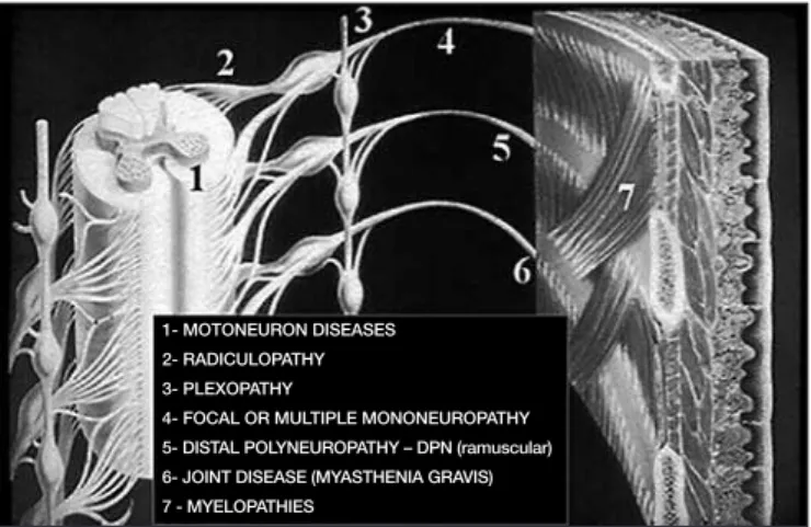
tions4 (Figure 1). ENMG is divided in two parts: nervous conduction study
or electroneurography (ENG)5 and needle electromyography (EMG).
ENMG is an important method for the diferential diagnosis of several muscular and nervous disorders. Computerized tomography (CT) and/or magnetic nuclear resonance (MRI) are just sophisticated pictures, whereas ENMG gives real-time information on what is going on in nerves and mus-cles and, together with imaging exams helps the accurate diagnosis of neuro-muscular disorders6.
Electrodiagnostic studies are essential to establish the accurate diagnosis of some diseases not visualized by imaging exams and, in general, one may con-sider the indication of ENMG when facing decreased sensitivity (hypoes-thesia); sensation of shock and tingling (pares(hypoes-thesia); decreased muscle mass (amyotrophy), cramps and/or fasciculations, or decreased or abolished deep relexes (myotactic hypo or arrelexia)7,8.
he electrodiagnostic study is used to supply accurate diagnosis; to locate the level of the injury; to reconcile treatment with diagnostic correction; to supply prognostic information4,5,9.
Soon after anamnesis and neurologic exam, the physician shall develop a diferential diagnosis to help identifying nervous and muscle segments to be tested (Figure 2).
Figure 1 is a summary of the electrodiagnostic study process.
Examples of most commonly found possible injured sites and diferential diagnosis are:
• Spinal cord anterior horn: amyotrophyc lateral sclerosis
• Roots: cervical or lumbosacral radiculopathy;
• Plexopathy: Parsonage Turner syndrome or thoracic outlet;
• Axonal: toxic and/or autoimmune neuropathy (axonal PRN – AMSAN);
• Unmyelinating: metabolic (diabetes mellitus), autoimmune (PRN – Guil-lain Barré syndrome), degenerative (CMT-I);
• Neuromuscular junction: myastenia gravis, Lambert-Eaton syndrome, botulism;
• Muscular: polymyositis, muscle dystrophies, periodic paralysis.
Aiming at gathering maximum information and perform ENMG with mini-mum discomfort for patients it is important to have in mind the temporal sequence of events happening when a nerve responds to an injury (Table 1). If the exam is to earlier performed there might be a false negative. Axo-nal reinnervation has a growth rhythm of approximately 1mm per day (ap-proximately 2.5cm per month) and if a serial study is being performed for prognosis, it will be necessary to space ENMG time according to axonal regeneration rate.
Table 1. Temporal sequence of post-traumatic electromyographic abnormalities
Abnormalities Onset time
Conduction block at injury site Immediate
Decreased EP amplitude on distal stimulation More than 7 days
Signs of denervation at rest 2 to 5 weeks
Reinnervation signs (partial injury) More than 4 to 8 weeks
EP = motor or sensory evoked potential.
In case of suspicion of myopathy, it is fundamental to collect muscle en-zymes before ENMG due to muscle trauma of electromyography needle insertion in different points and consequent secondary serum increase of muscle enzymes, in general of mild intensity. Still in case of suspicion of myopathy, the electromyographist shall indicate the best place for muscle biopsy10.
Due to multiple traumatic points at electromyography needle insertions, mandatorily the indicated muscle for biopsy shall not be submitted to needling to prevent false results of muscle biopsy. Indication of muscle biopsy in highly affected muscles with severe fibroadipose and/or muscles with questionable or minor EMG changes shall result in inconclusive pathologic changes at muscle biopsy (Table 2).
Neuroimaging Exams
There are many noninvasive modalities of human brain imaging studies. Some methods, such as CT and MRI, for their spatial resolution, supply details on anatomic structures; other methods, such as positron-emission CT (PET-CT) and functional magnetic resonance (fMRI), in addition to anatomic details also supply data on metabolic and functional measure-ments. In addition, imaging techniques, such as spectroscopy and diffusion tensor by MRI, allow the study of biochemical changes and brain connec-tivities with different temporal and spatial resolutions.
Neuroimaging methods versus pain neuroanatomy
Noninvasive imaging studies, especially PET and fMRI, have helped under-standing neural networks and pain pathophysiology, most of them focused on acute pain and few directed to understanding chronic pain associated to central or peripheral neurologic injuries11. These studies, focused on
deter-mining physiologic pain bases, have shown a pain processing pattern called “pain matrix”. Pain matrix involves different areas of the nervous system and of neural networks, which allow the differentiation of pain percep-tions. Although some networks are overlapped, NP processing seems to be different from acute pain11.
Most widely used modality for the study of pain has been fMRI, for its abil-ity to monitor in real time brain activabil-ity during cognitive stimuli and/or tasks. It supplies indirect evaluation of brain metabolism and function by measuring changes in brain oxygenation levels in vivo, by image enhance-ment called BOLD signal (blood-oxygen-level dependent)12.
fMRI resting state supplies information on brain functional connectivity areas. fMRI at rest has the advantage of supplying neuroimaging data of
Figure 2. Algorithm for electroneuromyography planning Paresthesia, hypoesthesia, weakness of extremities
Signs of peripheral nervous system injury: hypo e arrelexia,
hypotonia, amyotrophy, fasciculation, cramps
Neuropathic versus myopathic
Neuropathic
Polyneuropathy (distal diffuse predominance)
Motor Sensory
Signs of central nervous system injury: hyperrelexia,
hypertonia, hemi ou paraparesia, Babinski signal
Electroneuromyography is not indicated, except in cases of motoneuron disease associated
to pyramidal syndrome (amyotrophic lateral sclerosis)
Myopathic: Proximal weakness and lack of sensory signs
Mononeuropathy (sensory-motor signs with peripheral
nerve distribution)
Radicu-lopathy
Plexo-pathy
Nervous trunk nervo
Figure 1. The objective of the electrodiagnostic study is to determine whether there is some abnormality in addition to the injury site along peripheral nervous system pathway.
1- MOTONEURON DISEASES
2- RADICULOPATHY
3- PLEXOPATHY
4- FOCAL OR MULTIPLE MONONEUROPATHY
5- DISTAL POLYNEUROPATHY – DPN (ramuscular)
6- JOINT DISEASE (MYASTHENIA GRAVIS)
25
Additional tests to investigate neuropathic pain. he value of electroneuromyography for neuropathic pain
Rev Dor. São Paulo, 2016;17(Suppl 1):S23-6
individuals with chronic pain while they remain at rest inside the device. So, it allows the evaluation of the brain of chronic and neuropathic pain patients without the need for sensory or cognitive stimulation. It is believed that chronic pain changes the oscillation of some neural networks, espe-cially the default-mode network, which are more active at rest; protrusions and executive networks, which are more active during sensory and tasks stimulation, and networks related to sensory and motor processing12.
Neuroimaging studies for NP have been more often performed in patients with painful syndromes of unexplained origin, such as fibromyalgia, com-plex regional pain syndrome type I (CRP1) and in patients with chronic pain such as low back pain. All imaging studies have shown that chronic pain and its morbidities promote changes in different brain areas13.
Response to pain activation has been consistently reported in preferentially
some areas, being that no single area is responsible for chronic pain and its morbidities. These areas are: primary and secondary somatosensory cortex (S1 and S2), insular cortex (IC), anterior cyngulos cortex (ACC), motor cortex (MC), pre-frontal cortex (PFC), amygdale, thalamus, hippocampus and cerebellum11-13. The activation of lateral thalamus, S1, S2 and anterior
IC seems to be associated to pain sensory and discriminative aspects12.
ACC, posterior IC and limbic system components seem to be associated to emotional pain component. Some PET studies in patients with allodynia suggest that spontaneous NP is associated to thalamic activity, inferring an emotional pain dimension11.
The pre-frontal cortex seems to be related to the cognitive aspect of pain and somatosensory cortex and IC are responsible for pain intensity decod-ing. Some neuroimaging studies have also shown that in chronic pain situ-ations, such as fibromyalgia and chronic low back pain, there are changes in these areas, suggesting changes in pain intensity processing13.
Primary motor cortex and supplementary areas also play a role in chronic pain, such as those related to cerebellum changes, although these cerebellar changes are currently poorly understood11,12.
Imaging methods versus clinical neuropathic pain evaluation
Imaging evaluation of NP patients will depend on the type of pain and following a rational criterion for each case to prevent radiation and un-necessary costs.
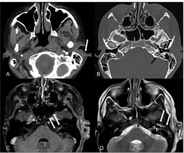
Neuropathic syndromes related to cranial and facial neuralgias, such as tri-geminal, facial and other cranial nerves neuralgia are in general diagnosed and evaluated by history and clinical evaluation. Imaging evaluations are necessary when classic symptoms of trigeminal neuralgia are not clear, in cases of trigeminal neuralgia in young patients or, for example, evaluation of facial nerve neuralgia of central origin. Brain MRI with enhancement and with emphasis on cranial nerves is the most indicated. Fifteen percent of patients with trigeminal neuralgia have altered imaging exams (CT or MRI), being caused by tumors, such as trigeminal Schwannoma (Figure 3) and multiple sclerosis14,15.
Although MRI provides a better evaluation of cranial nerves and is more sensitive to evaluate parenchymal injuries, CT is best indicated to deter-mine bone erosions, to evaluate bony labyrinth and tympanic cavity. Very often imaging exams are complementary to the clinical exam to determine injury extension and complications, such as in the following example of a patient with Ramsay Hunt syndrome (Figure 4).
Simple X-rays is the most common imaging modality for the initial evalu-ation of chronic low back pain. Its major objective is to detect gross/severe structural pathological changes. Simple X-rays findings are not specific; for example, osteoarthritis findings seen in X-rays of patients with radicu-lopathy may also be seen in totally asymptomatic patients. The opposite is also true; normal exams do not rule out the diagnosis of lumbosacral radiculopathy.
MRI is undoubtedly the best method to evaluate cervical, thoracic and lumbosacral radiculopathies, and plexopathies. However, due to its high cost and low availability, it should be rationally used being reserved as first choice for patients with progressive neurologic deficits, cauda equina syn-drome, suspicion of malignancy, inflammatory or infectious disease. In ad-dition, MRI may also be used to plan surgical treatment or modalities such as epidural steroid administration.
CT may also be used for the evaluation of radiculopathies. Although not being the method of choice to evaluate spinal cord and nerves, it allows the evaluation with good anatomic resolution (especially CT multislice modalities) of compressive causes of radiculopathy, spinal canal, foramina and even discs. It is especially important for postoperative evaluation after reconstruction with plates and screws, to evaluate integrity and looseness. Autonomic neuropathic syndromes are also investigated with brain or spine MRI, depending on the dysautonomia clinic. In addition, SPECT and PET exams may identify cardiac sympathetic dysfunction, such as in patients with diabetes mellitus types 1 and 2.
There are many cancer-related neuropathic syndromes. These neuropathies may result from one or more mechanisms, such as compression, inflam-mation, infiltration of nerves, trunks, plexuses, bones and meninges. All cancer patients with neuropathic syndromes should be evaluated with CT, MRI, PET and/or scintigraphy. In some cases, simple X-rays shall be ad-equate to identify skeletal injuries. MRI is more sensitive to detect early bone injuries and for medullary, radicular, plexuses and neural trunks
eval-Table 2. Muscles – peripheral nerves – spinal level
Muscles Nerves Segments
Trapezius Accessorius C3-C4
Diaphragm Phrenicus C3-C4
Rhomboid Dorsalis scapulae C5
Anterior serratus Thoracicus longus C5-C6-C7
Supraspinous Suprascapularis C5-C6
Infraspinatus Suprascapularis C5-C6
Deltoid Axillaris C5-C6
Brachial biceps Musculocutaneus C5-C6
Brachioradial Radialis C5-C6
Brachial triceps Radialis C6-C7
Supinator Radialis C6-C7
Extensor carpi radialis Radialis C6-C7
Pronator teres Medianus C6-C7
Flexor carpi radialis Medianus C6-C7
Extensor digitorum communis Radialis C7-C8
Extensor indicis proprius Radialis C8-T1
Flexor carpi ulnaris Ulnaris C8-T1
Flexus policis longus Medianus C8-T1
Opponens policis Medianus C7-C8
Abductor digiti quinti Ulnaris C8-T1
Interosseous dorsalis Ulnaris C8-T1
Flexor policis brevis Medianus C8-T1
Pronator quadratus Ulnaris C8-T1
Iliopsoas Femoralis L2, L3,L4
Abductor of the thigh Obturatorius L2, L3,L4
Quadriceps femoris Femoralis L2, L3,L4
Tibialis anterior Fibularis profundus L4, L5 Tensor fasciae latae Gluteus superior L5, S1
Biceps femoris Ischiadicus L5, S1
Semitendinosus and membranosus Ischiadicus L5, S1
Peroneus longus and brevis Fibularis supericialis L5, S1 Extensor hallucis longus Fibularis profundus L5, S1 Tibialis posterior Tibialis posterior L5, S1 Extensor digitorum brevis Tibialis profundus L5, S1
Gluteus maximus Gluteus inferior L5, S1
Abductor hallucis brevis Tibialis posterior L5, S1
Gastrocnemius medialis Tibialis posterior L5, S1
Bulbocavernosus Pudendus S2, S3, S4
26
Porto FH, Machado Porto GC and Brotto MW
Rev Dor. São Paulo, 2016;17(Suppl 1):S23-6
uation. CT is more sensitive than X-rays and although less sensitive than MRI for early bone injuries it is less expensive, more available and faster than MRI. Bone scintigraphy is more indicated to evaluate the extension of bone injuries along the body16.
Figure 3. Schwannoma of left trigeminal nerve
A: brain magnetic resonance, T2W1 showing injury with high signal, expansive, in the cis-ternal portion of the trigeminal nerve, enlarging Meckel’s cave, expanding to left cavernous sinus (white arrows). B: brain magnetic resonance, T1W1 with enhancement, showing intense and homogeneous enhancement of the injury (black arrows) with permeating cystic areas (white arrow).
Figure 4. Ramsay Hunt syndrome to the left.
A: brain computerized tomography with enhancement, soft tissues window, showing vesicles in pinna and external ear canal (white arrows). B: brain computerized tomography, bone win-dow, showing extension of vesicles to external ear canal, thickening of tympanic membrane and mastoid cells obliteration. C: brain computerized tomography, T1W1 with enhancement, shows enhancement and thickening of cisternal and canalicular portion of left facial nerve (white arrows). D: brain computerized tomography, T2W1 showing otomastoiditis.
OTHER ADDITIONAL EXAMS
Sensory quantification test (SQT)
SQT is used to measure sensory perception threshold for diferent nociceptive pathway modalities (warmth, cold, pain)1,3. By means of a thermodo placed on
the skin of the region to be tested (areas afected by pain and areas not afected as control), temperature is increased or decreased (1 to 4°C/s) until patients press a button when they feel a certain sensation, thus quantifying the four thermal thresholds of cold, pain by cold, warmth and pain by warmth. SQT is just occasionally used in the clinical practice for cost and applicability reasons.
Microneurography
his technique is more commonly used in research as compared to clinical practice. It consists of the insertion of a tungsten needle in the nerve to be studied3. he pattern of small ibers activity (C ibers) is evaluated by this
method. Microneurography is a time consuming and painful method dif-icult to be evaluated, reasons why it is seldom used in the clinical practice.
CONCLUSION
Additional exams, together with thorough history and neurologic exam fo-cused on neurologic propedeutics, may supply important data for NP diag-nosis and are often used in the clinical practice. It is critical for professionals involved with NP patients care to understand major additional exams and their indications.
REFERENCES
1. Bouhassira D, Attal N. Translational neuropathic pain research: a clinical perspective. Neuroscience. 2016; [Epub ahead of print].
2. Baron R, Binder A, Wasner G. Neuropathic pain: diagnosis, pathophysiological mechanisms, and treatment. Lancet Neurol. 2010;9(8):807-19.
3. Schestatsky P. Deinição diagnóstico e tratamento da dor neuropática. Rev HCPA 2008;28(3):177-87. 4. Aminof MJ. Electromyography in clinical practice. 2nd ed. New York: Churchill Livingstone; 1987.
5. Downie A W. Studies in nerve conduction. ln: Walton JN (ed.). Disorders of voluntary musck. 3nd
ed. Edinburgh: Churchill; 1974. 973-1002p.
6. Johnson EW. Practical electromyography – 4th ed. Lippincott Williams & Wilkins, 1997.
7. Kimura J. Nerve conduction and electromyography. In: Dick PJ, homas PK, Lambert EH, Bunge R, (eds.). Peripheral neuropathy. 2nd ed. Philadelphia: Saunders; 1984. 919-66p.
8. Kimura J. Electrodiagnostic in diseases of nerve and muscle: principles and practice. 2nd ed.
Philadel-phia: Davis; 1989.
9. Victor M, Ropper H. Adams and Victor’s – Principles of neurology. 7th ed. New York: McGraw-Hill;
2001.
10. WeissL. Easy EMG. Edited by Butterworth Heinemann, 2004.
11. Moisset X, Bouhassira D. Brain imaging of neuropathic pain. Neuroimage. 2007;37(Suppl 1):S80-8. 12. Geha PY, Apkarian AV. Brain imaging indings in neuropathic pain. Curr Pain Headache Rep.
2005;9(3):184-8
13. Katherine T Martucci, Pamela NG, Mackey S. Neuroimaging chronic pain: what have we learned and where are we going? Future Neurol. 2014;9(6):615-26.
14. Eller JL, Raslan AM, Burchiel KJ. Trigeminal neuralgia: deinition and classiication. Neurosurg Focus. 2005;18(5):E3.
15. Majoie CB, Hulsmans FJ, Castelijns JA, Verbeeten B, Tiren D, van Beek EJ, et al. Symptoms and signs related to the trigeminal nerve: diagnostic yield of MR imaging. Radiology. 1998;209(2):557-62. 16. Berger AM, Shuster Jr JL, Von Roenn JH. Principles and practice of Palliative Care and Supportive
Oncology, chapter 1: diicult pain syndromes: neuropathic pain, bone pain, visceral pain. 4th ed.
Philadelphia, USA: Wolters Kluwer Health; 2013. 960p.