Rev. bras.oftalmol. vol.76 número2
Texto
Imagem
Documentos relacionados
Bcl-2 and bax expression in retinal explants were identiied for evaluation the neuroprotective efect of conditioned medium from control MSCs (CM-MSCs), conditioned medium from
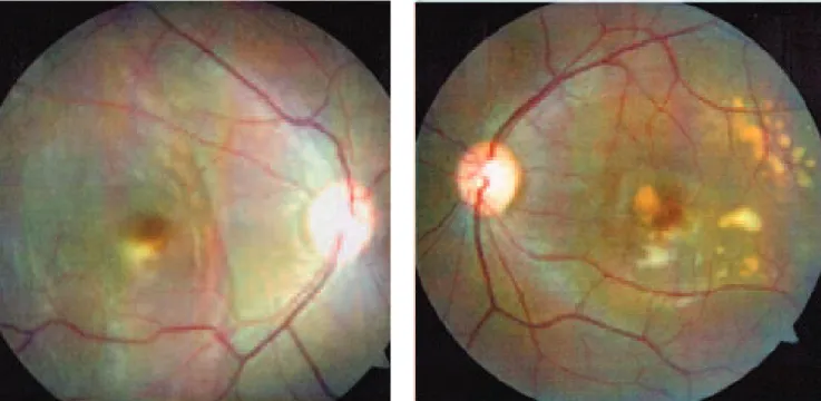
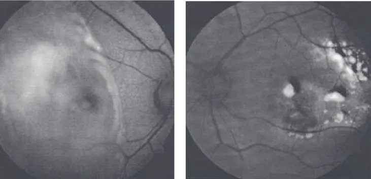
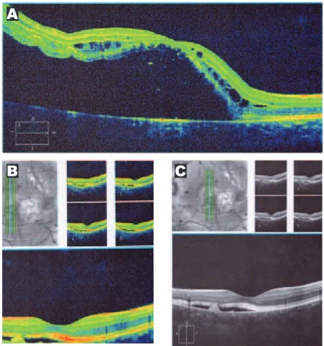
We report the case of a 38-year-old patient who presented with a clinical diagnosis of solar retinopathy in the left eye, no prior history of sun exposure, normal visual acuity
The lineal form is clinically seen as localized progressive corneal edema from the periphery to the center along with a line of keratic precipitates and associated with
The same patient at sixty days after surgery (advancing the left medial rectus) showing a residual limitation of left eye
The unusual localization and timing of this recurrence of granular corneal dystrophy after deep anterior lamellar keratoplasty suggests that corneal stromal keratocytes may play
The occurrence of Sjögren syndrome-like illness, reported as dry eye symptoms or signs, in patients having confirmed viral infections, such as human T-cell lymphotropic virus
However, due to diagnostic method limitations and analyzing the clinical findings and uniform response to the treatment, as well as all patients had been operated in two
des specific management issues such as cooperativism, civil society, marketing, contract negotiation, professional defense, career plan- ning, retirement, consumer protection
