doi: 10.1590/0037-8682-0361-2016
Case Report
Corresponding author: Profa. Ana Isabel Vieira Fernandes. e-mail: [email protected]
Received 14 September 2016 Accepted 9 August 2017
Cardiac tamponade in a patient with severe dengue fever
Ana Isabel Vieira Fernandes[1],[2], Ciro Leite Mendes[3], Raíssa Holmes Simões[1],
Ana Elisa Vieira Fernandes Silva[1], Clarissa Barros Madruga[1], Carlos Alexandre Antunes de Brito[4],
Lucio Roberto Castellano[2] and Marli Tenório Cordeiro[5]
[1]. Serviço de Doenças Infecciosas e Parasitárias, Hospital Universitário Lauro Wanderley, Universidade Federal da Paraíba, João Pessoa, PB, Brasil. [2]. Grupo de Estudos e Pesquisas em Imunologia Humana, Escola Técnica de Saúde, Universidade Federal da Paraíba, João Pessoa, PB, Brasil.
[3]. Unidade de Terapia Intensiva, Hospital Universitário Lauro Wanderley, Universidade Federal da Paraíba, João Pessoa, PB, Brasil. [4]. Departamento de Medicina Clínica, Universidade Federal de Pernambuco, Recife, Brasil.
[5]. Laboratório de Virologia e Terapia Experimental, Centro de Pesquisas Aggeu Magalhães, Fundação Oswaldo Cruz, Recife, PE, Brasil.
Abstract
A 26-year-old postpartum female presented with symptoms characteristic of dengue fever on the 16th day of puerperium. On
the third day of the illness, the patient presented a clinical picture consistent with shock. Tests determined primary infection
with dengue virus serotype 2. Cardiac tamponade was conirmed by echocardiography. This rare manifestation is described in a
patient without any associated comorbidity.
Keywords: Dengue. Severe dengue. Cardiac tamponade.
INTRODUCTION
Severe manifestations of dengue infections are associated with several complications, including massive bleeding, respiratory failure, liver failure, encephalopathy, myocardial, and other organ dysfunctions. In the last decade, atypical forms of dengue have been described, such as encephalopathy, liver disorders, including acute liver failure, and heart problems1,2.
Cardiac involvement in dengue is often limited and benign, but can lead to dengue shock syndrome, which can present as transient myocardial dysfunction3,4. In one fatal case of
dengue-associated myocarditis, large areas of muscular necrosis and intense interstitial edema were directly associated with dengue virus on the cardiac muscle5. When diagnosed and treated early,
myocarditis can resolve without sequelae6. However, the clinical
manifestation of myocarditis in conjunction with persistent
hypotension, despite adequate luid resuscitation, may lead to more aggressive luid resuscitation, resulting in large pleural
effusions and respiratory failure7.
Ultrasonography is useful in the prediction of dengue
severity, including the identiication of early signs of plasma
leakage, such as pleural effusion, ascites, perirenal fluid, subcapsular hepatic collection, and pericardial effusion8. In
severe cases, infection may be associated with hemodynamic consequences leading to refractory shock9,10. Pericarditis in
patients with dengue is extremely rare and usually associated with an extension of a myocardial lesion, rather than a direct deposit of immune complexes6,11. Small pericardial and pleural
effusions associated with severe plasma leakage have been described in patients with dengue hemorrhagic fever (DHF), but cardiac tamponade is an extremely rare in patients with dengue. We report a case of cardiac tamponade in a postpartum woman with primary dengue virus serotype-2 (DENV-2) in Brazil. This is the second case of cardiac tamponade in a patient with severe
dengue fever reported in literature and the irst well-documented
case in a patient without any associated comorbidity.
CASE REPORT
A 26-year-old African American woman who underwent a cesarean section presented with high fever, frontal headache, myalgia, and retro-orbital and bone pain on the 16th day
postpartum. The patient’s pregnancy progressed without complications, and she reported no comorbidities. The patient was admitted to the emergency room of a private hospital in João Pessoa, Paraíba State, Brazil. The patient received hydration,
and initial laboratory examination identiied hemoconcentration,
leukopenia, and mild thrombocytopenia (Table 1). The woman’s condition improved, and she was discharged with symptom severity guidance. Two days later, the patient returned to the hospital with dizziness, cold sweats, a prostration-related fainting episode, lid edema, nausea, vomiting, abdominal pain, diarrhea, mild uterine bleeding, and exanthema.
Exam RV BA At admission Days after admission
1 3 4 5 6 7 8 10 14
CBC
Hemoglobin 12 – 16g/dL 15.6 8.4 15.7 12.2 10.6 11 11.2 10.5 10.6 12.3 10.3
Hematocrit 4 – 5.2 millions/mm3 44.8 25.1 46.3 36.6 31.9 33.8 34,2 32 32,6 38,1 31
Leukocyte (x1,000) 4.2 – 11.0/mm3 3.7 4.4 10.8 8.0 9.0 10.4 11.3 7.9 8.7 10.8 7.7
Neutrophils 40 – 70% 70 65 70 83 80 82 78 79 76 69 69
Rods 0 – 5% 0 0 0 0 0 0 0 0 0 0 0
Platelets (x1,000) 150 - 450 thousand/mm3 169 115 133 170 203 316 304 287 239 324 323
Coagulation
APTT 10 – 14 10.2 10.2 10.2 10.2 10.4 - - - 11,3
-PTT 100% 100 100 100 100 97 - - - 84
-Biochemistry
AST 11 – 39U/L 29 38 - - 261 - - - - 83
ALT 11 – 45U/L 35 26 - - 143 - - - - 162
Albumin 3.5 – 5.5g/dL 1 - - 1.8 - - - - 5.2
C-reactive protein <5mg/L 6.4 - - - 87.2
TABLE 1
Evolution of laboratory tests for the patient with dengue following hospital admission.
RV: reference value; BA: before admission; CBC: cell blood count; APTT: activated partial thromboplastin time; PTT: prothrombin time test; AST: aspartate transaminase; ALT: alanine transaminase.
had an axillary temperature of 38.7°C, a heart rate of 130bpm, a thready pulse, cold sweats, a blood pressure of 80/40mmHg, and evidence of a positive tourniquet test. The patient also exhibited a diminished vesicular murmur at the base of the right lung, hypophonetic heart sounds, and jugular venous distention. Ancillary exams upon admission (the third day of illness) showed decreased platelet numbers and hemoconcentration, and serological tests were positive for immunoglobulin M (IgM), but negative for dengue virus immunoglobulin G (IgG), and positive for the dengue virus non-structural protein 1 (NS1) antigen. Reverse transcription polymerase chain reaction (RT-PCR) results were positive for the DENV-2 serotype.
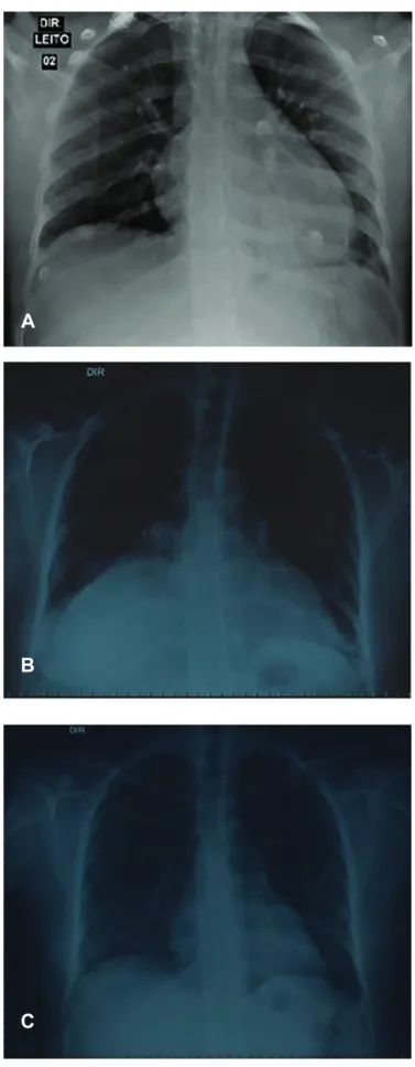
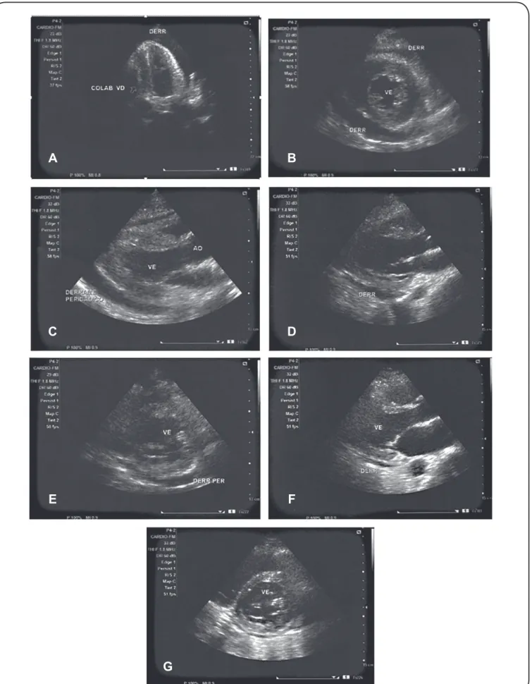
The patient was transferred to the Intensive Care Unit (ICU). with a blood pressure of 80/40mmHg and developed metabolic acidosis and persistent arterial hypotension, despite adequate volume expansion. Upon examination, the patient still exhibited hypophonetic heart sounds and jugular venous distention. She was dyspneic and put under alternating noninvasive ventilatory support. Chest radiography and abdominal ultrasound were unremarkable (Figure 1), showing relative hepatomegaly and increased uterine volume, both common during the postpartum period. The patient underwent an echocardiogram, which
conirmed the diagnosis of cardiac tamponade (Figure 2).
Pericardial luid was drained and analyzed and was negative for
bacterial or fungal structures. However, rare leukocytes were
identiied after 48 hours of bacterial culture.
The patient developed anasarca with a progressive increase in pleural effusion and ascites. On the third day in the ICU, the patient was administered noradrenaline and 400 mg/Kg/day immunoglobulin for three days. On the sixth day, norepinephrine was gradually withdrawn, and the patient developed anxiety and restlessness, requiring oxygen supplementation via a Venturi mask (50%). A diuretic was initiated. Echocardiography was performed, showing mild pericardial effusion and mitral regurgitation. On the eighth day, the patient showed an improvement in breathing patterns and was discharged from the ICU. In the ward, the patient exhibited an improvement in anxiety symptoms, occasional dry cough, rales in the right lung base, normophonetic heart sounds without jugular venous distention, a heart rate of 80bpm, and a blood pressure of 120/80 mmHg. She exhibited ascites and edema of the lower limbs and genitalia. Chest x-rays showed right inferior lobe atelectasis, small bilateral pleural effusion, and normal heart size. The patient underwent respiratory physiotherapy and was discharged without sequelae.
Ethical considerations
A
B
C
FIGURE 1 - Evolution of the chest radiograph. Increase in heart size and absence of pleural effusion (a). Atelectasis of the lower lobe of the right lung (b). Chest X-ray control to normal discharge from the hospital (c).
DISCUSSION
This is one of the few cases reported in literature of cardiac
tamponade in a patient with dengue fever and the irst
well-documented case in Brazil. In addition, this case is clinically important because it was a primary dengue infection of presumably mild intensity. Early in the course of disease, the patient presented with severe persistent vomiting and abdominal
pain, requiring luid resuscitation. On the third day, the patient
still had a fever and developed signs of shock, presenting with
large pleural effusion and signiicant vascular endothelium and
hypoalbuminemia impairment. During resuscitation, she received
a large volume of luids followed by vasoactive drugs. However,
since hypervolemia can cause an increase in effusion levels, the patient’s condition was reversed after the shock phase via diuretics and water restriction. In the present case, the pericardial effusion that progressed to cardiac tamponade occurred prior to the other effusions that increased after volume resuscitation.
In a previous case of cardiac tamponade in a female patient with dengue fever, the 59-year-old woman had lupus, which may predispose the person to serositis and stroke9. In the
present case, puerperium, during which the hypervolemic condition of pregnancy is maintained, may slowed the elimination of liquids observed three weeks after the patient gave birth. There is limited data on the association between DENV infection and cardiac tamponade. Previous reports on adult men based diagnoses on IgM/IgG serology, without any
conirmatory viral isolation and/or molecular identiication of
DENV serotypes5,12. Further, only one study tested for the NS1
viral protein, demonstrating undetectable circulating levels5.
The present case showed elevated NS1 levels and had positive
IgM results, but had negative IgG results with a conirmatory
RT-PCR for the DENV-2 serotype. Immunoglobin serology without any other diagnostic method could present challenges in the determination of the exact role played by the DENV-2 serotype. Previous studies have suggested that positive IgM and IgG results in patients with cardiac tamponade may indicate a DENV secondary infection. In the present case, the patient had negative IgG results, but positive IgM results for DENV, suggesting a primary DENV infection. In comparison with patients with secondary DENV infections, patients with primary DENV infections show fewer or no cardiac complications.
In India, retrospective studies evaluating dengue fever cases among the general population13 and adolescents14 have reported
the occurrence of myocarditis and pleural or pericardial effusion.
In the irst study13, 96 (56.8%) out of 169 patients did not present
positive results for any conirmatory test of DENV infection. For
these patients, referred to as the Sero-Negative Probable Dengue Fever group, only epidemiological information about a dengue outbreak in the studied area was provided by the authors. This was a limitation similar to that of the present case. In the second study14, DENV infection was described in children, not adults. In
literature, differences in the disease course of DENV have been well documented between children and adults, which may make the
comparison of the retrospective study to the present case dificult. In addition, both studies lacked conirmatory results to determine
FIGURE 2 - Echocardiogram demonstrating extensive pericardial effusion with cardiac tamponade and right ventricle collapsed (a-c). Control echocardiogram after pericardial drainage. Moderate pericardial effusion without evidence of cardiac tamponade (d-e). Echocardiogram control after four days of pericardial drainage. Small pericardial effusion (f-g). RV: right ventricle.
A
B
C
D
E
F
RT-PCR identiied the serotype as DENV-2. Although other
dengue serotypes may be associated with cardiac involvement, DENV-2 is believed to be the main cause of myocardial diseases10.
Here, no signs of myocarditis were found; however, the association
between speciic DENV serotypes and cardiac involvement needs to be clariied within a large cohort. The present case opens the
discussion of this complication of dengue infection.
Although the patient presented with severe symptoms, she
did not fulill the criteria for DHF because she maintained a
platelet count above 100 thousands/mm3, illustrating a gap in
the traditional classiication of the World Health Organization15.
In the revised classiication1, the case would meet the criteria
for severe dengue (SD) because of the development of large effusions in combination with respiratory failure and severe
organ dysfunction, exempliied here by cardiac tamponade. This
is, however, an extremely rare condition that by itself does not
justify a change in disease classiication.
Cardiac alterations in patients with dengue may be associated with shock dengue syndrome and should be diagnosed via echocardiography. Progression to cardiac tamponade in patients with dengue is a very rare condition that requires a high degree of suspicion and immediate intervention.
Acknowledgments
The authors would like to express their gratitude to the patient and to the staff and residents of the Infectious and Parasitic Diseases Service, Hospital Universitário Lauro Wanderley, Universidade Federal da Paraíba, João Pessoa, PB, Brasil.
Financial support
This work was supported by Conselho Nacional de Desenvolvimento Cientíico e Tecnológico (CNPq), Brazil, grant 576214/2008-6.
Conlict of interest
The authors declare that there are no conlicts of interest.
REFERENCES
1. World Health Organization (WHO). Dengue: guidelines for diagnosis, treatment, prevention and control: new edition. France: World Health Organization; 2009. 147p.
2. Weerakoon KG, Kularatne SA, Edussuriya DH, Kodikara SK, Gunatilake LP, Pinto VG, et al. Histopathological diagnosis of myocarditis in a dengue outbreak in Sri Lanka, 2009. BMC Research Notes. 2011;4:268.
3. Gupta VK, Gadpayle A. Subclinical cardiac involvement in dengue haemorrhagic fever. J Indian Acad Clin Med. 2010;11(2):107-11. 4. Miranda CH, Borges Mde C, Schmidt A, Pazin-Filho A, Rossi MA,
Ramos SG, et al. A case presentation of a fatal dengue myocarditis showing evidence for dengue virus-induced lesion. Eur eart J Acute Cardiovasc Care. 2013;2(2):127-30.
5. Miranda CH, Borges MC, Matsuno AK, Vilar FC, Gali LG, Volpe GJ, et al. Evaluation of cardiac involvement during dengue viral infection. Clin infect Dis. 2013;57(6):812-9.
6. Wiwanitkit V. Dengue cardiac infection, A Brief Review. Acta Cardiol Sin. 2008;24:226.
7. Pesaro AE, D'Amico E, Aranha LFC. Dengue: cardiac manifestations and implications in antithrombotic treatment. Arq Bras Cardiol. 2007;89(2):e12-5.
8. Oliveira RVB, Rios LTM, Branco MRFC, Braga Jr LL, Nascimento JMS, Silva GF, et al. Valor da ultrassonograia em crianças com suspeita de febre hemorrágica do dengue: revisão da literatura. Radiol Bras. 2010;43(6):401-7.
9. Kumar S, Iuga A, Jean R. Cardiac tamponade in a patient with dengue fever and lupus nephritis: a case report. J Intensive Care Med. 2010;25(3):175-8. Epub 2010/01/22. doi: 10.1177/0885066609358955. PubMed PMID: 20089524.
10. Kularatne SA, Pathirage MM, Kumarasiri PV, Gunasena S, Mahindawanse SI. Cardiac complications of a dengue fever outbreak in Sri Lanka, 2005. Trans R Soc Trop Med Hyg. 2007;101(8):804-8. 11. Nagaratnam N, Siripala K, de Silva N. Arbovirus (dengue type) as a cause of acute myocarditis and pericarditis. Br Heart J. 1973;35(2):204-6.
12. Bendwal S, Malviya K, Jatav OP, Malviya K. Cardiac tamponade presenting as early manifestation in dengue fever. J Assoc Physicians India. 2014;62(3):257-9.
13. Motla M, Manaktala S, Gupta V, Aggarwal M, Bhoi SK, Aggarwal P, et al. Sonographic evidence of ascites, pleura-pericardial effusion and gallbladder wall edema for dengue fever. Prehosp Disaster Med. 2011;26(5):335-41.
14. Pothapregada S, Kamalakannan B, Thulasingam M. Clinical proile of atypical manifestations of dengue fever. Indian J Pediatr. 2016;83(6):493-9.