Ductal carcinoma
in situ
of the breast: Evaluation of main
presentations on magnetic resonance imaging compared with
findings on mammogram and histology
GUSTAVO MACHADO BADAN1*, DÉCIO ROVEDA JÚNIOR2, SEBASTIÃO PAITO3, EDUARDODE FARIA CASTRO FLEURY4, BIANCA MARAGNO5,
MARIO SÉRGIO DANTASDO AMARAL CAMPOS6, CARLOS ALBERTO PECCI FERREIRA7, FELIPE AUGUSTO TROCOLI FERREIRA8
1Coordinator at the FEMME Breast Intervention Sector, Laboratório da Mulher. Assistant Physician of the Diagnostic Imaging Service, Santa Casa de Misericórdia de São Paulo, São Paulo, SP, Brazil
2PhD in Health Sciences from Santa Casa de São Paulo. Coordinator at the FEMME Breast Radiology Sector, Laboratório da Mulher. Assistant Physician of the Diagnostic Imaging Service, Santa Casa de Misericórdia de
São Paulo, São Paulo, SP, Brazil
3Full Professor of the Faculdade de Ciências Médicas da Santa Casa de São Paulo. Head of the Gynecology Clinic, Santa Casa de Misericórdia de São Paulo, São Paulo, SP, Brazil 4Professor, PhD in Health Sciences from Santa Casa de São Paulo. Assistant Physician of the Diagnostic Imaging Service, Santa Casa de Misericórdia de São Paulo, São Paulo, SP, Brazil 5Specialist in Breast Imaging – MD, Member of the Breast Radiology Team at Santa Casa de São Paulo and the FEMME, Laboratório da Mulher, São Paulo, SP, Brazil
6Coordinator of the FEMME Mammography Service, Laboratório da Mulher. Assistant Physician of the Diagnostic Imaging Service, Santa Casa de Misericórdia de São Paulo, São Paulo, SP, Brazil
7Coordinator of the Breast Imaging Service, Department of Internal Medicine, Faculdade de Ciências Médicas da Santa Casa de São Paulo. Mammographer at FEMME, Laboratório da Mulher, São Paulo, SP, Brazil 8Specialist in General and Breast Radiology – Assistant Physician of the Diagnostic Imaging Service at Santa Casa de São Paulo and the FEMME, Laboratório da Mulher, São Paulo, SP, Brazil
S
UMMARYStudy conducted at Santa Casa de Misericórdia de São Paulo, Serviço de Diagnóstico por Imagem, São Paulo, SP, Brazil
Article received: 4/18/2015
Accepted for publication: 5/16/2015
*Correspondence:
Address: Rua Marquês de Itu, 381, 5o andar, Santa Cecília
São Paulo, SP – Brazil Postal code: 01223-001 [email protected]
http://dx.doi.org/10.1590/1806-9282.62.05.421
Objective: The purpose of this study was to evaluate the various morphologies and kinetic characteristics of the ductal carcinoma in situ (DCIS) on breast mag-netic resonance imaging (MRI) exam, to establish which are the most prevalent and to determine the effectiveness of the method in the detection of DCIS.
Method: A prospective observational study, starting in May 2014. We evaluat-ed 25 consecutive patients with suspicious or highly suspicious microcalciica-tions on mammography screening, BI-RADS categories 4 and 5, who underwent breast MRI and then surgery with proven diagnosis of pure DCIS. Surgery was considered the gold standard for correlation between histologic indings and ra-diological indings obtained on MRI.
Results: The most frequent morphological characteristic of DCIS on MRI was
non-mass-like enhancement (NMLE), p<0.001, observed in 22/25 (88%) patients (95CI 72.5-100). Of these, segmental distribution was the most prevalent, rep-resented by 9/22 (40.91%) cases (95CI 17.4-64.4), p=0.306, and a clumped inter-nal enhancement pattern was most commonly characterized in DCIS, observed in 13/22 (50.09%) cases.
Conclusion: DCIS has a wide variety of imaging features on MRI and being able to recognize these lesions is crucial. Its most common morphological presenta-tion is non-mass-like enhancement, while segmental distribupresenta-tion and a clumped internal enhancement pattern are the most common presentations. Faced with the combined analysis of these indings, percutaneous core needle biopsy (core biopsy) or vacuum-assisted biopsy (VAB) should be encouraged.
Keywords: breast neoplasms, non-iniltrating intraductal carcinoma,
mam-mography, magnetic resonance imaging, large-core needle biopsy.
I
NTRODUCTIONDuctal carcinoma in situ (DCIS) is a type of non-invasive breast malignancy. The pathological indings are charac-terized by proliferation of malignant epithelial cells in the terminal ductal lobular unit, with no evidence of in-vasion through the basement membrane. They may
af-fect more than one breast lobule, or occur bilaterally; and if detected before invasion, the chances of cure reach ap-proximately 100%.1,2
silent for a long time or never leave the duct, while those of highest degree exhibit high growth rates, high mitot-ic indmitot-ices, and in 30-50% of times progress to high-grade carcinoma.3
Its main presentation is grouped microcalciications, seen on mammography, which helped to establish this imaging exam as a screening method for breast cancer.4
After the introduction of mammographic screening, the incidence of DCIS has increased from 2 to 20% of all diagnosed cases,5,6 contributing to the reduction of the mortality rate for breast cancer.6-8 However, not all DCIS calcify, and mammographic sensitivity ranges from 27 to 80%.5,9,10
The limitations of mammography have raised inter-est in the use of other imaging methods for the detec-tion of DCIS. Magnetic resonance imaging (MRI) has a sensitivity in the diagnosis of carcinoma of 86-96%11,12 and some series of studies have shown sensitivity for DCIS detection using this method ranging from 20 to 95%.13-27
Due to the wide variation observed in sensitivity rates, demonstrating the most common presentations of this condition in Brazilian patients derived from a screening program in a Teaching Hospital is justiied.
The hypothesis is that as MRI allows to evaluate the functional part of the lesions by injecting the paramag-netic contrast, and as DCIS progresses with increased per-meability of the basement membrane and protease in its pathophysiology,28 the vast majority of DCIS detected us-ing this method features non-nodular enhancement, and about 90% of the cases are characterized by microcalcii-cations on mammography.29
Our study aims to evaluate the various morphologies and kinetic characteristics of DCIS on breast MRI, estab-lish which are the most prevalent, and determine the ef-fectiveness of the method in detecting DCIS.
M
ETHODSample
Prospective observational study approved by the Institu-tional Ethics and Research Committee and held by the Diagnostic Imaging Service of Santa Casa de Misericór-dia de São Paulo. 110 consecutive patients were evaluat-ed over a period of 24 months starting in May 2014. The participants presented suspicious or highly suspicious microcalciications on mammographic screening, classi-ied as BI-RADS category 4 and 5. They underwent breast percutaneous thick needle (core) or vacuum assisted (mam-motomy) biopsy for laboratory diagnosis, and underwent breast MRI prior to biopsy.
Six cases that did not undergo biopsy due to follow--up loss were excluded. Other two were deleted because they performed MRI only after biopsy, contrary to the methodology of the study, and also three cases were elim-inated for lack of access to their surgical results.
99 cases remained and underwent breast biopsy. Of these, 56 had histological results consistent with benign lesions. The other 43 were treated with surgery, as they either demonstrated increased risk for malignancy, i.e. atypical ductal hyperplasia (ADH), or were cases positive for malignancy.
In all cases, microcalciications were observed on ra-diographs of post-biopsy fragments, ensuring that the target of diagnostic investigation was reached.
Cases negative for malignancy were monitored dur-ing visits for examinations over a period of 2 years, as rec-ommended by the BI-RADS lexicon. They were consid-ered true negatives in the presence of lesion stability. Dur-ing this period, there were no cases with increased num-ber of calciications and in need of new breast biopsy.
Cases that met all inclusion criteria, which had mam-mographic changes of microcalciications classiied as BI--RADS categories 4 and 5, who underwent breast MRI and subsequent biopsy, and with histological results of DCIS obtained after surgery, were the basis of our study.
Therefore, surgery was considered the gold standard for correlation between histologic indings and radiolog-ical indings obtained using MRI.
The mean interval between mammograms and breast MRI was 21 days (range 3-60 days).
Mammography technique
The mammograms were performed in full-ield digital mammography (DR) Selenia/Lorad (Hologic, Bedford, U.S.) model, with standard mediolateral-oblique and cra-niocaudal views bilaterally and digital zooming of areas of microcalciications.
Breast MRI technique
MRI examinations of breasts were performed with the patient in prone position, using equipment with 1.5 Tes-la and a 4-channel dedicated breast coil (MRI Philips An-chieva, Eindhoven, Netherlands). The examination con-sists of protocol sequences for conventional MRI breast, including acquisition of dynamic sequences with image subtraction, and 3D reconstruction.
dynam-ic study divided into ive phases with image acquisition every 60 seconds after administration of the paramagnet-ic agent. Gadolinium was used as contrast (Gadovist®) at a dose of 0.1 mL/kg (Bayer Schering Pharma AG, Ber-lin, Germany), administered intravenously, often as “bo-lus” and either followed by saline injection or not. Last-ly, subtraction of post and pre-contrast series is performed.
Interpretation of mammograms and breast MRIs
The mammograms and breast MRI were interpreted by two radiologists specialized in breast imaging with at least 10 years of experience. Image interpretation was carried out by correlating clinical data and other imaging stud-ies, including mammography and ultrasound, when avail-able, as recommended by the BI-RADS lexicon.
The images of the scans were acquired from the com-munication system (PACS, Agfa HealthCare, Mortsel, Bel-gium) and analyzed on high resolution monitors with 5 megapixels for mammograms and 3 megapixel monitors for breast MRI (BARCO NV, Kortrijk, Belgium) with soft-ware that allows manual windowing and optimization of image parameters.
The tests were analyzed and classiied according to the ifth edition of BI-RADS (0 – need evaluation of ad-ditional tests; 1 – negative radiographic indings; 2 – be-nign indings; 3 – probably bebe-nign indings, recommend-ing reassessment at 6 months; 4 – suspicious indrecommend-ings; 5 – highly suspicious indings, recommending histological correlation; 6 – proven positive radiographic indings for breast cancer).
Lesions referred for biopsy in this study included sus-picious and highly sussus-picious microcalciications (BI--RADS 4 and 5), regardless of the indings featured in
breast MRI.
Biopsy methods
Biopsies of microcalciications were guided by stereotaxy done on a dedicated table with a high-resolution digital camera (Lorad Multicare Platinum, Hologic, Bedford, U.S.). The success of the procedure was measured by the presence of microcalciications in radiographs of the frag-ments, ensuring that the target was correctly achieved.
The protocol used in the service restricts the use of needles for vacuum-assisted biopsy (mammotomy) for cases of microcalciications with area smaller than 1 cm, due to its limited availability and high inancial cost. 9-gauge needles (Suros System, Hologic, Bedford, U.S.) were used, and 11 fragments were collected. Cases involv-ing microcalciications greater than one centimeter in length underwent core biopsy by using 12-gauge needles
(SACN, Biopsy Needle, Medical Device Technologies) cou-pled to an automated biopsy gun (Magnum, BARD, Cov-ington, U.S.) with advancement of 2.2 cm.
Analysis of the indings on breast MRI
The breast MRI studies were interpreted by the two radi-ologists specialized in breast imaging. For each case, the presence or absence of indings on breast MRI related to the topography of microcalciications found on mam-mography was reported. All lesions with enhancement after contrast administration were classiied as nodules or non-nodular enhancements, or both, and their mor-phological and kinetic characteristics were described cording to BI-RADS lexicon. Nodules were reported ac-cording to shape (oval, round or irregular), margin (limited, irregular or spiculated) and internal enhance-ment patterns (homogeneous, heterogeneous or periph-eral enhancement), while non-nodular enhancements were classiied according to distribution (diffuse, region-al, segmentregion-al, linear, focal) and internal enhancement patterns (homogeneous, heterogeneous or clumped en-hancement). The lesions were measured on ilm images using software and digital calipers.
The indings of breast MRI were correlated with the histological result obtained from surgery to ensure that there was no diagnostic underestimation of biopsies and this was the gold standard for analyzing the variety of DCIS presentations to the method.
Statistical analysis
The strength of association between the variables stud-ied and DCIS presentations on MRI were evaluated us-ing Fisher’s exact test, Pearson’s chi-square test, and in-tervals with 95% conidence (95CI).
A p-value below 0.05 was considered statistically sig-niicant.
R
ESULTSOf the 43 cases treated with surgery, ive were ADH, 13 were invasive ductal carcinoma (IDC), and 25 DCIS, which constituted the study population. This group of patients presented age range 33-84 years (mean 56.6 years, medi-an 55 years).
Furthermore, there was no statistically signiicant difference among the patterns of morphological charac-teristics of DCIS characterized on MRI, manifesting most often as non-nodular enhancements (p<0.001), observed
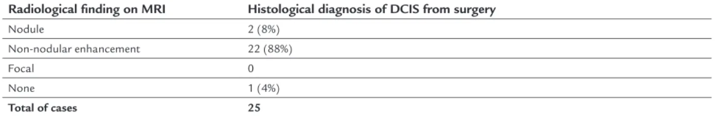
in 22/25 (88%) cases (95CI 72.5-100 ) (Figures 1 and 2). Less commonly characterized by nodules in 2/25 (8%) of the cases and in 1/25 (4%) of the cases, there were no ind-ings on MRI (Table 1).
FIGURE 1 64 year-old woman with DCIS. A: Mammography shows pleomorphic microcalcifications grouped at the junction of the upper quadrants (JUQ) in the left breast. B: MRI – T1 axial post-contrast sequence with image subtraction reveals non-nodular linear enhancement on the microcalcifications’ topography (arrow). C: Histopathological confirmation of moderate grade DCIS after surgery.
FIGURE 2 53 year-old woman with DCIS. A: Zoomed mammogram shows amorphous and clustered microcalcifications in retroareolar region
of the right breast. B: MRI – T1 axial post-contrast sequence with image subtraction reveals non-nodular segmental enhancement on the microcalcifications’ topography (arrow). C: Histopathological confirmation of high grade DCIS after surgery.
TABLE 1 Correlation between histopathological diagnosis of DCIS obtained from surgery and radiological findings on breast MRI.
Radiological finding on MRI Histological diagnosis of DCIS from surgery
Nodule 2 (8%)
Non-nodular enhancement 22 (88%)
Focal 0
None 1 (4%)
Total of cases 25
MRI: magnetic resonance imaging; DCIS: ductal carcinoma in situ. A
A
B C
D A
A
B C
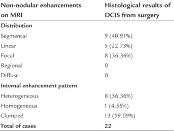
Table 2 summarizes the patterns of distribution and internal enhancement of non-nodular enhancements on MRI, with histological result of DCIS obtained by surgery. Segmental distribution was the most prevalent among pure DCIS, representing 40.91% of lesions (95CI 17.4-64.4), p=0.306. Clumped internal enhancement was the most fre-quently observed in DCIS. This inding was related to DCIS in 13/22 (50.09%) of the cases, followed by heterogeneous enhancement in 8/22 (36.36%) of the cases.
TABLE 2 Patterns of distribution and internal
enhancement of non-nodular enhancements on MRI and histopathological results of DCIS obtained from surgery.
Non-nodular enhancements on MRI
Histological results of DCIS from surgery
Distribution
Segmental Linear Focal Regional Diffuse
9 (40.91%) 5 (22.73%) 8 (36.36%) 0 0
Internal enhancement pattern
Heterogeneous Homogeneous Clumped
8 (36.36%) 1 (4.55%) 13 (59.09%)
Total of cases 22
MRI: magnetic resonance imaging; DCIS: ductal carcinoma in situ.
Of the 25 cases of DCIS, 15 had a high nuclear grade; nine had intermediate grade; and only one did not appear on MRI and presented nuclear grade 1. There was one sta-tistically signiicant difference between the detection of DCIS of high and intermediate nuclear grade compared with low-grade (p<0.005).
D
ISCUSSIONAlthough the detection of DCIS has increased signiicant-ly with the spread of mammographic screening programs, some authors believe that having a more accurate assess-ment of the extent of disease is essential to the success of conservative surgery. Patients with positive margins af-ter surgery, as well as patients with other synchronous foci of DCIS have an increased risk of local disease recur-rence and it can occur in 50% of cases in the form of in-vading carcinoma, with 20% of these cases progressing to distant metastases in 10 years.30
In order to improve the diagnostic accuracy of DCIS, MRI can be used as a tool and recent studies demonstrate that MRI sensitivity is greater than that of
mammogra-phy (92 vs. 56% respectively),27,31 reinforcing the impor-tance of being able to identify the different forms of the disease by method.
The purpose of this study was to characterize the ra-diological indings of MRI that are more associated with DCIS based on the ifth edition of BI-RADS lexicon for MRI. The lexicon provides names and deinition for each imaging inding and improves standardization in MRI reports, facilitating the comparison of studies based on similar terminology.
According to scientiic research, the most common manifestation of DCIS on MRI is non-nodular enhance-ment (60-81% of cases), while it is less commonly char-acterized as nodular (14-41% of cases) or focal (1-12% of cases).26,32,33 In our study, 22 of the 25 cases of pure DCIS (88%) were non-nodular enhancements and two cases were nodules (8%). Focal enhancements were not found on MRI in the topography of suspicious microcalciica-tions detected on mammogram; therefore, there was no association between this morphological pattern and DCIS. These results diverge slightly from those expect-ed in the literature, probably due to the small sampling. Among the non-nodular enhancements, segmental distribution was the most frequent inding associated with DCIS (Table 2), in line with international articles26,32,33 Secondly, focal distribution was observed, with 36.36% of cases, close to the upper limit expected from the liter-ature. Interestingly, linear distribution was close to that expected according to the studies by Jansen et al.32 (24%) and higher than expected by Rosen et al.26 (6%) and Chan et al.33 (7%). This result can be attributed to the fact that our study does not employ the term ductal distribution, which is no longer used in the latest edition of BI-RADS, incorporating this pattern to linear distribution. Table 3 correlated the results of our study with those expected in the literature.
TABLE 3 Comparison of distribution patterns of non-nodular enhancements on MRI in cases positive for DCIS in the study and the expected results according to the literature.
Distribution of non- -nodular enhancements
Present study
Literature review26,32,33
Segmental 40.91% 14 – 77%
Linear 22.73% 6 – 24%
Focal 36.36% 16 – 33%
surgery, the clumped internal enhancement pattern was present in 50.09% of the cases. This result is in accordance with the literature (41-64%),26,32,33 where it is also the most observed inding.
In our study, 24/25 (96%) of the cases of DCIS pre-sented intermediate or high nuclear grade, and only one (4%) showed low nuclear grade and was not diagnosed by the method (false negative). There was a signiicant asso-ciation between intermediate or high nuclear grade and detection on MRI compared to low grade (p<0.005). These results were not observed by Menell et al.,10 but reproduce the results obtained by Kuhl et al.27 The data suggest that MRI might predict the aggressiveness of DCIS when char-acterized by the method, and a clinically inert DCIS when there are no radiological indings.
As limitations, MRI was assessed only in patients with mammographic changes BI-RADS categories 4 or 5, com-posed of suspicious or highly suspicious microcalciica-tions and the valorization of MRI indings may have been inluenced by the presence of microcalciications in the same topography.
C
ONCLUSIONDCIS has a wide variety of imaging features on MRI and being able to recognize these lesions is crucial. Our study showed that MRI can detect the presence of DCIS in 96% of the cases. Its most common morphological presenta-tion is non-mass-like enhancement, while segmental dis-tribution and a clumped internal enhancement pattern are the most common presentations. Faced with the com-bined analysis of these indings, percutaneous core nee-dle biopsy (core biopsy) or vacuum-assisted biopsy (VAB) should be encouraged.
R
ESUMOCarcinoma ductal in situ mamário: avaliação das princi-pais apresentações à ressonância magnética em correla-ção com os achados da mamograia e histologia
Objetivo: avaliar as várias morfologias e características cinéticas do carcinoma ductal in situ (CDIS) ao exame de ressonância magnética (RM) de mama, estabelecer as mais prevalentes e determinar a eicácia do método na detec-ção do CDIS.
Método: estudo prospectivo e observacional, com início em 2011 e duração de 24 meses. Foram avaliadas 25 pa-cientes consecutivas que apresentaram microcalciicações suspeitas ou altamente suspeitas ao exame mamográico de rastreamento, categorias 4 e 5 de BI-RADS, que
reali-zaram RM mamária e, posteriormente, foram submeti-das à cirurgia com resultado comprovado de CDIS puro. A cirurgia foi considerada padrão-ouro para correlação
entre os resultados histológicos e os achados radiológi-cos obtidos à RM.
Resultados: a característica morfológica do CDIS mais frequente à RM foi o realce não nodular (p<0,001), ob-servada em 22/25 (88%) casos (IC 95% 72,5-100). Dentre estes, a distribuição segmentar foi a mais prevalente, re-presentada por 9/22 (40,91%) casos (IC 95% 17,4-64,4), p=0,306, e o realce interno tipo clumped foi o padrão mais frequentemente caracterizado no CDIS, observado em 13/22 (50,09%) casos.
Conclusão: o CDIS tem uma grande variedade de
carac-terísticas imaginológicas à RM e é fundamental reconhe-cê-las. A apresentação morfológica mais comum é o real-ce não nodular, sendo a distribuição segmentar e o padrão interno de realce tipo clumped as apresentações mais fre-quentes. Diante da análise combinada desses achados, a biópsia percutânea por agulha grossa (core biopsy) ou as-sistida a vácuo (mamotomia) deve ser encorajada.
Palavras-chave: neoplasias da mama, carcinoma
intra-ductal não iniltrante, mamograia, imagem por resso-nância magnética, biópsia com agulha de grande calibre.
R
EFERENCES1. Brown PW, Silverman J, Owens E, Tabor DC, Terz JJ, Lawrence W Jr. Intraductal “noniniltrating” carcinoma of the breast. Arch Surg. 1976; 111(10):1063-7.
2. Dershaw DD, Abramson A, Kinne DW. Ductal carcinoma in situ: mammographic findings and clinical implications. Radiology. 1989; 170(2):411-5.
3. Guidi AJ, Schnitt SJ, Fischer L, Tognazzi K, Harris JR, Dvorak HF, et al. Vascular permeability factor (vascular endothelial growth factor) expression and angiogenesis in patients with ductal carcinoma in situ of the breast. Cancer. 1997; 80(10):1945-53.
4. Leonard GD, Swain SM. Ductal carcinoma in situ, complexities and challenges. J Natl Cancer Inst. 2004; 96(12):906-20.
5. Ernster VL, Ballard-Barbash R, Barlow WE, Zheng Y, Weaver DL, Cutter G, et al. Detection of ductal carcinoma in situ in women undergoing screening mammography. J Natl Cancer Inst. 2002; 94(20):1546-54.
6. Badan GM, Roveda Júnior D, Ferreira CAP, Noronha Júnior, OA. Auditoria interna completa do serviço de mamograia em uma instituição de referência em imaginologia mamária. Radiol Bras. 2014; 47(2):74-8.
7. Tabár L, Vitak B, Chen TH, Yen AM, Cohen A, Tot T, et al. Swedish two-county trial: impact of mammographic screening on breast cancer mortality during 3 decades. Radiology. 2011; 260(3):658-63.
8. Kopans DB, Smith RA, Duffy SW. Mammographic screening and “overdiagnosis”. Radiology. 2011; 260(3):616-20.
9. Berg WA, Gutierrez L, NessAiver MS, Carter WB, Bhargavan M, Lewis RS, et al. Diagnostic accuracy of mammography, clinical examination, US, and MR imaging in preoperative assessment of breast cancer. Radiology. 2004; 233(3): 830-49.
11. Scheidhauer K, Walter C, Seemann MD. FDG PET and other modalities in the primary diagnosis of suspicious breast lesions. Eur J Nucl Med Mol Imaging. 2004; 31 Suppl 1:S70-9.
12. Peters NH, Borel Rinkes IH, Zuithoff NP, Mali WP, Moons KG, Peeters PH. Meta-analysis of MR imaging in the diagnosis of breast lesions. Radiology. 2008; 246(1):116-24.
13. Orel SG, Schnall MD, Powell CM, Hochman MG, Solin LJ, Fowble BL, et al. Staging of suspected breast cancer: effect of MR imaging and MR-guided biopsy. Radiology. 1995; 196(1):115-22.
14. Gilles R, Meunier M, Lucidarme O, Zafrani B, Guinebretière JM, Tardivon AA, et al. Clustered breast microcalciications: evaluation by dynamic contrast-enhanced subtraction MRI. J Comput Assist Tomogr. 1996; 20(1):9-14. 15. Gilles R, Guinebretière JM, Lucidarme O, Cluzel P, Janaud G, Finet JF, et al.
Nonpalpable breast tumors: diagnosis with contrast-enhanced subtraction dynamic MR imaging. Radiology. 1994; 191(3):625-31.
16. Stomper PC, Herman S, Klippenstein DL, Winston JS, Edge SB, Arredondo MA, et al. Suspect breast lesions: indings at dynamic gadolinium-enhanced MR imaging correlated with mammographic and pathologic features. Radiology. 1995; 197(2):387-95.
17. Boetes C, Mus RDM, Holland R, Barentsz JO, Strijk SP, Wobbes T, et al. Breast tumors: comparative accuracy of MR imaging relative to mammography and US for demonstrating extent. Radiology. 1995; 197(3):743-47.
18. Fobben ES, Rubin CZ, Kalisher L, Dembner A, Seltzer MH, Santoro EJ. Breast MR imaging with commercially available techniques: radiologic-pathologic correlation. Radiology. 1995; 196(1):143-52.
19. Teifke A, Hlawatsch A, Beier T, Werner Vomweg T, Schadmand S, Schmidt M, et al. Undetected malignancies of the breast: dynamic contrast enhanced MR imaging at 10T. Radiology. 2002; 224(3):881-8.
20. Orel SG, Mendonca MH, Reynolds C, Schnall MD, Solin LJ, Sullivan DC. MR imaging of ductal carcinoma in situ. Radiology. 1997; 202(2):413-20. 21. Gilles R, Zafrani B, Guinebretière JM, Meunier M, Lucidarme O, Tardivon
AA, et al. Ductal carcinoma in situ: MR imaging-histopathologic correlation. Radiology. 1995; 196(2):415-9.
22. Soderstrom CE, Harms SE, Copit DS, Evans WP, Savino DA, Krakos PA, et al. Three-dimensional RODEO breast MR imaging of lesions containing ductal carcinoma in situ. Radiology. 1996; 201(2):427-32.
23. Boetes C, Strijk SP, Holland R, Barentsz JO, Van Der Sluis RF, Ruijs JHJ. False-negative MR imaging of malignant breast tumors. Eur Radiol. 1997; 7(8):1231-4.
24. Viehweg P, Lampe D, Buchmann J, Heywang-Kobrunner SH. In situ and minimally invasive breast cancer: morphologic and kinetic features on contrast enhanced MR imaging. MAGMA. 2000; 11(3):129-37.
25. Westerhof JP, Fischer U, Moritz JD, Oestmann JW. MR imaging of mammographically detected clustered calciications: is there any value? Radiology. 1998; 207(3):675-81.
26. Rosen EL, Smith-Foley SA, DeMartini WB, Eby PR, Peacock S, Lehman CD. BI-RADS MRI enhancement characteristics of ductal carcinoma in situ. Breast J. 2007; 13(6):545-50.
27. Kuhl CK, Schrading S, Bieling HB, Wardelmann E, Leutner CC, Koenig R, et al. MRI for diagnosis of pure ductal carcinoma in situ: a prospective observational study. Lancet. 2007; 370(9586):485-92.
28. Kuhl CK. Why do purely intraductal cancers enhance on breast MR images? Radiology. 2009; 253(2):281-3.
29. Dershaw DD, Abramson A, Kinne DW. Ductal carcinoma in situ: mammographic indings and clinical implications. Radiology. 1989; 170(2):411-5.
30. Bijker N, Peterse JL, Duchateau L, Julien JP, Fentiman IS, Duval C, et al. Risk factors for recurrence and metastasis after breast-conserving therapy for ductal carcinoma-in-situ: analysis of European Organization for Research and Treatment of Cancer Trial 10853. J Clin Oncol. 2001; 19(8):2263-71.
31. Lehman CD. Magnetic resonance imaging in the evaluation of ductal carcinoma in situ. J Natl Cancer Inst Monogr. 2010; 2010(41):150-1. 32. Jansen SA, Newstead GM, Abe H, Shimauchi A, Schmidt RA, Karczmar GS.
Pure ductal carcinoma in situ: kinetic and morphologic MR characteristics compared with mammographic appearance and nuclear grade. Radiology. 2007; 245(3):684-91.