J Bras Pneumol. 2007;33(3):355-357 355
Case Report
Tuberculosis of the thymus*
Mauro Tadeu Ajaj Saieg1, Fabíola del Carlo Bernardi2, Roberto Gonçalves3, Márcio Botter4, Roberto Saad Junior5, Geanete Pozzan6
Abstract
Tumors of the anterior mediastinum include several entities with different radiological and clinical manifestations, constituting a heterogeneous group of congenital, inflammatory, and neoplastic conditions. Among these lesions, the most common primary tumor of the mediastinum is thymoma, nearly followed by germ cell tumors and lymphomas. Tuberculosis of the thymus, an extremely rare condition, typically involves the mediastinal lymph nodes. We present, in this study, pathological, radiological, and clinical findings of one case of tuberculosis of the thymus in an 18-year-old patient who presented thoracic pain, dyspnea upon minimal effort, and progressive worsening of the symptoms in one week. The chest X-ray showed a large mass in the mediastinum, and computed tomography scans indicated that it was located anteriorly. The patient was submitted to surgery in order to excise the mass. Microscopy revealed a massive inflammatory response and granulomas in the thymic tissue. Ziehl-Neelsen staining for acid-fast bacilli yielded positive results, and a diagnosis of tuberculosis was made. Surgeons and pathologists should remain alert for this condition and should include it in the differential diagnosis of mediastinal masses.
Keywords: Thymus; Tuberculosis; Thoracic surgery; Pathology.
* Study carried out at the Faculdade de Ciências Médicas da Santa Casa de São Paulo – FCMSCSP - Santa Casa School of Medical Sciences in São Paulo – São Paulo (SP) Brazil.
1. PhD student in Pathology at the Faculdade de Medicina da Universidade de São Paulo – FMUSP, University of São Paulo School of Medicine – São Paulo (SP) Brazil.
2. Assistant Professor in the Department of Pathology of the Faculdade de Ciências Médicas da Santa Casa de São Paulo – FCMSCSP, Santa Casa School of Medical Sciences in São Paulo – São Paulo (SP) Brazil.
3. Postgraduate student in Surgery at the Faculdade de Ciências Médicas da Santa Casa de São Paulo – FCMSCSP, Santa Casa School of Medical Sciences in São Paulo – São Paulo (SP) Brazil.
4. Attending Physician in the Department of Surgery of the Santa Casa Hospital of São Paulo, São Paulo (SP) Brazil.
5. Full Professor in the Department of Surgery of the Faculdade de Ciências Médicas da Santa Casa de São Paulo – FCMSCSP, Santa Casa School of Medical Sciences in São Paulo – São Paulo (SP) Brazil.
6. Assistant Professor in the Department of Pathology of the Faculdade de Ciências Médicas da Santa Casa de São Paulo – FCMSCSP, Santa Casa School of Medical Sciences in São Paulo – São Paulo (SP) Brazil.
Correspondence to: Mauro Tadeu Ajaj Saieg. Av. dos Jamaris, 291, apto. 101, Moema, CEP 04078-000, São Paulo, SP, Brasil. Phone 55 11 5051-8714. E-mail: [email protected]
Submitted: 4 April 2006. Accepted, after review: 1 June 2006.
Introduction
The mediastinum is located in the central portion of the chest, between the two pleural cavities, above the diaphragm, and below the upper chest. It is divided into anterior, medium, and posterior portions in order to locate and classify tumors and other entities that affect this region.
The anterior mediastinum is defined as the region posterior to the sternum and anterior to the heart and the brachiocephalic vessels. It extends from the upper portion of the chest to the diaphragm, and contains the thymus, fat, and lymph nodes.
Tumors of the anterior mediastinum include various enti-ties with different radiological and clinical manifestations,
with cystic and solid patterns, being a heterogeneous group of congenital, inflammatory, and neoplastic conditions. Neural tumors, thymomas, and benign cysts account for 60% of the surgically treated lesions, whereas lymphomas, germ cell tumors, and inflammatory diseases account for 30%. The remaining 10% include vascular lesions, princi-pally aortic aneurysms.(1)
Among these lesions, the most common primary tumor of the mediastinum is thymoma, followed by germ cell tumors and lymphomas.
356 Saieg MTA, Bernardi FC, Gonçalves R, Botter M, Saad Jr R, Pozzan G
J Bras Pneumol. 2007;33(3):355-357
be affected in patients with TB.(2) Only four cases
of thymic TB have been reported in the literature, of which two were reported in the last 30 years.(3,4)
However, only one pf those cases tested positive for acid-fast bacilli.(3) In the present study, we present
pathological, radiological, and clinical findings of a case of TB of the thymus in an 18-year-old patient.
Case report
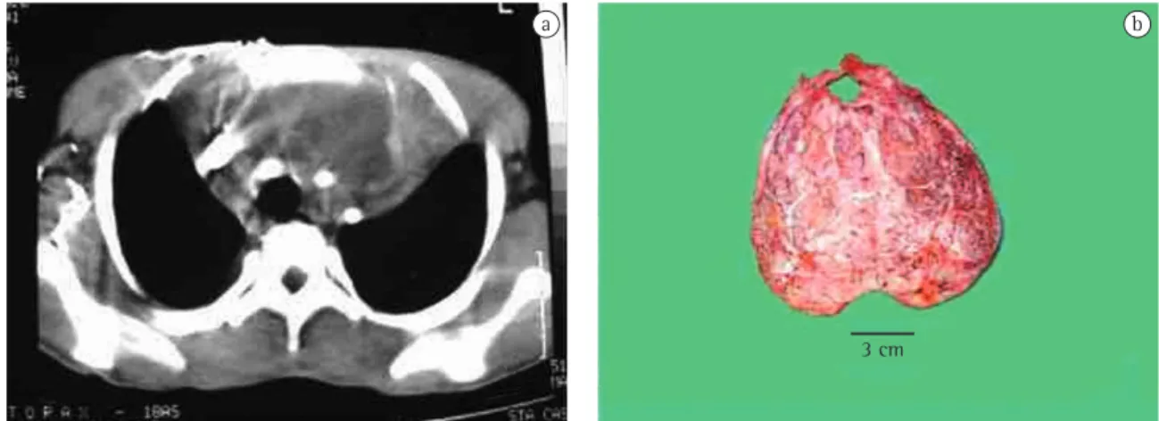
An 18-year-old male patient presented with chest pain, dyspnea upon moderate exertion, and progressive worsening for one year. The patient was admitted to the hospital with respiratory failure, sudden worsening, evolving to require intubation and ventilatory support some hours after admission to the emergency room. He had a personal history of heart disease since childhood. The physical exam-ination of the chest revealed bilateral crackles in the lung bases. The chest X-ray showed a large tumor in the mediastinum, and the computed tomog-raphy scan showed that the lesion was located in the anterior mediastinum, was well delimited, and had not invaded the adjacent pulmonary structures (Figure 1a).
Due to the emergency of the patient’s health status and the necessity of an immediate resolu-tion, we opted for surgical treatment, even without a previous request of biopsy.
The surgical incision was a left posterior lateral thoracotomy, providing ample access to the tumor
and allowing its complete excision. The tumor affected half of the left thoracic cavity and part of the right thoracic cavity, as well as the ante-rior mediastinum. It also partially involved the left pulmonary hilum and the mediastinal structures.
Macroscopically, the tumor measured 20.0 × 1.0 × 4.0 cm, had whitish and spongy areas in the cutoff surface, as well as cavities and foci of necrosis. There was no evidence of areas with macro-scopic appearance of thymus. However, the tumor shape resembled that of the organ (Figure 1b).
Microscopically, there was an intense inflam-matory reaction, with accumulation of granulomas formed by giant cells surrounding areas of caseous necrosis, with a ring of epithelioid cells and lymphocytes. The Ziehl-Neelsen (ZN) staining tested positive for acid-fast bacilli in the examined tissue. Therefore, the final diagnosis of TB was made (Figure 2a). The polymerase chain reaction tests were not performed during the surgical procedure. Due to the high incidence of TB in Brazil, the microscopy results, together with the ZN staining for acid-fast bacilli, suffice to make the diagnosis. The tissue was fundamentally composed of lymphoid tissue, with areas that resembled the Hassall’s corpuscles. The immunohistochemical reaction for AE1/AE3 showed that these areas were epithelial, confirming the diagnosis of thymus (Figure 2b).
Evolution was satisfactory. The patient remained in the intensive care unit for only two days after surgery. He was discharged from hospital after ten
Figure 1 - a) Computed tomography scan of the chest showing the lesion in the anterior mediastinum; and b) Surgical
sample with macroscopic similarity to the thymus.
a b
Tuberculosis of the thymus
J Bras Pneumol. 2007;33(3):355-357 357
days. The TB treatment was started on postopera-tive day 3 and continued for six months.
Discussion
A diagnosis of TB of the thymus is extremely rare, even considering the fact that the incidence of TB has increased in parallel with the increased prev-alence of acquired immunodeficiency syndrome. This is the fifth case reported in the literature, and the second in this decade. Using the ZN histochem-ical technique, we identified acid-fast bacilli within the granulomas. In addition to the inflammatory process, characterized by the presence of granu-lomas, there was no microscopic evidence of thymic tissue. However, there was evidence of Hassall’s corpuscles, which constitute a specific marker for
Figure 2 - a) Positive Ziehl-Neelsen staining showing
acid-fast bacilli (arrow); and b) immunohistochemistry for AE1/AE3, showing epithelial cells amid the lymphoid tissue.
epithelial elements, in the AE1/AE3 immunohisto-chemical tests. The presence of these components, in combination with typical granulomas and bacilli, led us to the diagnosis of TB of the thymus.
As in the case presented by another group of authors in 1997, (3)our case presented radiological
findings compatible with a solid tumor, which, together with the lack of clinical and laboratorial findings of TB, suggested a lesion of neoplastic etiology, possibly a germ cell tumor, lymphoma or thymoma, and surgery was therefore indicated.
The excision of these masses in the mediastinum poses a dilemma for the surgeon, since computed tomography is useful in the evaluation of the extent of these lesions but does not aid the differential diagnosis. In these cases, mediastinoscopy or thora-coscopy, accompanied by biopsy, can be of great value in making the diagnosis of these mediastinal tumors. In our case, the urgency of the condition did not allow these methods to be applied. In other cases, (3) the diagnosis has been made through fine
needle aspiration. However, the role of this method is controversial. Since the treatment of tumors of the anterior varies according to the diagnostic hypoth-esis, intra-operative frozen section biopsies are important for establishing the differential diagnosis of these various entities and determining their treat-ment, considering that surgery is the treatment of choice for thymomas and benign germ cell tumors, and clinical therapy is the treatment of choice for lymphomas and malignant germ cell tumors.(5,6)
Therefore, surgeons and pathologists should remain alert for TB of the thymus and should include this entity in the differential diagnosis of mediastinal masses.
References
1. Strollo DC, Rosado de Christenson ML, Jett JR. Primary Mediastinal Tumors. Part 1: Tumors of the anterior mediastinum. Chest. 1997;112(2):511-22.
2. Duprez A, Cordier R, Schmitz P. Tuberculoma of the thymus. First case of surgical excision. J Urol Nephrol (Paris). 1962; 44:115-20
3. Simmers TA, Jie C and Sie MC. Thymic tuberculosis: a case report. Neth J Med. 1997;51(2):87-90.
4. FitzGerald JM, Mayo JR, Miller RR, Jamieson WR, Baumgartner F. Tuberculosis of the thymus. Chest. 1992; 102(5):1604-5.
5. Ferguson MK, Lee E, Skinner DB, Little AG. Selective operative approach for diagnosis and treatment of anterior mediastinal masses. Ann Thorac Surg. 1987;44(6):583-6. 6. Trastek VF. Management of mediastinal tumors. Ann Thorac