Revista da
ASSOCIAÇÃO MÉDICA BRASILEIRA
w w w . r a m b . o r g . b r
Review article
Association between nonalcoholic fatty liver disease
and coronary artery disease
夽
Consuelo P. Vilar
a,b, Helma P. Cotrim
a,∗, Gesira S. Florentino
a,b, Cibelle P.V. Barreto
b,
André Vinicius A. Florentino
b, Gerson Bragagnoli
a,b, Paulo A. Schwingel
aaPostgraduate Program in Medicine and Health, Faculdade de Medicina da Bahia (FMB), Universidade Federal da Bahia (UFBA),
Salvador, BA, Brazil
bMedical School, Universidade Federal de Campina Grande (UFCG), Campina Grande, PB, Brazil
a r t i c l e
i n f o
Article history:
Received 27 September 2012 Accepted 23 November 2012 Available online 16 May 2013
Keywords: Fatty liver
Cardiovascular diseases Nonalcoholic fatty liver disease Coronary artery disease
a b s t r a c t
Objective:Although some investigations have shown a relationship between nonalcoholic fatty liver disease (NAFLD) and cardiovascular diseases, there are few studies analyzing the relationship between NAFLD and coronary artery disease (CAD). The aim of this article was to review the relationship between NAFLD and CAD and the methods of diagnosis used to assess such relationship.
Methods:A review was performed using search engines of indexed scientific material, including MEDLINE (by PubMed), Web of Science, IBECS, and LILACS, to identify articles published in Portuguese, English, and Spanish until August, 2012. The studies were eligible if they included the following data: place and year of publication, prevalence and methods used to diagnose NAFLD (ultrasound, computed tomography, nuclear magnetic resonance, or biopsy) and CAD (coronary angiography, or computed tomography), and the exclusion of patients due to alcohol consumption greater than 20 g/day.
Results:Ten articles were selected, most of which were cross-sectional studies. The studies mostly observed the association between NAFLD and the presence and severity of CAD. Conclusion:The analysis of the review showed that evaluating the existence of NAFLD in patients with CAD from its subclinical form up to the symptomatic clinical form is important due to the higher risk of acute myocardial infarction and consequent increase of mortality. © 2013 Elsevier Editora Ltda. All rights reserved.
夽Study conducted at Universidade Federal de Campina Grande, Campina Grande, PB, Brazil.
∗ Corresponding author: Programa de Pós-graduac¸ão em Medicina e Saúde, Complexo Hospital Universitário Professor Edgard Santos,
Rua Augusto Viana, s/n, 5oandar, Canela, Salvador, BA, 40110-060, Brazil.
E-mail: [email protected] (H.P. Cotrim).
Associac¸ão entre doenc¸a hepática gordurosa não-alcoólica e doenc¸a
arterial coronariana
Palavras-chave:
Doenc¸a hepática gordurosa Doenc¸as cardiovasculares Doenc¸a hepática gordurosa não alcoólica
Doenc¸a arterial coronariana
r e s u m o
Objetivo: Embora algumas investigac¸ões demonstrem uma associac¸ão entre a doenc¸a hep-ática gordurosa não-alcoólica (DHGNA) e doenc¸as cardiovasculares, existem poucos estudos analisando a relac¸ão entre DHGNA e doenc¸a arterial coronariana (DAC). O objetivo deste artigo foi realizar uma revisão sobre a associac¸ão entre DHGNA e CAD e os métodos diag-nósticos usados para avaliar esta associac¸ão.
Métodos: Foi realizada uma revisão da literatura utilizando métodos de busca de material científico indexado, incluindo MEDLINE (através do PubMed), Web of Science, IBECS e LILACS, para identificar artigos publicados em português, inglês e espanhol até agosto de 2012. Os estudos eram elegíveis se incluíam os seguintes dados: local e ano de publicac¸ão, prevalência e os métodos utilizados para o diagnóstico da DHGNA (ultrassonografia, tomografia com-putadorizada, ressonância nuclear magnética ou biópsia) e DAC (angiografia coronária ou tomografia computadorizada), e a exclusão de pacientes com consumo de álcool maior do que 20 g/dia.
Resultados: Dez artigos foram selecionados, predominando os estudos de corte transversal. Na maioria dos estudos foi observada a associac¸ão entre DHGNA e a presenc¸a e gravidade da DAC.
Conclusão: A análise da revisão mostra que é importante avaliar a existência de DHGNA em pacientes com DAC desde sua forma subclínica até a forma clínica sintomática, devido ao maior risco de infarto agudo do miocárdio e consequente aumento da mortalidade.
© 2013 Elsevier Editora Ltda. Todos os direitos reservados.
Introduction
Some investigations have shown a relationship between nonalcoholic fatty liver disease (NAFLD) and cardiovascular diseases (CVDs),1–3 with the latter being one of the main causes of morbidity and mortality in these patients. Although fatty liver is related to factors (such as dyslipidemia, cen-tral obesity, diabetes, and metabolic syndrome [MS]) that may cause CVDs including coronary artery disease (CAD), there are few studies analyzing the relationship between NAFLD and CAD, and they present controversial results.
Different methods can be applied to diagnose NAFLD and CAD. NAFLD can be diagnosed by ultrasound (US), computed tomography (CT), nuclear magnetic resonance (NMR), and liver biopsy. A recent meta-analysis showed that US has a sen-sitivity from 73.3% to 90.5% and a specificity from 69.6% to 85.2%. CT has a sensitivity from 46.1% to 72% and a speci-ficity from 88.1% to 94.6%, but its cost is higher. NMR has a sensitivity from 82.0% to 97.4% and a specificity from 76.1% to 95.3%, and as a noninvasive technique, NMR is good at diagnosing, especially cases of steatosis < 25%.4However, due to its high cost, NMR is not available to most patients. Liver biopsy is considered the gold standard for diagnosing steato-sis, but it is an invasive method that may cause bleeding. US is relatively precise for the diagnosis of NAFLD; and since it is low-cost, risk-free, and widely available, it has been the most used method.
Although angiography is the gold standard exam for CAD diagnosis, the coronary calcification score identified by mul-tislice computed tomography (MSCT) has been proposed as a potential method to improve the risk discrimination with-out invasive intervention, because it visualizes not only the
coronary artery stenosis but also the characteristics of the plaque.5Thus, this method can be used to diagnose subclinical CAD.
The aim of this article was to review the relationship between NAFLD and CAD and the methods of diagnosis used to assess their relationship.
Methods
and CAD. The studies were eligible if they had included the following data: place and year of publication, prevalence and methods used to diagnose NAFLD (US, CT, NMR, or biopsy) and CAD (angiography or tomography), exclusion of patients due to alcohol consumption higher than 20 g/day, and other causes of fatty liver.
Results
Nineteen articles involving the relationship of CAD and NAFLD or steatosis were identified, and nine were excluded: three6–8 for not fulfilling the NAFLD criteria, five for using other meth-ods to diagnose CAD,9–13and one for not citing the diagnostic method for CAD.14
However, a global interpretation of the investigations selected (Table 1) was not allowed because they differ with respect to the design of the study, the size and selected sam-ples, inclusion and exclusion criteria of patient in each study (Table 2), methods used to diagnose NAFLD and CAD, and the confounding factors included in the analysis. Thus, to evaluate the association of NAFLD and CAD, the studies were grouped according to the method used for CAD diagnosis.
Analysis of the selected articles
Type of study
The articles selected had different methods. The
major-ity were characterized as prospective cross-sectional
studies15,19,22 there was also one case-control study,20 two retrospective cross-sectional studies,21,24 and a prospective cohort study.23
Locale of the studies and sample size
Nine of the selected studies involved research with Asian patients. The samples varied from 6120 to 4,023 patients.24 In the investigations using the prospective cross-sectional model,15–19,22a total of 2,585 patients were evaluated (Table 1).
Inclusion criteria used for the selection of samples
The studies used the following inclusion criteria: clinical suspicion of CAD;15,16,18 clinical suspicion of CAD in hospi-talized patients;22 MS;17 evaluation of risk factors for CVD and cancer;19 low-intermediate risk for CAD and the pres-ence of NAFLD without any other hepatobiliary disease;20the first coronary angiography (CAG);15check-up for hepatobili-ary disease and CAD in asymptomatic individuals;21 adults who received health evaluation24and age≥18 years21,23who underwent CAG.23
Exclusion criteria
There were varied criteria (Table 2) and most investigations listed only alcohol consumption > 20 g/day and the presence of other liver diseases as common exclusion criteria.
Studies with CAD diagnosis by MSCT
Five investigations assessed CAD by MSCT,16,19–21,24 but the CAD criteria varied among the investigations. Most of these studies19,21,24 evaluated the coronary artery calcium (CAC) score. Two of these investigations21,24 quantified the total calcium score according to a scoring system pro-posed by Agatston et al.25 In two articles,19,21 a CAC score > 100 was considered to indicate a moderate-high risk of CAD. In another,24 CAC presence (CAC > 0) was con-sidered as evidence of calcification. Two studies analyzed the characteristics of coronary lesions in MSCT: Akabame et al.16 classified the lesions of coronary arteries as calcified plaques, non-calcified plaques, low-density plaques, and pos-itive remodeling vessels, and they observed that all major arteries had a diameter > 2.0 mm using enhanced images. The lipid pool was defined as having a plaque density of < 60 Hounsfield units (HU), and positive remodeling as having a remodeling index (RI) > 1.1. A calcified plaque was considered severe if > 180 HU, and as mild if < 180 HU. Assy et al.20used the degree of stenosis of the coronary artery (> 50%) as the CAD criterion. The plaques were classified as calcified or non-calcified on a segmental basis according to plaque features, including volume, attenuation, and calcification pattern. A calcified lesion was defined as a minimum of two pixels (area, 0.52 mm2) with a minimum attenuation of 130 HU.
Methods and diagnostic criteria of NAFLD: In two of these studies, steatosis was diagnosed by CT16,20. In the study by Akabame et al.,16hepatic and splenic attenuation values were measured on non-contrast CT scans using 16 circular region-of-interest (ROI) cursors in the liver and four in the spleen. The calculation of the relationship between the liver and spleen was made by the division between the average value of the liver attenuation (16 points) and the average value of the splenic attenuation (4 points). The cut-off value for the liver to spleen ratio to diagnose NAFLD was defined as < 1.1. Assy et al.20defined hepatic steatosis as the result of an attenua-tion of≥-10 HU (calculated as the liver attenuation minus the spleen attenuation).
Two studies used US for diagnosis of steatosis. In the study by Jung et al.,19 subjects were diagnosed with steatosis if at least two of the following three findings were present: increased liver echogenicity, deep attenuation, and vascular blurring. In Kim et al.24steatosis diagnosis was made based on ultrasound feature characteristics consisting of “bright liver” and evident contrast between hepatic and renal parenchyma, vessel blurring, focal sparing, and narrowing of the lumen of the hepatic veins.
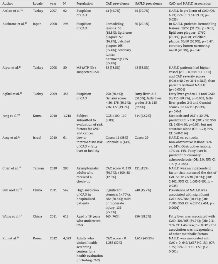
Table 1 – Selected studies about the association of CAD and NAFLD.
Author Locale year N Population CAD prevalence NAFLD prevalence CAD and NAFLD association
Arslan et al.15 Turkey 2007 92 Suspicion
of CAD
43 (46.7%) 65 (70.7%) NAFLD is predictor of CAD (OR:
6.73; 95% CI: 1.14-39.61; p= 0.035)
Akabame et al.16 Japan 2008 298 Suspicion
of CAD
Remodeling lesions: 56 (18.8%); lipid core plaques: 50 (16.8%); calcified plaque: 165 (55.4%); coronary lumen
narrowing: 165 (55.4%)
60 (20.1%) In NAFLD patients: Remodeling
lesions: 19/60 (31.7%), p = 0.01; lipid core plaques: 17/60 (28.3%), p= 0.01; calcified plaque: 36/60 (60.0%), p = 0.47; coronary lumen narrowing: 47/60 (78.3%), p = 0.47
Alper et al.17 Turkey 2008 80 MS (ATP III) +
suspected CAD
63 (78.8%) 43 (53.8%) NAFLD patients had higher
vessel (2.5±0.9vs.1.1±1.0) and CAD severity scores (90.0±40.0vs36.4±28.9), than patients without NAFLD (p < 0.0001)
Ac¸ikel et al.18 Turkey 2009 355 Suspicion
of CAD
250 (70.4%); Gensini score:
≤36: 178 (50.1%);
>36: 177 (49.9%)
Fatty liver: 215 (60.5%); fatty liver grades 2-3: 115 (32.4%)
Fatty liver grades 2-3 and CAD: 93/115 (80.9%), p = 0.003; fatty liver grades 2-3 and Gensini score > 36: 67/115 (58.3%), p = 0.041
Jung et al.19 Korea 2010 1,218 Subject
submitted to evaluation of risk factors for CVD and cancer
CCS >100: 110 (9.0%)
514 (42.2%) Steatosis and ALT > 30 U/L predict CCS > 100 (OR: 2.12; 95% CI: 1.08-4.20; p<0.05), but not steatosis alone (OR: 1.24; 95% CI: 0.68-2.26)
Assy et al.20 Israel 2010 61 Low or
intermediate risk of CAD + fatty liver or healthy
Cases: 11 (38%) Controls: 4 (14%)
Cases: 29 NAFLDvs.controls:
non-obstructive lesions: 38%
vs.14%; Obstructive lesions: 10%vs.14%. Fatty liver is predictor of coronary
atherosclerosis (OR: 2.0; 95% CI: 1-4; p < 0.04)
Chen et al.21 Taiwan 2010 295 Asymptomatic
adults who received a check-up
CAC score: 0: 179 (60.7%); >100: 38 (12.9%)
121 (41%) NAFLD was an independent
factor that increased the risk of CAC >100: 23/38 (60.5%); (OR: 2.462; 95% CI: 1.065-5.691; p = 0.035)
Sun and Lu22 China 2011 542 High suspicion
of CAD in hospitalized patients
Significant stenosis (≥70%): 382 (70.5%); mild or moderate injury: 136 (25.1%)
248 (45.7%) Prevalence of NAFLD was
associated with significant CAD: 222/382 (58.1%); (OR: 7.585; 95% CI: 4.617-12.461; p < 0.0001)
Wong et al.23 China 2011 612 Aged≥18 years
who underwent CAG
465 (76%) 356 (58.2%) Fatty liver was associated with
CAD: 301/465 (64.7%); (OR: 2.31; 95% CI: 1.46-3.64; p < 0.001); the association was independent of other metabolic factors
Kim et al.24 Korea 2012 4,023 Adults who
visited health screening centers for a health evaluation (including CAC)
CAC score > 0: 1,286 (32%)
1,617 (40.2%) NAFLD was associated with
CAC > 0: 649/1,617 (40.1%); (OR: 1.35; 95% CI: 1.15-1.59; p < 0.001)
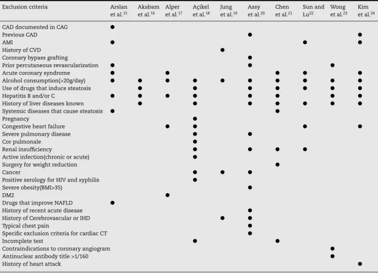
Table 2 – Exclusion criteria used in the selected studies.
Exclusion criteria Arslan
et al.15
Akabam et al.16
Alper et al.17
Ac¸ikel et al.18
Jung et al.19
Assy et al.20
Chen et al.21
Sun and Lu22
Wong et al.23
Kim et al.24
CAD documented in CAG 䊉
Previous CAD 䊉 䊉
AMI 䊉 䊉 䊉
History of CVD 䊉
Coronary bypass grafting 䊉
Prior percutaneous revascularization 䊉 䊉 䊉
Acute coronary syndrome 䊉 䊉 䊉 䊉 䊉
Alcohol consumption(>20g/day) 䊉 䊉 䊉 䊉 䊉 䊉 䊉 䊉 䊉 䊉
Use of drugs that induce steatosis 䊉 䊉 䊉 䊉 䊉 䊉 䊉
Hepatitis B and/or C 䊉 䊉 䊉 䊉 䊉 䊉 䊉 䊉
History of liver diseases known 䊉 䊉 䊉 䊉 䊉 䊉 䊉
Systemic diseases that cause steatosis 䊉 䊉
Pregnancy 䊉
Congestive heart failure 䊉 䊉 䊉 䊉
Severe pulmonary disease 䊉 䊉
Cor pulmonale 䊉
Renal insufficiency 䊉 䊉 䊉 䊉
Active infection(chronic or acute) 䊉
Surgery for weight reduction 䊉
Cancer 䊉 䊉 䊉
Positive serology for HIV and syphilis 䊉
Severe obesity(BMI>35) 䊉
DM2 䊉
Drugs that improve NAFLD 䊉
History of recent acute disease 䊉
History of Cerebrovascular or IHD 䊉 䊉
Typical chest pain 䊉
Specific exclusion criteria for cardiac CT 䊉
Incomplete test 䊉 䊉
Contraindications to coronary angiogram 䊉
Antinuclear antibody title >1/160 䊉
History of heart attack 䊉
NOTE: AMI, acute myocardial infarction, BMI, body mass index; DM2, diabetes mellitus type 2; CAD, coronary artery disease; CAG, coronary
angiography; CVD, cardiovascular disease; CT, computed tomography; HIV, Human immunodeficiency virus; IHD, Ischemic heart disease; NAFLD, nonalcoholic fatty liver disease.
segment and the intrahepatic vessels or invisibility of the diaphragm.
Regarding the prevalences of CAD and NAFLD, in the inves-tigation that evaluated the total calcium score (CAC), 9% to 12.9% of the individuals had a moderate-high risk of CAD (CAC > 100), while the NAFLD prevalence ranged from 20.1% to 42.2% (Table 1).
Regarding the relationship between CAD and NAFLD, in those investigations that used MSCT to diagnose CAD, Chen et al.21observed a prevalence of NAFLD of 41% (121/295), while a moderate-high risk of CAD (CAC > 100) was observed in 12.9% (38/295). The prevalence of NAFLD increased as the CAC score increased (p = 0.003). The results indicated that NAFLD is related to a moderate-high risk of coronary artery disease (CAC >100), but NAFLD is not guaranteed to be an independent risk factor or an epiphenomenon of CAD. In Jung et al.,19hepatic steatosis was found in 42.2% (514/1,218), and coronary calcium score (CCS) > 100 with moderate-high risk of CVD was found in 9% (110/1,218) of subjects. An association was observed among the simultaneous presence of steatosis and elevated alanine aminotransferase (odds ratio [OR] = 2.12; 95% CI: 1.08-4.20; p < 0.05) and CSS >100, but not with steatosis alone (OR = 1.24; 95% CI: 0.68-2.26). In the study by Kim et al.,24the presence of
NAFLD was 40.2% (1,617/4,023) and that of CAC > 0 was 32% (1,286/4,023). In the univariate analysis, the presence of CAC (score > 0) was significantly associated with NAFLD. Increasing CAC scores (0, < 10, 10-100,≥100) were associated with higher prevalence of NAFLD (OR, 1.84; 95% CI: 1.61-2.10; p < 0.001).
Akabame et al.16observed the existence of a relationship between NAFLD and the presence of remodeling lesions (OR = 2.41; 95% CI: 1.24-4.67; p = 0.0009) and lesions with a lipid core (OR = 2.29; 95% CI: 1.15-4.56; p = 0.0018), but they did not observe a correlation with calcified plaques or luminal stenosis. Assy et al.20 found relationship of NAFLD with a higher prevalence of calcified and non-calcified plaques and nonstenotic lesions (38%vs. 14%). Although the presence of obstructive lesions was more frequent in the controls than in the patients with NAFLD (14% vs. 10%), the multivariate analysis showed the association of NAFLD with more severe coronary atherosclerosis.
Studies with CAD diagnosis by CAG
coronary artery.15,22,23Two studies17,22assessed CAD severity by the number of vessels affected (vessel score), the degree of coronary artery stenosis, and by use of the Gensini sever-ity score.26The vessel scores ranged from 0 to 3, depending on the vessels involved. Significant stenosis was defined as a reduction of 70% or greater in the luminal diameter in any view compared with the nearest normal segment. The Gensini score considers the number of vessels affected, the impor-tance
of these vessels, the degree of stenosis, and its localiza-tion in the artery. The cut-off was set as the average value of Gensini score = 36. The patients were divided into two groups, those with a Gensini score≤ 36 points (absent or mild coronary atherosclerosis) and those with a Gensini score >36 points (medium to severe coronary atherosclerosis). One study18 considered CAD if the stenosis was ≥ 50% in the epicardial coronary arteries or their major branches. In this investigation, to assess the severity of the coronary atheroscle-rosis, a modified Gensini score and the number of vessels affected (one, two, or three vessels) were used.
Regarding the methods and diagnostic criteria of NAFLD, four studies used US for NAFLD15,17,18,23 diagnosis. Arslan et al.15defined the presence of hepatic steatosis as the dif-fuse increase in the echogenicity of the liver compared with the kidney according to the conventional criteria. In Ac¸ikel et al.,18the right kidney echogenicity was used to determine the echogenicity of the hepatic parenchyma, and the liver was considered normal if it presented echogenicity equal to that of the kidney (degree 0). Fatty infiltration of the liver was described in three levels: mild (degree 1), when there was a minimum diffuse increase in the hepatic echogenicity, con-tours of the diaphragm and intrahepatic vessels with normal appearance; average (degree 2), when there was a moderate diffuse increase in the hepatic echogenicity and a slight deteri-oration in the image of the vessels of the liver and diaphragm; and severe (degree 3), when there was an apparent increase in the echogenicity. The posterior segment of the right lobe of the liver was difficult to visualize, and the structure of the intra-hepatic vessels and contours of the diaphragm were smeared or not viewed.
Alper et al.17described the US criteria to diagnose steatosis, including the hyperechoic appearance of the liver parenchyma with fine, tightly packed echoes and posterior beam atten-uation. Steatosis was classified as mild, moderate, or severe when using the following parameters: normal liver – a nor-mal hepatic echotexture and nornor-mal beam attenuation; mild hepatic steatosis –presence of a minimum increase in the echogenicity of the liver parenchyma, with a slight decrease in the definition of the portal vein walls, and minimal or no posterior beam attenuation; severe steatosis – a grossly increased hepatic parenchyma echotexture, allowing only for the visualization of the main portal vein walls and a strikingly increased posterior beam attenuation; moderate steatosis – the characteristics of these parameters (hepatic echogenicity, portal venous definition, and beam attenuation) fall between mild and severe. In the case of a confounding coexistence, the grade was assigned according to the most predominantly abnormal finding. The study by Wong et al.23 was based on ultrasonographic features of diffusely increased liver echo-genicity greater than that of the kidney or spleen, vascular
blurring, and deep attenuation of the ultrasound signal. In the study by Sun and Lu,22NAFLD was investigated by CT. They used the same criteria as Chen et al.21
CAD and NAFLD prevalence: CAD prevalence by CAG var-ied from 46.7% to 95.6%. In these patients, NAFLD prevalence varied from 32.4% to 70.7% (Table 1)
Regarding the relationship between CAD and NAFLD, Arslan et al.15found a prevalence of NAFLD of 70.7% (65/92), while significant CAD was observed in 46.7% (43/92) of the patients. The probability of detecting the presence of CAD was 6.73 times higher in patients with NAFLD than in patients without it (p = 0.035). The presence of NAFLD was inde-pendently related to the presence and extent of CAD. Alper et al.17observed NAFLD and CAD in 53.8% (43/80) and 78.8% (63/80), respectively, of the patients with MS. Patients with NAFLD had significantly higher scores for affected vessels (2.5±0.9vs. 1.0±1.0) and for the severity of CAD as evaluated by the Gensini score (90.2±40.0vs. 36.4±28.9) than patients without NAFLD (p < 0.001). The presence of NAFLD, the degree of NAFLD, and the patient’s age were significantly correlated with the severity score of CAD. Ac¸ikel et al.18found a preva-lence of NAFLD of 32.4% (115/355), while CAD was present in 70.4% (250/355) of the patients. They concluded that the presence of steatosis in the US and its severity may repre-sent an independent effect in both the presence and severity of CAD. Sun and Lu22showed that the prevalence of CAD was 95.6% (518/542) and significant CAD was 70.5% (382/542), while that of NAFLD was 45.7% (248/542). Patients with NAFLD had significantly higher vessel scores (1.5±0.6vs.1.4±08, p = 0.001) and more severe CAD scores. Wong et al.23found fatty liver prevalence of 58.2% (356/612) while significant CAD was observed in 76.0% (465/612) of subjects. Their study concluded that fatty liver is associated with CAD independently of other metabolic factors. However, fatty liver cannot predict cardio-vascular mortality and morbidity in patients with established CAD.
Evaluation of other risk factors
Two studies15,21evaluated the relationship between CAD and risk factors, three16,17,20made a similar evaluation concern-ing NAFLD, and five evaluated the risk factors associated with these two conditions.18,19,22–24
NAFLD was related to age in three studies,19,22,24 but this relationship was not observed in four others.16,17,20,23
NAFLD was also related to DM,22–24 male gender,23,24
waist circumference,17,20,22–24 smoking,24 obesity,22 hypertension,23,24MS,22higher levels of triglycerides,17,20,22–24 ALT,20,24 AST,24 GGT,24 TC24 homeostasis model assess-ment (HOMA),20 lower HDL-cholesterol levels,17,22–24 and Gensini score.17,22 There was a relationship with BMI in five studies16,17,22–24but not in a different investigation.20NAFLD severity(grades 2-3) was associated with the male gender, dyslipidemia, BMI, obesity, MS, triglycerides, and Gensini score (p < 0.05).18
There were no relevant differences between patients
with and without NAFLD regarding male gender16,17,22
smoking16,17,22,23hypertension,16,17,22DM,16,20dyslipidemia16 family history of CAD17,22 biochemical parameters of glu-cose during fasting,16,17 TC,17,22,23 HDL-cholesterol,20 LDL-cholesterol,17,20,22AST,22and ALT.17,22
Discussion
Due to differences concerning the selection criteria of the sam-ples, methods, and parameters for CAD and steatosis in most of the investigations, it is difficult to compare the results. In investigations that used MSCT to diagnose CAD, two stud-ies analyzed the characteristics of coronary plaques and their association with NAFLD and found different results. While Assy et al., in a small study, found an association of NAFLD with coronary stenosis of at least 50%, the results of Akabame et al. indicate that patients with NAFLD might have a risk factor for vulnerable plaque rather than coronary stenosis. This result, therefore, suggested that NAFLD was related to the initial phase of CAD, but not with CAD severity. These observations highlighted the importance of evaluating NAFLD in individuals with subclinical CAD to establish strategies to prevent the evolution of the disease. The study by Chen et al.21identified a correlation between NAFLD and CAC > 100. However, in the selection of the 295 participants, 1,391 individ-uals were excluded. In this study, individindivid-uals aged≥18 were included, and the exams, which are expensive, were paid by the patients. This may have caused the selection of individuals with fewer risk factors and higher socioeconomic class. Jung et al.19considered CCS > 100 as moderate to severe risk of CAD, and identified a correlation among steatosis and elevated ala-nine aminotransferase and CAD. However, their sample only excluded patients with alcohol intake > 20 g/day and positive virus B and C. They did not exclude other secondary causes of steatosis such as autoimmune diseases and use of hepa-totoxic drugs, which may constitute an important bias. Kim et al. considered CAC values > zero as positive, but most of the patients have positive values between 0 and 100 and there-fore, low or very low risk for CAD. It was a retrospective study involving a large number of subjects conducted in two health screening centers. Due to the study design, information about patients may have been affected.
Regarding the studies involving angiography to evaluate the relationship between NAFLD and CAD, such relationship may be a consequence of the selection. The studies considered CAD to be the presence of stenosis≥50%, representing the
existence of moderate to severe obstruction of the coronary arteries. Nevertheless, these studies do not consider the initial phases of the disease when obstructions < 50% are observed, confirming the presence of mild CAD. Taking into account that coronary occlusion and myocardial ischemia may frequently be due to mild or moderate stenosis, the identification of such levels of obstruction in patients with NAFLD might be impor-tant for the risk stratification and therapeutic orientation, in addition to demonstrating such relationship more reliably. In the study by Arslan et al., NAFLD was observed in 70.7% of the patients, while coronary disease was present in only 46.7%, since only patients submitted to the first angiography could take part, and those individuals with previously diagnosed CAD were excluded. Wong et al.23also evaluated the effects of the presence of NAFLD for outcomes, and found no associ-ation of NAFLD and cardiovascular mortality in patients with confirmed CAD. These researchers concluded that NAFLD cor-relates with incident CAD, but cannot be used as a prognostic marker in patients with established CAD. In the latter case, the prognosis may be governed by other factors. Their study included patients referred for CAG by causes other than CAD. Some comments can be made concerning the investiga-tions that used the Gensini score,17,18,22which was used to establish the severity criterion because it evaluates lesions from mild to 100% obstruction. The score is determined according to the importance of the vessel affected, and has a cut-off value of 36 (Gensini score≤36 points: absent or mild coronary atherosclerosis; > 36 points: moderate to severe coro-nary atherosclerosis). Acikel et al.,18used the Gensini score in a different form, and no justification for such a modifi-cation was presented, although the results were similar to other studies17,22. In the study by Sun and Lu,22the preva-lence of CAD was 95.6%, with 70.5% (382/542) of the individuals presenting significant stenosis, taking into account the hos-pitalization of the patients. In their study, a Gensini score > 36 was established, indicating moderate to severe coronary atherosclerosis. However, even in the comparison between patients with significant and non-significant CAD, the average Gensini score (23.2±12.1vs. 10.1±7.0, p < 0.001) was lower than the cut-off point. When the relationship of NAFLD with the CAD severity score was evaluated, the average value was again lower than the cut-off point, although the average score was higher in patients with NAFLD compared with patients without NAFLD (24.5±12.6vs. 14.9±10.4, p < 0.001).
Finally, all selected studies involved Asian patients, and this should be noted because they have different epidemio-logical characteristics, lifestyles, and eating habits compared with Western individuals. The contribution of these charac-teristics to the association between CAD and NAFLD should not be neglected.
Conclusion
the studies were performed in Asian patients, and further tri-als using other study designs are needed, especially involving Western patients.
Conflicts of interest
The authors declare no conflicts of interest.
r e f e r e n c e s
1. Brea A, Mosquera D, Martin E, Arizti A, Cordero JL, Ros E. Nonalcoholic fatty liver disease is associated with carotid atherosclerosis: case-control study. Arterioscler Thromb Vasc Biol. 2005;25:1045–50.
2. Hamaguchi M, Kojima T, Takeda N, Nagata C, Takeda J, Sarui H, et al. Nonalcoholic fatty liver disease is a novel predictor of cardiovascular disease. World J Gastroenterol.
2007;13:1579–84.
3. Targher G, Arcaro G. Non-alcoholic fatty liver disease and increased risk of cardiovascular disease. Atherosclerosis. 2007;191:235–40.
4. Bohte AE, van Werven JR, Bipat S, Stoker J. The diagnostic accuracy of US, CT, MRI and (1)H-MRS for the evaluation of hepatic steatosis compared with liver biopsy: a meta-analysis. Eur Radiol. 2011;21:87–97.
5. Motoyama S, Kondo T, Anno H, Sugiura A, Ito Y, Mori K, et al. Atherosclerotic plaque characterization by 0.5-mm-slice multislice computed tomographic imaging: comparison with intravascular ultrasound. Circ J. 2007;71:363–6.
6. McKimmie RL, Daniel KR, Carr JJ, Bowden DW, Freedman BI, Register TC, et al. Hepatic steatosis and subclinical cardiovascular disease in a cohort enriched for type 2 diabetes: the Diabetes Heart Study. Am J Gastroenterol. 2008;103:3029–35.
7. Mirbagheri SA, Abouzari M, Rashidi A. Independent association between sonographic fatty liver and ischemic heart disease confirmed by coronary angiography: preliminary results of an ongoing study. Gastroenterology. 2007;132. A814-A.
8. Santos RD, Nasir K, Conceicao RD, Sarwar A, Carvalho JAM, Blumenthal RS. Hepatic steatosis is associated with a greater prevalence of coronary artery calcification in asymptomatic men. Atherosclerosis. 2007;194:517–9.
9. Agarwal AK, Jain V, Singla S, Baruah BP, Arya V, Yadav R, et al. Prevalence of non-alcoholic fatty liver disease and its correlation with coronary risk factors in patients with type 2 diabetes. J Assoc Physicians India. 2011;59:351–4.
10. Lin YC, Lo HM, Chen JD. Sonographic fatty liver, overweight and ischemic heart disease. World J Gastroenterol. 2005;11:4838–42.
11. Lu H, Zeng L, Liang B, Shu X, Xie D. High prevalence of coronary heart disease in type 2 diabetic patients with non-alcoholic fatty liver disease. Arch Med Res. 2009;40:571–5.
12. Targher G, Bertolini L, Rodella S, Tessari R, Zenari L, Lippi G, et al. Nonalcoholic fatty liver disease is independently associated with an increased incidence of cardiovascular events in type 2 diabetic patients. Diabetes Care. 2007;30:2119–21.
13. Targher G, Bertolini L, Padovani R, Rodella S, Zoppini G, Pichiri I, et al. Prevalence of non-alcoholic fatty liver disease and its association with cardiovascular disease in patients with type 1 diabetes. J Hepatol. 2010;53:713–8.
14. Thiruvagounder M, Khan S, Sheriff DS. Non-alcoholic fatty liver disease (NAFLD): is it an emerging risk factor for coronary artery disease? Preliminary study in a local Indian population. Sultan Qaboos Univ Med J. 2010;10:221–6. 15. Arslan U, Turkoglu S, Balcioglu S, Tavil Y, Karakan T, Cengel A.
Association between nonalcoholic fatty liver disease and coronary artery disease. Coron Artery Dis. 2007;18:433–6. 16. Akabame S, Hamaguchi M, Tomiyasu K, Tanaka M,
Kobayashi-Takenaka Y, Nakano K, et al. Evaluation of vulnerable coronary plaques and non-alcoholic fatty liver disease (NAFLD) by 64-detector multislice computed tomography (MSCT). Circ J. 2008;72:618–25.
17. Alper AT, Hasdemir H, Sahin S, Onturk E, Akyol A, Nurkalem Z, et al. The relationship between nonalcoholic fatty liver disease and the severity of coronary artery disease in patients with metabolic syndrome. Turk Kardiyol Dern Ars.
2008;36:376–81.
18. Acikel M, Sunay S, Koplay M, Gundogdu F, Karakelleoglu S. Evaluation of ultrasonographic fatty liver and severity of coronary atherosclerosis, and obesity in patients undergoing coronary angiography. Anadolu Kardiyol Derg. 2009;9:273–9. 19. Jung DH, Lee YJ, Ahn HY, Shim JY, Lee HR. Relationship
of hepatic steatosis and alanine aminotransferase with coronary calcification. Clin Chem Lab Med. 2010;48:1829–34. 20. Assy N, Djibre A, Farah R, Grosovsk M, Marmor A. Presence
of coronary plaques in patients with nonalcoholic fatty liver disease. Radiology. 2010;254:393–400.
21. Chen CH, Nien CK, Yang CC, Yeh YH. Association between nonalcoholic fatty liver disease and coronary artery calcification. Dig Dis Sci. 2010;55:1752–60.
22. Sun L, Lu S. Association between non-alcoholic fatty liver disease and coronary artery disease severity. Chin Med J. 2011;124:867–72.
23. Wong VW, Wong GL, Yip GW, Lo AO, Limquiaco J, Chu WC, et al. Coronary artery disease and cardiovascular outcomes in patients with non-alcoholic fatty liver disease. Gut. 2011;60:1721–7.
24. Kim D, Choi SY, Park EH, Lee W, Kang JH, Kim W, et al. Nonalcoholic fatty liver disease is associated with coronary artery calcification. Hepatology. 2012;56:605–13.
25. Agatston AS, Janowitz WR, Hildner FJ, Zusmer NR, Viamonte Jr M, Detrano R. Quantification of coronary artery calcium using ultrafast computed tomography. J Am Col Cardiol. 1990;15:827–32.