Melo-Reis, PR.
a, Andrade, LS.
c, Silva, CB.
a,
Araújo, LMM.
a, Pereira, MS.
a, Mrue, F.
band Chen-Chen, L.
c*
aDepartamento de Biomedicina, Laboratório de Estudos Experimentais e Biotecnológicos – LEB, Área V,
Campus I, Pontifícia Universidade Católica de Goiás – PUCGo Rua 232, no. 128, 3° andar, sala 6, CEP 74605-140, Goiânia-GO, Brazil
bDepartamento de Medicina, Área IV, Bloco I,
Campus I, Pontifícia Universidade Católica de Goiás – PUCGo, Av. Universitária, 1440, Setor Universitário, CEP 74605-010, Goiânia, GO, Brazil
cDepartamento de Biologia Geral, Instituto de Ciências Biológicas,
Campus-Samambaia, Universidade Federal de Goiás – UFG, CEP 74001-970, Goiânia, GO, Brazil
*e-mail: chenleego@yahoo.com.br
Received September 1, 2008 – Accepted January 5, 2009 – Distributed February 28, 2010 (With 3 figures)
Abstract
Synadenium umbellatum Pax, popularly known as “cola-nota”, is a medicinal plant that grows in tropical regions.
Latex of this plant is used to treat various diseases such as diabetes mellitus, Hansen´s disease, tripanosomiases, leukemia and several malignant tumors. In the present study, the angiogenic activity of S. umbellatum latex was
evaluated using the chick embryo chorioallantoic membrane (CAM) assay. Results showed significant increase of the vascular net (p < 0.05) compared to the negative control (H2O). The histological analysis was in accordance with the results obtained. In conclusion, our data indicate that S. umbellatum latex, under the conditions of this research,
presented angiogenic effect.
Keywords: Angiogenesis, latex, chick chorioallantoic membrane, Synadenium umbellatum.
Atividade angiogênica do látex de
Synadenium umbellatum
Pax
Resumo
Synadenium umbellatum Pax, popularmente conhecida como “cola-nota”, é uma planta medicinal que cresce em
regiões tropicais. O látex desta planta tem sido utilizado no tratamento de várias doenças, como diabetes mellitus, hanseníase, tripanossomíases, leucemia e vários tumores malignos. No presente estudo, a atividade angiogênica do látex de S. umbellatum foi avaliada pelo ensaio da membrana corio-alantóide (MCA) de ovo embrionado de galinha. Os resultados mostraram aumento significativo da rede vascular (p < 0.05) em relação ao controle negativo (H2O). A análise histológica está em concordância com os resultados obtidos. Em conclusão, os dados indicaram que, nas condições experimentais deste estudo, o látex de S. umbellatum exibiu efeito angiogênico.
Palavras-chave: Angiogênese, látex, membrana corio-alantóide de galinha, Synadenium umbellatum.
1. Introduction
Angiogenesis, the formation of new blood vessels from a preexisting vasculature, is a process involving the proliferation and migration of endothelial cells (ECs) and occurs during normal wound healing. Angiogenesis involves a series of coordinated events: proliferation of ECs, migration to distal sites, cell realignment, vessel formation, and production of a new basement membrane (Folkman, 2003). Revascularisation may be beneficial in the recovery from injuries such as ischaemic stroke (Krupinski et al., 2003; Slevin et al., 2005), but might be
detrimental in promoting tumor growth and metastasis, diabetic retinopathy, and atherosclerosis (Slevin et al., 2006).
plants have shown angiogenic actions. The extracts of
Ginkgo biloba, Aloe vera, Angelica sinensis, Dalbergia odorifera, Epimedium sagittatum, Patrinia villosa and Trichosanthes kirilowii enhanced angiogenesis in vivo
(Juarez et al., 2000; Choi et al., 2002; Wang et al.,
2004).
Synadenium umbellatum Pax (Euphorbiaceae),
popularly known as “cola-nota”, “avelós”, “milagrosa”, “cancerola”, is a medicinal plant that grows in tropical regions, both in America and Africa. The latex of this plant is used against various diseases such as diabetes mellitus, Hansen´s disease, tripanosomiases, leukemia, and several malignant tumors (Ortêncio, 1997). The mutagenic, cytotoxic, antitumoral and antiangiogenic action of the leaves of this plant have been already iden-tified (Valadares et al., 2007; Nogueira et al., 2008). In
folk medicine, the latex of plants belonging to the ge-nus Synadenium has been considered caustic and
tox-ic. Studies carried out with Synadenium grantii Hook
showed the presence of toxic substances and proteolytic enzymes in its latex (Govindappa et al., 1987, Jäger
et al., 1996; Menonn et al., 2002). Also, other species
of this genus demonstrated anti-inflammatory activity (Jäger et al., 1996).
In the present study, we aimed at evaluating the an-giogenic activity of S. umbellatum latex using the chick embryo chorioallantoic membrane (CAM) assay.
2. Materials and Methods
2.1. Synadenium umbellatum latex
S. umbellatum latexwas collected in Goiânia, in the state of Goiás, Brazil, in November 2007. A voucher specimen was deposited at the Herbarium of the Federal University of Goiás under the number 40.006/UFG. The sap was extracted through incisions in the trunk, at the height of 100 cm (3.28 feet) in relation to the soil. The secretory cells drained and 1.0 mL of this latex was col-lected directly in a sterile plastic syringe immediately transferred to a container of sterile glass container with 9 mL of sterile distilled water. This material was stocked at 4 °C for a maximum period of 30 days (Mendonça, 2004, Mrué, 1997).
The density of the pure latex was 1 g.mL–1. Later, it
was diluted with distilled water to obtain the concentra-tions of 10 and 20 mg.mL–1.
2.2. Fertilised chicken eggs
We obtained 100 fertile chicken eggs (Galilus domesticus) lineage Rhoss from the Zootechnics
Department of the Catholic University of Goiás, Brazil, to be used in this experiment.
2.3. Drugs and reagents
We used the following drugs and reagents in this study: sterile H2O (Halex Istar Indústria Farmacêutica Ltda), 4 mg.mL–1 dexamethasone solution (C
22H29FO5)
(Aché Laboratórios Farmacêuticos S.A –lot no 2668),
latex biomembrane (Biocure) (Pele Nova Biotecnologia lot no 04080100), Leishman dye (Doles Reagentes),
for-maldehyde 37% (Rioquímica Ltda, lot no 0402296), and
paraffin (Petrobras).
2.4. Experimental design
We incubated five groups of 20 fertilised chicken eggs at 37 °C in a humidified atmosphere (60-70% rela-tive humidity). On day 5 of incubation a circular window was opened in the large end of the eggshell, the mem-brane was removed, and the eggs were returned to the incubator. Filter paper disks were soaked up with 3 µL of an aqueous solution of S. umbellatum latex at 10 mg.mL–1
(30 µg) and 20 mg.mL–1 (60 µg) of S. umbellatum and
were placed on top of growing CAM at day 13 of incuba-tion under sterile condiincuba-tions. Positive (Biocure), negative (3 µL water) and inhibitor (12 µg dexametasone) con-trols were included.
The angiogenic response was evaluated 72 hours after the treatments. CAMs were fixed in formaldehyde solution (3.7%) for 5 minutes, cut with curves blunt scis-sors and maintained in Petri dishes in the presence of formaldehyde solution.
2.5. Obtaining images and automated measure of the angiogenesis
Through a digital camera (Sony Cyber-shot 6.0 mega pixels) CAM pictures were taken on a white background, at 640 X 480 pixels and 24-bit RGB.
Analysis and quantification of the neoformed vas-cular net were made through the captured images. The percentage area of each assay was determined using the programs Gimp for Windows (version 2.0.5) and Image J
(version 1.28). The images were prepared so that the sat-uration, light and contrast allowed a better resolution of the blood vessels which were quantified in correspond-ing pixels. The amount of selected pixels is proportional to the level of vascularisation of the captured image field (Doukas, 2006a; 2006b; Blat et al., 2004; Mendonça, 2004; Mansur et al., 2006).
2.6. Histological analysis
CAM of the fertilised chicken eggs with vascular neo-formed network was fixed in 10% formaldehyde solution and embedded in paraffin. After that,sections were cut from each block, stained with hematoxylin-eosin (HE) and examined in a light microscopy. CAM pictures were obtained using a JVC TK1270 camera coupled to a microscope and the images were captured by plate Pinnacle Studio AV/DV Deluxe.
2.7. Statistical analysis
In order to analyse the angiogenic activity of
3. Results
The results obtained from the neoformed vascu-lar net were analysed using two different processes. In the first one, the percentage of vascular net area for
S. umbellatum latex and the different controls were
cal-culated and the images of vascular net were shown. In the second, histological analysis of the neoformed vas-cular net were carried out.
Table 1 presents the results of vascularisation per-centage in CAM after treatment using two different con-centrations of S. umbellatum latex and the controls.
In the treatments using S. umbellatum latex at
the concentrations of 10 and 20 mg.mL–1 of latex of
S. umbellatum, the vascularisation percentage means
were 49.1 and 52.9 respectively, while the negative con-trol (water) was 32.6. The two concentrations of latex exhibited a significant increase of vascular net percent-age compared both to the negative control (P < 0.05) and the inhibitor (P < 0.05). We did not observe a significant difference among the two doses of latex and the posi-tive control (P > 0.05). The inhibitor (dexamethasone) showed a considerable reduction compared to the nega-tive control (H2O), although we did not find a signifi-cant difference between these controls using the Kruskal Wallis test (P > 0.05).
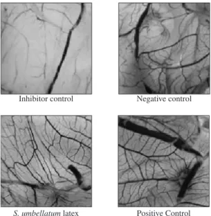
The images of the vascular net of the group treated with the latex (10 mg.mL–1) and the different control
groups of controls are shown in Figure 1. In this figure, it is possible to observe a clear difference in formation of vascular net among the group treated with latex and the different control groups. A larger vascularisation was observed in the positive control group as well as in the group treated with S. umbellatum latex. The
vasculari-sation was smaller in the negative control and inhibitor groups. The pure latex was also tested, but it destroyed CAM completely and killed the chick embryo.
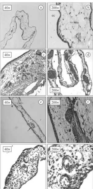
Figure 2 shows the images of histological analysis, exhibiting the formation of the vascular networks in the different controls and in the group treated with 10 mg.L–1
of S. umbellatum latex.
Figure 3 presents a detail of Figure 2h, showing the formation of the blood vessels and inflammatory elements caused by the treatment using S.umbellatum
latex.
4. Discussion
For centuries, plants have been widely used as food and for medicinal purposes in different cultures. In the last few years, the interest in plant medicines has in-creased worldwide.
Figure 1. Formation of the vascular network in different controls and in the treatment using S. Umbellatum latex.
Table 1. Vascularisation percentage obtained with treatment of S. umbellatum latex and different controls.
S. umbellatum latex Vascularisation percentage Mean ± SD
10 mg.mL–1
(3 µL)
47.2, 52.9, 46.5, 62.4, 54.4, 52.5, 51.6, 57.8, 56.3, 43.5, 58.6, 48.6, 50.2, 47.2, 58.4, 46.2, 55.7, 51.9, 49.1, 42.4
49.1 ± 5.58#
20 mg.mL–1
(3 µL)
60.5, 57.1, 48.9, 50.7, 55.6, 54.8, 47.9, 52.4, 58.3, 60.1, 46.5, 56.4, 49.3, 54.7, 55.2, 51.6, 46.3, 50.7, 52.1, 48.3
52.9 ± 4.35#
H2O (3 µL) (negative control)
36.3, 32.5, 38.5, 33.6, 29.1, 37.2, 28.7, 35.6, 33.2, 30.1, 28.5, 27.9, 32.1, 35.5, 30.2, 33.7, 32.8, 28.2, 33.7, 34.6
32.6 ± 3.19*
Dexamethasone (12 µg) (inhibitor)
9.6, 14.7, 14.3, 15.6, 12.4, 11.9, 10.8, 9.5, 9.1, 12.9, 13.5, 12.1, 11.3, 9.8, 10.3, 9.3, 14.1, 12.5, 10.2, 11.5
11.8 ± 1.96*
Biocure (positive control)
55.8, 52.3, 58.6, 53.8, 57.4, 50.9, 58.7, 59,2 54.3, 53.2, 57.1, 54.6, 58.9, 51.5, 57.6, 56.3, 54.9, 52.6, 55.5, 60.3
55.7 ± 2.77#
Same symbols (p > 0.05) Different symbols (p < 0.05).
All the results were compared to controls groups by Kruskal-Wallis one way ANOVA on ranks and followed by multiple comparison procedure.
Figure 3. Treatment using S.umbellatum latex (Detail of Figure 2h) Note the presence of fibroconjunctive tissues, blood vessels well formed, nuclear erythrocytes within the lumen of new vasculature and inflammatory elements.
a b
e f
c d
g h
Figure 2. Paraffin sections stained with hematoxylin-eosin. Control inhibitor (dexamethasone) shows a few conjunctive tissue cells and also few blood vessels (a and b). Positive control (Hevea brasiliensis latex biomembrane) shows some newly formed blood vessels and inflammatory elements (c and d) and, in detail, well formed blood vessels and many nuclear erythrocytes (d). For the negative control (distilled water), the arrows show few blood vessels structures (e and f). Treatment using S. umbellatum latex shows well organ-ised vessels, replete of nuclear erythrocytes and inflamma-tory elements (g and h): ec = ectoderm ms = mesoderm en = endoderm.
titumoral and antiangiogenic effects of this plant. Also the latex of plants belonging to the genus Synadenium is
a common source of folk medicine, mainly to treat can-cer (Ortêncio, 1997) and some of its biological activi-ties have been identified (Afonso-Cardoso et al., 2007; Rogério et al., 2007; Premaratna et al., 1984). Thus,
we aimed at evaluating the angiogenic activity of the
S. umbellatum latexon chick embryo.
The CAM assay has been widely used as an in vivo model to study the angiogenic activity of various agents, e.g. growth factors, cytokines, hormones, drugs, tissue extracts and implanted tissue grafts (Zwadlo-Klarwasser et al., 2001). Toxicity of drugs on chick embryos can be evaluated by embryo death or adverse effects on CAM, including inflammation and neovascularization (Vargas et al., 2007).
The results obtained in this study demonstrated that the treatments using 10 and 20 mg.mL–1S. umbellatum
latex showed a significant increase of percentage area of vascular net in fertilized chicken eggs compared to the negative and inhibitor control groups (P < 0.05). However, there was not a significant increase in latex in-duction of neoformed vascular net when compared to the positive control (P > 0.05). The angiogenic activity was measured by counting the number of blood vessels in a given area (Staton et al., 2004). There was a significant increase of vascularisation in the positive control and in the group treated with S. umbellatum latex compared to
the negative and inhibitor controls (P < 0.05) (Figure 1). The different formations of vascular net evaluated by histological analysis in the different controls and in group treated with the latex of the plant(Figure 2) are in accordance with the digital images presented. We can observe an evident inhibition of blood vessels by dex-amethasone (inhibitor control) since these areas showed poor vascularisation (Figures 2a and 2b). We also ob-served poor blood vascular structures in the negative con-trol (Figures 2e and 2f). The positive concon-trol (Figures 2c and 2d) and the treatments using S. umbellatum latex Because of the immense flora existing all over the
world along with cultural aspects, the use of plants in the form of crude extracts, infusions, or plasters has been revived as a usual practice to treat common diseases (Marques and Farah, 2009).
an-(Figures 2g and 2h) presented a relevant increase in vascular net as well as infiltrated inflammatory cells (Figure 3 – detail of Figure 2h).
All the results herein obtained using CAM assays in vascular net (percentage of vascularization, digital images, and histological analysis) allow to infer that
S. umbellatum latex stimulated the growth of new
ves-sels in CAM.
We observed in our experiment that even a small quantity of the pure latex killed the chick embryo (re-sults not shown here) proving it is very toxic. Vargas et al. (2007) showed that toxic substances can induce inflammatory response. As S. umbellatum latex is toxic,
it probably stimulated inflammatory responses which permitted the migration of neutrophils and macrophages cells shown in Figure 3.
It has already been pointed out in the literature that the inflammatory cells are important to activate factors such as cytokines, interleukins (IL-1, IL-2 and IL-8), vascular endothelial growth factor (VEGF), and plate-let activating factor. These are endothelial cell-specific growth factors and have an important role in the initia-tion and amplificainitia-tion of inflammatory response (Zijlstra et al., 2006), and consequently in activating angiogenic factors (Donà et al. 2003; May et al., 2008), since all of them induce the growth of the pre-existing vessels and neoformation of others in CAM.
5. Conclusion
In the present research, the angiogenic activity of
S. umbellatum latex was evaluated using the CAM
as-say and the results showed that it presents angiogenic activity.
Acknowledgements — This work was supported by Fundação de Amparo à Pesquisa do Estado de Goiás (FAPEG).
References
AFONSO-CARDOSO, SR., RODRIGUES, FH., GOMES, MA., SILVA, AG., ROCHA, A., GUIMARÃES, AH., CANDELORO, I., FAVORETO Jr., S., FERREIRA, MS. and SOUZA, MA., 2007. Protective effect of lectin from Synadenium carinatum on Leishmania amazonensis infection in BALB/c mice. Korean Journalof Parasitology, vol. 45, no. 4, p. 255-266.
CHOI, S., KIM, KW., CHOI, JS., HAN, ST., PARK, YI., LEE, SK., KIM, JS. and CHUNG, MH., 2002. Angiogenic activity of beta-sitosterol in the ischaemia/reper fusion-damaged brain of Mongolian gerbil. Planta Medica,vol. 68, no. 4, p. 330-335. DONÀ, M., DELL’AICA, I., CALABRESE, F., BENELLI, R., MORINI, M., ALBINI, A. and GARBISA, S., 2003. Neutrophil Restraint by Green Tea: Inhibition of Inflammation, Associated Angiogenesis, and Pulmonary Fibrosis. Journalof Immunology, vol. 170, no. 8, p. 4335-4341.
FOLKMAN, J., 2003. Fundamental concepts of the angiogenic process. Current Molecular Medicine, vol. 3, no. 7, p. 643-651. GOVINDAPPA, T., GOVARDHAN, L., JYOTHY, PS. and VEERABHADRAPPA, PS., 1987. Purification and
characterisation of acetylcholinesterase isozymes from the latex of Synadenium grantii Hook, “f”. Indian Journal of Biochemistryand Biophysics, vol. 24, no. 4, p. 209-217. GURIB-FAKIM, A., 2006. Medicinal plants: traditions of yesterday and drugs of tomorrow. Molecular Aspects of Medicine, vol. 27, no. 1, p. 1-93.
JÄGER, AK., HUTCHINGS, A. and STADEN, J. Van, 1996. Screening of Zulu medicinal plants for prostaglandin-synthesis inhibitors. Journal of Ethnopharmacology, vol. 52, no. 2, p. 95-100.
JUAREZ, CP., MUIÑO, JC., GUGLIELMONE, H., SAMBUELLI, R., ECHENIQUE, JR., HERNÁNDEZ, M. and LUNA, JD., 2000. Experimental retinopathy of prematurity: angiostatic inhibition by nimodipine, ginkgobiloba, and dipyridamole, and response to different growth factors. European Journal of Ophthalmology, vol. 10, no. 1, p. 51-59. KRUPINSKI, J., STROEMER, P., SLEVIN, M., MARTI, E., KUMAR, P. and RUBIO, F., 2003. Three-dimensional structure and survival of newly formed blood vessels after focal cerebral ischemia. Neuroreport, vol. 14, no. 8, p. 1171-1176.
MARQUES, V. and FARAH, A., 2009. Chlorogenic acids and related compounds in medicinal plants and infusions. Food Chemistry, vol. 113, no. 4, p. 1370-1376.
MAY, AE., SEIZER, P. and GAWAZ, M., 2008. Platelets: inflammatory firebugs of vascular walls. Arteriosclerosis, Thrombosis, andVascular Biology, vol. 28, no. 3, p. 5-10. MENDONÇA, RJ., 2004. Caracterização biológica de uma fração angiogênica do látex natural da seringueira: Hevea brasiliensis. Ribeirão Preto: Universidade de São Paulo. [Dissertação de Mestrado].
MENONN, M., VITHAYATHIL, PJ., RAJU, SM. and RAMADOSS, CS., 2002. Isolation and characterization of proteolytic enzymes from the latex of Synadenium grantii Hook, “f ”. Plant Science, vol. 163, no. 1, p. 131-139. MRUÉ, F., 1997. Substituição do esôfago cervical por prótese biossintética de látex: estudo experimental em cães. Ribeirão Preto: Universidade de São Paulo. [Dissertação de Mestrado]. NOGUEIRA, IAL., LEÃO, ABB., VIEIRA, MS., BENFICA, PL., CUNHA, LC. and VALADARES, MC., 2008. Antitumoral and antiangiogenic activity of Synadenium umbellatum Pax. Journal ofEthnopharmacology, vol. 120, no. 3, p. 474-478. ORTÊNCIO, WB., 1997. Medicina popular do Centro-Oeste. 2 ed. Brasília: Thesaurus. p. 59.
PREMARATNA, A., SHDAKSHARASWAMY, M. and NANJAPPA, S., 1984. Some biological properties of Synadenium grantil lectin. Indian Journal of Pathologyand Microbiology, vol. 27, no. 2, p. 91-97.
following ischaemic stroke: strategies towards neuroprotection. Journal of Cellular and Molecular Medicine, vol. 9, no. 1, p. 85-102.
SLEVIN, M., KUMAR, P., GAFFNEY, J., KUMAR, S. and KRUPINSKI, J., 2006. Can angiogenesis be exploited to improve stroke outcome? Mechanisms and therapeutic potential. Clinical Science, vol. 111, no. 3, p. 171-183.
STATON, CA., STRIBBLING, SM., TAZZYMAN, S., HUGHES, R., BROWN, NJ. and LEWIS, CE., 2004. Current methods for assaying angiogenesis in vitro and in vivo. International Journal of ExperimentalPathology, vol. 85, no. 5, p. 233-248.
VALADARES, MC., CASTRO, NC. and CUNHA, LC., 2007. Synadenium umbellatum: citotoxicidade e danos ao DNA de células da medula óssea de camundongos. RevistaBrasileira de CiênciasFarmacêuticas, vol. 43, no. 4, p. 631-638.
VARGAS, A., ZEISSER-LABOUÈBE, M., LANGE, N., GURNY, R. and DELIE, F., 2007. The chick embryo and its
chorioallantoic membrane (CAM) for the in vivo evaluation of drug delivery systems. Advanced Drug Delivery Reviews, vol. 59, no. 11, p. 1162-1176.
WANG, S., ZHENG, Z., WENG, Y., YU, Y., ZHANG, D., FAN, W., DAI, R. and HU, Z., 2004. Angiogenesis and anti-angiogenesis activity of Chinese medicinal herbal extracts. Life Sciences, vol. 74, no. 20, p. 2467-2478.
ZIJLSTRA, A., SEANDEL, M., KUPRIYANOVA, TA., PARTRIDGE, JJ., MADSEN, MA., HAHN-DANTONA, EA., QUIGLEY, JP. and DERYUGINA, EI., 2006. Proangiogenic role of neutrophil-like inflammatory heterophils during neovascularization induced by growth factors and human tumor cells. Blood, vol. 107, no. 1, p. 317-327.