(1) Division of Gastroenterology, Hepatitis Section, Federal University of Sao Paulo, Sao Paulo, SP, Brazil. (2) Department of Pathology, Federal University of Sao Paulo, Sao Paulo, SP, Brazil.
Correspondence to: Patricia da Silva Fucuta Pereira, Rua Athayde Baptista Jerico 55, Jardim Vivendas, 15090-465 Sao Jose do Rio Preto, SP, Brazil. Tel.: 5576-4050; Fax:
+55-11-CHRONIC HEPATITIS C: HEPATIC IRON CONTENT DOES NOT CORRELATE WITH RESPONSE TO
ANTIVIRAL THERAPY
Patricia da Silva Fucuta PEREIRA(1), Ivonete Sandra de SOUZA E SILVA(1), Silvia Naomi de Oliveira UEHARA(1), Christini Takemi EMORI(1), Valéria Pereira LANZONI(2), Antonio Eduardo Benedito SILVA(1) & Maria Lucia Gomes FERRAZ(1)
SUMMARY
The complex interaction between hepatitis C virus infection, iron homeostasis and the response to antiviral treatment remains controversial. The aim of this study was to evaluate the inluence of hepatic iron concentration (HIC) on the sustained virological response (SVR) to antiviral therapy in patients with chronic hepatitis C. A total of 50 patients who underwent pretreatment liver biopsy with assessment of HIC by graphite furnace atomic absorption spectroscopy and were subsequently submitted to antiviral treatment with interferon/peginterferon and ribavirin were included in the study. Patients with alcoholism, history of multiple blood transfusion, chronic kidney disease, hemolytic anemia and parenteral iron therapy were excluded. The iron related markers and HIC were compared between those who achieved an SVR and non-responders (NR) patients. The mean age was 45.7 years and the proportion of patients’
gender was not different between SVR and NR patients. The median serum iron was 138 and 134 µg/dL (p = 0.9), the median serum
ferritin was 152.5 and 179.5 ng/mL (p = 0.87) and the median HIC was 9.9 and 8.2 µmol/g dry tissue (p = 0.51), for SVR and NR
patients, respectively. Thus, hepatic iron concentration, determined by a reliable quantitative method, was not a negative predictive factor of SVR in patients with chronic hepatitis C presenting mild to moderate hepatic iron accumulation.
KEYWORDS: Iron; Hepatitis C virus; Interferon; Peginterferon; Ribavirin; Liver biopsy.
INTRODUCTION
Iron is an essential element for all living organisms and the complex interaction between iron homeostasis and human viral infections remains a challenging issue13.
The association between hepatitis C virus (HCV) infection and altered serum iron proile or hepatic iron deposits has been evaluated in several studies6,9,36, but the prevalence of hepatic iron overload, its relationship
with the severity of disease and its inluence on the response to antiviral treatment are still controversial14,15,27,34,35.
Despite marked advances in the treatment of HCV infection, sustained elimination of the virus is not achieved in almost half the patients even with the current combined treatment of peginterferon
and ribavirin18. Several viral and host factors such as HCV genotype,
age, gender and degree of ibrosis are associated with the response to treatment, but the impact of hepatic iron deposits remains uncertain. Some studies have indicated that increased hepatic iron is one of the negative predictive factors of response to treatment2,7,24,29,33,40, but this
fact has not been conirmed in other investigations1,12,20,22,31. However, it
must be pointed out that most of these studies had not applied adequate methods of quantifying hepatic iron.
The objective of the present study was to evaluate the inluence of
hepatic iron concentration (HIC) on the response to combined treatment with interferon/peginterferon and ribavirin in patients with chronic hepatitis C using a very accurate method of assessing hepatic iron content.
PATIENTS AND METHODS
This retrospective study identiied patients with chronic hepatitis C (anti-HCV and HCV RNA positive) seen at the Hepatitis Outpatient Clinic of the Federal University of Sao Paulo, who were consecutively submitted to a pretreatment liver biopsy between May 2000 and August
2001 and evaluated regarding HIC38. Among these patients, those who
received antiviral treatment with interferon/peginterferon and ribavirin were included in the present study after giving their written informed consent. The protocol conformed to the ethical guidelines of the 1975 Helsinki Declaration and was approved by the local Ethical Committee.
Exclusion criteria were alcohol consumption > 20 g/day, co-infection with HIV or HBV, immunosuppression, previous antiviral therapy, a history of multiple blood transfusions (> 6 units), chronic kidney disease, hemolytic anemia and parenteral iron therapy.
an automatized kinetic method and were reported as the number of times the upper limit of normal (ULN). Serum iron (normal value: 37-145 µg/dL for women and 59-158 µg/dL for men) was measured
by a colorimetric method(Olympus AU 640 Analyser, Tokyo, Japan)
and serum ferritin (normal value: 5-200 ng/mL for women and 22-415 ng/mL for men) was assayed by a chemiluminescence immunoassay (Access 2 Immunoassay System, Beckman Coulter Inc., Fullerton, CA, USA). Serum transferrin (normal value: 200-400 mg/dL) was determined by immunoturbidimetry (Olympus AU 640 Analyser, Tokyo,
Japan) and its saturation was calculated using the following formula39:
iron/transferrin x 71.2.
HCV RNA was detected by qualitative PCR using Amplicor kits (Cobas Amplicor HCV Test, Roche Diagnostic System, USA; detection limit: 50 IU/mL) and HCV genotyping was performed by sequencing
the NS5B region of HCV30.
Evaluation of hepatic iron concentration in liver tissue: The quantiication of iron in liver tissue was performed according to the
modiied method described by OLYNYK et al.28. To measure HIC, fresh
liver specimens placed in a clean tube were dried to constant weight in a vacuum oven at 52 ºC. The dried samples were then weighed and placed in a tube containing 1 mL concentrated nitric acid and 1 mL ion-free water. Next the samples were digested in a microwave digestion system (MDS 2000, USA). A blank and samples of bovine liver used as a reference material (1577b) were treated similarly. The resulting solutions were transferred to volumetric lasks and made up with ion-free water. All digested samples were diluted 1:8 in 0.2% nitric acid and a 10-µL volume was injected into the furnace for measurement. Iron concentration was determined by graphite furnace atomic absorption spectroscopy using a Varian Perkin-Elmer SIMAA-6000 spectrometer (USA). The results were expressed as µmol/g dry tissue weight. The hepatic iron index (HII), deined as the HIC to age ratio3, was also calculated.
Antiviral treatment of chronic hepatitis C: Treatment was indicated for patients with a degree of ibrosis ≥ 2 (Metavir classiication4)
in liver biopsies and consisted of combined administration of standard
interferon (3MU three times weekly) or peginterferon (α2b 1.5 µg/kg
or α2a 180 µg once-weekly) plus ribavirin (body weight < 75 kg: 1.0g/
day; ≥ 75 kg: 1.25g/day) for 24 to 48 weeks, according to the genotype.
Comparative analysis according to the response to treatment:
The response to antiviral treatment was evaluated by the detection of HCV RNA in serum by qualitative PCR at the end of treatment and six months thereafter. Patients who tested HCV RNA negative in the sixth month post-treatment were classiied as responders (sustained virological response, SVR). All other patients, including those who relapsed, were classiied as non-responders (NR). The patients were compared regarding hepatic iron concentration (HIC) and serum iron proile according to the response to treatment (SVR or NR).
Statistical analysis: The results were irst reported descriptively and then the association between variables of interest was investigated. Continuous variables were compared by the Student t-test or Mann-Whitney test when appropriate. Categorical variables were compared using the chi-square test. A p value of less than 0.05 was considered statistically signiicant. Statistical analysis was performed by PASW Statistics 17.0 (SPSS Inc., Chicago, IL).
RESULTS
Among the 96 patients evaluated regarding HIC (previously reported results38), 50 underwent antiviral treatment for chronic hepatitis C (39
with interferon + ribavirin and 11 with peginterferon + ribavirin) and were included in the present study. The mean age was 45.7 years (28-70), the mean duration of HCV infection was 20 years and there was predominance of male gender (60%). Hepatitis C virus genotyping was performed for 35 patients and resulted in type 1 in 27 (77%) and type 3 in eight (23%). Sustained virological response was achieved in 18 patients (36%). Among patients infected with HCV genotype 1 and 3, the rates of sustained virological response were 26% and 25%, respectively (p > 0.05).Proportion of patients’ gender was not signiicantly different between SVR and NR patients (female/male: 7/11 vs 13/19, respectively, p = 0.90).
The demographic and clinical characteristics of the patients are shown in Table 1.
Table 1
Demographic and clinical characteristics of the sample studied
Characteristics n = 50
Sex
Male/female 30/20
Age (years) 45.7 ± 10.2
Duration of HCV infection (years) 20.4 ± 8.8
HCV genotype (%)
Type 1 77
Type 3 23
Histology
Fibrosis stage 3/4 Metavir (%) 20
Hemoglobin (g/dL) 14.8 ± 1.1
Hematocrit (%) 45.0 ± 3.2
Platelet count (x104/mm3) 18.5 ± 6.0
ALT (xULN) 2.4 (1.6-3.4)
AST (xULN) 1.8 (1.5-2.8)
Serum iron (µg/dL) 136.5 (104.0-162.2)
Serum ferritin (ng/mL) 164.0 (86.9-352.0)
Serum transferrin (mg/dL) 310.6 ± 50.1
Transferrin saturation (%) 33.5 ± 13.2
HIC (µmol/g dry tissue) 8.4 (4.8-13.3)
Hepatic iron index 0.18 (0.09-0.36)
No difference was observed in serum iron, serum ferritin and transferrin saturation between responders and non-responders. HIC did not inluence the SVR rate (Fig. 1). Table 2 shows the serum and hepatic iron proile of responders and non-responders patients.
DISCUSSION
Chronic hepatitis C has been associated with the accumulation of iron in the liver, which could interfere with the response to antiviral treatment. However, studies evaluating the effect of iron on treatment in this population are extremely conlicting in terms of the method used for the determination of hepatic iron content, the adopted therapeutic regimen and the evaluation of response to treatment.
In the present investigation, the gold standard for the evaluation of HIC was adopted, i.e., quantiication of iron in liver tissue specimens
and the adopted antiviral therapy included either standard interferon or peginterferon, both with ribavirin.The results demonstrated that severe iron overload was a rare event among HCV-infected patients and HIC was not associated with the SVR to treatment with interferon/ peginterferon plus ribavirin. In fact, hepatic iron content has been shown to be low in most patients infected with HCV8,17,25. In the present study,
patients with possible other causative factors of iron deposition, such as multiple blood transfusion and hemolytic anemia were excluded. Thus, our results reflect the HIC in patients infected with HCV
without confounding factors of secondary iron overload.Moreover,
the proportion of patients’ gender was similar between responders and non-responders and thus it is unlikely that the distinct serum and hepatic iron proiles between men and women have inluenced the present analysis. The lack of association between HCV genotypes and the rate of SVR observed in our study is likely due to the low number of patients with this analysis available.
Early studies regarding HIC and monotherapy with standard interferon have only evaluated the impact on the biochemical response.
VAN THIEL et al.40 studied 79 patients treated with interferon for six
months and showed that the biochemical response (deined as complete normalization of ALT) or partial response (deined as a > 50% reduction in ALT levels six months after the end of treatment) was worse in patients
with a higher HIC. Soon thereafter, in 1995, BARTON et al.2found that
the pattern of iron distribution in the liver was signiicantly different between patients with and without a complete biochemical response, also
deined as normalization of ALT. OLYNYK et al.29 reported higher HIC
in biochemical non-responders to interferon therapy for six months than in those with good response. Similar indings were reported by PIPERNO
et al.33, who observed an association between higher hepatic iron content
and a poor biochemical response in patients with chronic hepatitis C treated with standard interferon for 12 months. It should be emphasized that 80% of the patients with elevated HIC presented genotype 1b. Also in that study, iron depletion by phlebotomy performed on 16 patients subsequently treated with interferon did not improve the response to treatment despite improvement of ALT levels.
On the other hand, studies evaluating virological response and hepatic iron determined by histological semiquantiication have reported conlicting data. The results obtained in a group of patients with low
levels (< 1mEq/mL) of HCV RNA1 treated with interferon for 24 weeks,
showed higher iron-staining score in virological responders than in non-responders and the investigators hypothesized that an optimal range of iron concentration may be beneicial for immune response. In contrast,
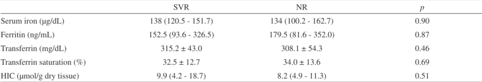
Table 2
Serum and hepatic iron proiles in patients presenting a sustained virological response (SVR) and in non-responders (NR)
SVR NR p
Serum iron (µg/dL) 138 (120.5 - 151.7) 134 (100.2 - 162.7) 0.90
Ferritin (ng/mL) 152.5 (93.6 - 326.5) 179.5 (81.6 - 352.0) 0.87
Transferrin (mg/dL) 315.2 ± 43.0 308.1 ± 54.3 0.46
Transferrin saturation (%) 32.5 ± 12.7 34.0 ± 13.6 0.69
HIC (µmol/g dry tissue) 9.9 (4.2 - 18.7) 8.2 (4.9 - 11.3) 0.51
Quantitative variables are expressed as mean ± SD or median (interquartile range). HIC, hepatic iron concentration.
Fig. 1 - Comparison of hepatic iron concentration between patients presenting sustained virological response and non-responders.
FUJITA et al.16 observed a signiicantly higher histological total iron score
in patients not responding to combined interferon and ribavirin and LIN
et al.24 found that positive hepatic iron stain predicted non-response to
peginterferon and ribavirin therapy.
However, when hepatic iron levels were actually measured, recent publications12,22,31 did not ind any inluence of HIC on the SVR in patients
undergoing combined therapy with standard interferon and ribavirin.
To the best of our knowledge, only one study20 has previously
evaluated the role of hepatic iron concentration (measured by atomic absorption spectrophotometry) in relation to the eficacy of chronic
hepatitis C therapy schedule including peginterferon.In agreement
with our results, HOFER et al.20 observed no difference in tissue iron
concentration between patients who achieved an SVR and non-responders both submitted to combined interferon/peginterferon and ribavirin treatment.
Even in cases of iron overload, as in thalassemic patients, two studies11,37 found no impact of excessive iron deposition on the SVR
rate. In addition, demonstrating the eficacy of combined treatment with
peginterferon and ribavirin in young thalassemic patients, one study21
showed an SVR in 62.5% of cases, a rate similar to that observed in non-thalassemic patients.
The role of HFE genotype on hepatic iron in chronic hepatitis C is
another interesting area of investigation. This issue has been addressed in several studies and controversial indings have been reported32.
CORENGIA et al.8 studied 206 patients with chronic hepatitis C and
found higher frequency of genotypes containing HFE mutations in
the subgroup of patients with hepatic iron accumulation, whereas no significant difference was observed between the subgroup of patients without hepatic iron deposits and control subjects. They also
demonstrated a signiicant association between HFE genotypes and
iron deposition in hepatocellular compartment, but not in sinusoidal and portal areas, suggesting that the cause of hepatic iron accumulation in chronic hepatitis C could not be simply the release of iron from necrotic hepatocytes or the inlammatory-mediated perturbation of iron traficking, when iron would accumulate preferentially in Kupffer cells.
But in another study, KAZEMI-SHIRAZI et al.23 assessed the HFE
mutations in 184 patients with chronic hepatitis C and 487 controls and found no difference in hepatic iron content among patients with or without
HFE mutations. In hepatitis C and control group, 32% and 29% of patients
carried either mutations (C282Y and H63D), respectively. Histological iron grades 3 or 4 (Perls’ Prussian blue stain) were found in 23.4% and 15.7% of chronic hepatitis C patients with and without mutation of the
HFE gene, respectively, p = 0.36.
Interestingly, recent data5 from patients with advanced ibrosis (part
of the HALT-C study) showed that the presence of mutations in the
HFE gene (especially the most common H63D mutation) resulted in
a higher chance of an SVR during retreatment with peginterferon and ribavirin, with the observation of a direct correlation with ferritin and semiquantiied histological iron levels. Although we did not investigate mutations of hereditary hemochromatosis in this study, none of the patients presented an HII suggestive of this disease, i.e., higher than 1.93.
In addition, there is evidence indicating that the prevalence of this disease
among patients infected with HCV does not differ from that found in the general population23,26.
The lack of a correlation between HIC and response of patients with hepatitis C to antiviral treatment agrees with the majority of the results
regarding phlebotomy as coadjuvant treatment7,10,19. DI BISCEGLIE et
al.10 showed that in patients with hepatitis C not responding to interferon
monotherapy iron depletion by phlebotomy did not result in a higher SVR rate during retreatment, despite an association with improved liver injury manifested by a decrease in transaminases and improved liver
histopathology after phlebotomy. Similarly, in the study of HERRERA19
iron depletion by phlebotomy did not improve SVR in the retreatment of patients who were previously non-responders. But on the contrary, in
the study of CARLO et al.7 83 patients were randomly assigned to be
submitted to iron depletion before interferon monotherapy and a better complete response (biochemical and virological) was achieved in the phlebotomy group.
In conclusion, hepatic iron concentration determined by a reliable quantitative method, was not a negative predictive factor of SVR to antiviral treatment with interferon/peginterferon and ribavirin in patients with chronic hepatitis C presenting mild to moderate hepatic iron accumulation.
RESUMO
Hepatite C crônica: concentração hepática de ferro não é correlacionada com a resposta ao tratamento antiviral
A complexa interação entre infecção pelo vírus da hepatite C, homeostase do ferro e resposta ao tratamento antiviral permanece controversa. O objetivo deste estudo foi avaliar a influência da concentração hepática de ferro (CHF) na resposta virológica sustentada (RVS) à terapia antiviral na hepatite C crônica. Foram incluídos 50 pacientes que foram submetidos à biopsia hepática pré-tratamento com determinação da CHF por espectrofotometria de absorção atômica com forno de graite e tratados posteriormente com interferon/peginterferon e ribavirina. Pacientes com alcoolismo, história de múltiplas transfusões sanguíneas, doença renal crônica, anemia hemolítica e terapia com
ferro parenteral foram excluídos.O peril de ferro sérico e a CHF foram
comparados entre aqueles que atingiram RVS e os não-respondedores (NR). A média de idade dos pacientes foi 45,7 anos e não houve diferença na proporção de homens e mulheres entre os grupos RVS e NR. A mediana do ferro sérico foi 138 and 134 µg/dL (p = 0.9), a
mediana da ferritina sérica foi 152,5 e 179,5 ng/mL (p = 0,87) e a CHF
mediana foi 9,9 e 8,2 µmol/g de tecido seco (p = 0,51), para pacientes
com RVS e NR, respectivamente. Concluindo, a concentração hepática de ferro, determinada por um método quantitativo coniável, não foi um fator preditivo negativo de RVS em pacientes com hepatite C crônica e acúmulo de ferro hepático leve a moderado.
ACKNOWLEDGMENTS
REFERENCES
1. AKIYOSHI, F.; SATA, M.; UCHIMURA, Y.; SUZUKI, H. & TANIKAWA, K. - Hepatic iron stainings in chronic hepatitis C patients with low HCV RNAlevels: a predictive marker for IFN therapy. Amer. J. Gastroent., 92: 1463-1466, 1997.
2. BARTON, A.L.; BANNER, B.F.; CABLE, E.E. & BONKOVSKY, H.L. - Distribution of iron in the liver predicts the response of chronic hepatitis C infection to interferon therapy. Amer. J. clin. Path., 103: 419-424, 1995.
3. BASSETT, M.L.; HALLIDAY, J.W. & POWELL, L.W. - Value of hepatic iron measurements in early hemochromatosis and determination of the critical iron level associated with ibrosis. Hepatology, 6: 24-29, 1986.
4. BEDOSSA, P. & POYNARD, T. - An algorithm for the grading of activity in chronic hepatitis C. The Metavir Cooperative Study Group. Hepatology, 24: 289-293, 1996. 5. BONKOVSKY, H.L.; NAISHADHAM, D.; LAMBRECHT, R.W. et al. - Roles of iron and
HFE mutations on severity and response to therapy during retreatment of advanced chronic hepatitis C. Gastroenterology, 131: 1440-1451, 2006.
6. BOUCHER, E.; BOURIENNE, A.; ADAMS, P. et al. - Liver iron concentration and distribution in chronic hepatitis C before and after interferon treatment. Gut, 41: 115-120, 1997.
7. CARLO, C.; DANIELA, P. & GIANCARLO, C. - Iron depletion and response to interferon in chronic hepatitis C. Hepatogastroenterology, 50: 1467-1471, 2003.
8. CORENGIA, C.; GALIMBERTI, S.; BOVO, G. et al. - Iron accumulation in chronic hepatitis C: relation of hepatic iron distribution, HFE genotype, and disease course. Amer. J. clin. Path., 124:846-853, 2005.
9. DI BISCEGLIE, A.M.; AXIOTIS, C.A.; HOOFNAGLE, J.H. & BACON, B.R. - Measurements of iron status in patients with chronic hepatitis. Gastroenterology, 102: 2108-2113, 1992.
10. DI BISCEGLIE, A.M.; BONKOVSKY, H.L.; CHOPRA, S. et al. - Iron reduction as an adjuvant to interferon therapy in patients with chronic hepatitis C who have previously not responded to interferon: a multicenter, prospective, randomized, controlled trial. Hepatology, 32: 135-138, 2000.
11. DI MARCO, V.; LO IACONO, O.; ALMASIO, P. et al. - Long-term eficacy of alpha-interferon in beta-thalassemics with chronic hepatitis C. Blood, 90: 2207-2212, 1997. 12. DISTANTE, S.; BJORO, K.; HELLUM, K.B. et al. - Raised serum ferritin predicts non-response to interferon and ribavirin treatment in patients with chronic hepatitis C infection. Liver, 22: 269-275, 2002.
13. DRAKESMITH, H. & PRENTICE, A. - Viral infection and iron metabolism. Nature Rev. Microbiol., 6: 541-552, 2008.
14. FABRIS, C.; TONIUTTO, P.; SCOTT, C. et al. - Serum iron indices as a measure of iron deposits in chronic hepatitis C. Clin. Chem. Acta, 304: 49-55, 2001.
15. FARINATI, F.; CARDIN, R.; DE MARIA, N. et al. - Iron storage, lipid peroxidation and glutathione turnover in chronic anti-HCV positive hepatitis. J. Hepat., 22: 449-456, 1995.
16. FUJITA, N.; SUGIMOTO, R.; URAWA, N. et al. - Hepatic iron accumulation is associated with disease progression and resistance to interferon/ribavirin combination therapy in chronic hepatitis C. J. Gastroent. Hepat., 22: 1886-1893, 2007.
17. GUYADER, D.; THIROUARD, A.S.; ERDTMANN, L. et al. - Liver iron is a surrogate marker of severe ibrosis in chronic hepatitis C. J. Hepat., 46: 587-595, 2007. 18. HEATHCOTE, E. - Antiviral therapy: chronic hepatitis C. Review. J. viral Hepatitis,
14: 82-88, 2007.
19. HERRERA, J.L. - Iron depletion is not effective in inducing a virologic response in patients with chronic hepatitis C who failed to respond to interferon therapy. Amer. J. Gastroent., 94: 3571-3575, 1999.
20. HOFER, H.; OSTERREICHER, C.; JESSNER, W. et al. - Hepatic iron concentration does not predict response to standard and pegylated-ifn/ribavirin therapy in patients with chronic hepatitis C. J. Hepat., 40: 1018-1022, 2004.
21. INATI, A.; TAHER, A.; GHORRA, S. et al. - Eficacy and tolerability of peginterferon alpha-2a with or without ribavirin in thalassaemia major patients with chronic hepatitis C virus infection. Brit. J. Haemat., 130: 644-646, 2005.
22. JURCZYK, K.; KARPINSKA, E.; WAWRZYNOWICZ-SYCZEWSKA, M. et al. - State of the iron metabolism in patients with chronic hepatitis C type C does not inluence antiviral treatment with interferon and ribavirin. Hepatogastroenterology, 55: 557-561, 2008.
23. KAZEMI-SHIRAZI, L.; DATZ, C.; MAIER-DOBERSBERGER, T. et al. - The relation of iron status and hemochromatosis gene mutations in patients with chronic hepatitis C. Gastroenterology, 116: 127-134, 1999.
24. LIN, T.J.; LIAO, L.Y.; LIN, C.L.; CHANG, T.A. & LIU, S.O. - Hepatic iron inluences responses to combination therapy with peginterferon alfa and ribavirin in chronic hepatitis C. Hepatogastroenterology, 55: 1412-1415, 2008.
25. MARTINELLI, A.L.; RAMALHO, L.N. & ZUCOLOTO, S. - Hepatic stellate cells in hepatitis C patients: relationship with liver iron deposits and severity of liver disease. J. Gastroent. Hepat., 19: 91-98, 2004.
26. MARTINELLI, A.L.; ZAGO, M.A.; ROSELINO, A.M. et al. - Porphyria cutanea tarda in Brazilian patients: association with hemochromatosis C282Y mutation and hepatitis C virus infection. Amer. J. Gastroent., 95: 3516-3521, 2000.
27. METWALLY, M.A.; ZEIN, C.O. & ZEIN, N.N. - Clinical signiicance of hepatic iron deposition and serum iron values in patients with chronic hepatitis C infection. Amer. J. Gastroent., 99: 286-291, 2004.
28. OLYNYK, J.; WILLIAMS, P.; FUDGE, A. et al. - Fine-needle aspiration biopsy for the measurement of hepatic iron concentration. Hepatology, 15: 502-506, 1992. 29. OLYNYK, J.K.; REDDY, K.R.; DI BISCEGLIE, A.M. et al. - Hepatic iron concentration
as a predictor of response to interferon alfa therapy in chronic hepatitis C. Gastroenterology, 108: 1104-1109, 1995.
30. PEREZ, R.M.; FERRAZ, M.L.; FIGUEIREDO, M.S. et al. - Unexpected distribution of hepatitis C virus genotypes in patients on hemodialysis and kidney transplant recipients. J. med. Virol., 69: 489-494, 2003.
31. PIANKO, S.; McHUTCHISON, J.G.; GORDON, S.C. et al. - Hepatic iron concentration does not inluence response to therapy with interferon plus ribavirin in chronic HCV infection. J. Interferon Cytokine Res., 22: 483-489, 2002.
32. PIPERNO, A.; SAMPIETRO, M.; D’ALBA, R. et al. - Iron stores, response to alpha-interferon therapy, and effects of iron depletion in chronic hepatitis C. Liver, 16: 248-254, 1996.
33. RIGAMONTI, C.; ADORNO, S.; MADULI, E. et al. - Gender and liver ibrosis in chronic hepatitis: the role of iron status. Aliment. Pharmacol. Ther., 21: 1445-1451, 2005. 34. RIGAMONTI, C.; ANDORNO, S.; MADULI, E. et al. - Iron, hepatic stellate cells and ibrosis in chronic hepatitis C. Europ. J. clin. Invest., 32(suppl. 1): 28-35, 2002. 35. SHAN, Y.; LAMBRECHT, R. & BONKOVSKY, H. - Association of hepatitis C virus
infection with serum iron status: analysis of data from the third national health and nutrition examination survey. Clin. infect. Dis., 40: 834-841, 2005.
37. SILVA, I.S.; PEREZ, R.M.; OLIVEIRA, P.V. et al. - Iron overload in patients with chronic hepatitis C virus infection: clinical and histological study. J. Gastroent. Hepat., 20: 243-248, 2005.
38. TURATI, C.; TURSINI, P.; FRANZINI, C. et al. - Saturazione della transferrina del siero e sovraccarico di ferro. Biochim. clin., 21: 10-15, 1997.
39. VAN THIEL, D.H.; FRIEDLANDER, L.; FAGIUOLI, S. et al. - Response to interferon alpha therapy is inluenced by the iron content of the liver. J. Hepat., 20: 410-415, 1994.