Postangioplasty restenosis: a practical
model in the porcine carotid artery
Serviços de 1Cardiologia, 2Cirurgia Vascular, 3Patologia and
4Oncologia (South-American Office for Anticancer Drug Development), Hospital de Clínicas de Porto Alegre and 5Hospital de Clínicas Veterinárias, Universidade Federal do Rio Grande do Sul, Porto Alegre, RS, Brasil P.R.A. Caramori1,
E.E. Eggers2, A.P.F. Silva-Filho5, D.M. Uchoa3, F. Jung1, A.C. Zago1, C.T.S. Cerski3, G. Schwartsmann4 and A.J. Zago1
Abstract
Transluminal coronary angioplasty is a routine therapeutic interven-tion in coronary heart disease. Despite the high rate of primary success, restenosis continues to be its major limitation. Porcine mod-els have been considered to be the most adequate experimental modmod-els for studying restenosis. One limitation of porcine models is the need for radiological guidance and the expenses involved. The objective of the present study was to adapt an experimental model of angioplasty in the porcine carotid artery that does not require radiological equipment. Eight animals were used to develop the technique of balloon injury to the common carotid artery by dissection without radiological guid-ance. This technique was then employed in six other animals. Under anesthesia, the left common carotid artery was dissected and incised at the carotid sinus for insertion of an over-the-wire angioplasty balloon towards the aorta. Overstretch injury of the carotid artery was per-formed under direct visualization. After 30 days, the arteries were excised and pressure-fixated. Uninjured carotid arteries from 3 addi-tional animals were used as controls. A decreased luminal area associ-ated with intimal hyperplasia and medial reaction was observed in all injured arteries. Immunohistochemistry identified the intimal hyper-plastic cells as smooth muscle cells. Computerized morphometry of the ballooned segments revealed the following mean areas: lumen 2.12 mm2 (± 1.09), intima 0.22 mm2 (± 0.08), media 3.47 mm2 (± 0.67), and adventitia 1.11 mm2 (± 0.34). Our experimental model of porcine carotid angioplasty without radiological guidance induced a vascular wall reaction and permitted the quantification of this response. This porcine model may facilitate the study of vascular injury and its response to pharmacological interventions.
Correspondence
P.R.A. Caramori
Cardiovascular Clinical Research Laboratory, Mount Sinai Hospital 600 University Avenue, Suite 1609 Toronto, Ontario M5G 1X5 Canada
E-mail: caramori@outer-net.com Research supported by CNPq and Fundo de Incentivo a Pesquisa of the Hospital de Clínicas of Porto Alegre. Dr. P.R.A. Caramori is currently a Clinical Research Fellow of the Cardiovascular Clinical Research Laboratory, University of Toronto, Canada.
Received September 10, 1996 Accepted July 21, 1997
Key words •Arterial injury •Intimal hyperplasia •Restenosis
•Percutaneous transluminal coronary angioplasty
Percutaneous coronary angioplasty is rou-tinely used for the treatment of coronary heart disease. Every year, more than 300,000 angioplasties are carried out in the USA and more than 10,000 in Brazil (1). Unfortu-nately, some of these procedures are per-formed for the second or third time on the
angiographic follow-up on a large number of patients, which implies risks and high oper-ating costs. Studies on the pathophysiology of restenosis, and the assessment of strate-gies aimed at inhibiting this process may be performed on animal models, which allow for the selection of interventions most likely to be effective in subsequent human trials.
The major goal of these experimental mod-els is the morphometric analysis of histologi-cal sections of the injured arteries (3). The porcine model of restenosis has shown the most consistent similarities with human reste-nosis (3). The histological aspect of the intimal hyperplasia and the results of experiments using the porcine common carotid artery (4), or coronary arteries (5), have been consistent with those observed in humans. One limitation of porcine models is the need for radiological guidance and the expenses that it incurs. The objective of the present study was to adapt an experimental model of balloon overstretch vascular injury of the porcine common carotid artery that dispenses with radiological equip-ment. This model should permit investigation of the pathophysiology of restenosis and as-sessment of pharmacological strategies for its prevention.
Fourteen crossbred pigs (Yorkshire-Hampshire-Landrace) obtained from local breeders with an approximate age of 3 to 4 months and weighing 20 to 35 kg were used. The animals were kept in captivity and fed a commercial chow, without cholesterol or lipid supplementation. Husbandry was car-ried out under supervision of a veterinarian, according to international norms (6).
Adaptation of the model. To develop the technique of angioplasty of the common ca-rotid artery by dissection, 8 animals were used. One animal was submitted to carotid angiography to assess the options for a surgi-cal approach. No radiologisurgi-cal guidance was used in the other procedures. Three different approaches were evaluated. Initially, an at-tempt was made to introduce the balloon into the common carotid artery through the
exter-nal carotid artery. This access was unsuc-cessful because of unfavorable anatomy. Specifically, the external carotid was found to be represented by multiple small-caliber branches, frequently leaving the common carotid at a right angle. Next, the direct incision of the proximal portion of the com-mon carotid artery close to the thoracic inlet was evaluated. This second approach had restrictions due to an insufficient surgical field. Finally, a cranial approach to the com-mon carotid, at its bifurcation, was techni-cally feasible and considered to be the ideal choice. All stages foreseen for the model were tested and perfected during this period, including i) sedation and anesthesia tech-niques, ii) histological fixing in a pressure perfusion system, and iii) morphometric a-nalysis using a computerized system.
Tissue preparation. After 30 days, the animals were sacrificed, and the carotid ar-teries were removed and fixed by pressure perfusion (100 mmHg) for 15 min in a solu-tion of 2% glutaraldehyde and 1% parafor-maldehyde in sodium phosphate buffer, pH 7.25 (7), followed by overnight immersion fixation. The balloon-injured portion of each artery was divided into five equally spaced annular segments, subsequently embedded in paraffin, prepared for histological sec-tioning, and stained using the Verhoeff and Masson trichrome techniques.
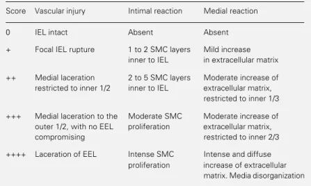
Histological analysis. Three aspects were evaluated by light microscopy, using an a-dapted semiquantitative graduation system, and the respective scores were recorded for each histological section: intensity of the vascular injury caused by dilatation (8), inti-mal reaction to injury (9), and medial reac-tion to injury (9) (scores 0 to ++++, Table 1). The presence of arterial thrombosis was also recorded. All measurements were independ-ently made by two observers. In any case of discrepancy, consensus was obtained. The left carotid arteries of three animals matched for weight and age, that did not undergo angioplasty, were used for qualitative com-parison. Immunoperoxidase staining for α-γ smooth muscle actin was carried out using HHF-35 monoclonal antibody (Draco, Carpinteria, CA).
Morphometric analysis. Histological im-ages were obtained by light microscopy (Unimac, Meiji, Japan), and digitized using a video camera (CK 3800, Meiji, Japan) and an analog-digital interface (Targa plus 16, Truevision, Indianapolis, IN). The borders of the arterial wall layers were manually traced using commercially available soft-ware (Aldus PhotoStyler 2.0, Aldus Corp., Seattle, WA), operated by a single independ-ent observer. The segmindepend-ents that showed the smallest luminal area in each artery were used for morphometric analysis. Planimetry was performed with the Imagescale software (Electronic Imagery, Pompano Beach, FL).
The arterial luminal area was obtained by direct measurement of the area delimited by the endothelium. The intimal area was cal-culated by subtracting the luminal area from the area delimited by the internal elastic lamina (IEL area). The medial area was de-termined by subtracting the IEL area from the area delimited by the external elastic lamina (EEL area). The adventitia was de-fined as the area between the EEL and the edge of the periadventitial adipose tissue. A millimeter scale was digitized by the same process and used for calibration. Morpho-metric analysis was validated by an inde-pendent laboratory using the Byltanalise KS300 system (Kontron Electronic, Eching, Germany). Inter-observer variability was less than 5%.
One animal died during the procedure. Five animals and a total of 25 carotid artery segments were available for analysis. Semiquantitative microscopic analysis (Table 1) showed focal loss of continuity of the IEL in multiple points in 3 animals (score +). In 2 animals, overstretch injury caused partial laceration of the media (score ++),
associ-Table 1 - Semiquantitative scoring system for histological analysis of vascular injury and reaction.
Vascular injury scores were adapted from Schwartz et al. (8). Intimal and medial reaction scores were adapted from Karas et al. (9). IEL, Internal elastic lamina; EEL, external elastic lamina; SMC, smooth muscle cell.
Score Vascular injury Intimal reaction Medial reaction
0 IEL intact Absent Absent
+ Focal IEL rupture 1 to 2 SMC layers Mild increase
inner to IEL in extracellular matrix
++ Medial laceration 2 to 5 SMC layers Moderate increase of
restricted to inner 1/2 inner to IEL extracellular matrix, restricted to inner 1/3
+++ Medial laceration to the Moderate SMC Moderate increase of
outer 1/2, with no EEL proliferation extracellular matrix,
compromising restricted to inner 2/3
++++ Laceration of EEL Intense SMC Intense and diffuse
ated with mural thrombosis in one case and occlusive thrombosis in the other. In the latter animal, the occlusive thrombosis did not allow the identification of the internal elastic lamina in large parts of the arterial circumference, hindering definition of the intima. When compared to control arteries, all balloon-injured segments showed a marked decrease in lumen diameter, thick-ening of the intima and evidence of medial remodeling (Figure 1). Intimal hyperplasia was present in all balloon-injured arteries at varying intensity (score range + to ++++). The intima was predominantly formed by spindle-shaped cells surrounded by variable amounts of extracellular matrix. The great majority of these cells were positive for α and γ smooth muscle actin by immunostain-ing, which identified them as smooth muscle cells. In the media, cells next to the internal elastic lamina frequently exhibited histologi-cal characteristics of activated smooth muscle cells. Some of these cells were located at sites where the internal elastic lamina was discontinued, suggesting that they might be migrating to the intima. The media showed minimal to moderate signs of reaction (score range + to +++). In one balloon-injured ar-tery the media was normal. In the animals
that underwent angioplasty, the computer-ized quantitative analysis of the digitcomputer-ized histological images showed a mean area of the vascular lumen of 2.12 mm2 (± 1.09).
The average area was 0.22 mm2 (± 0.08) for
the intima, 3.47 mm2 (± 0.67) for the media,
and 1.11 mm2 (± 0.34) for the adventitia.
This experimental model of porcine ca-rotid angioplasty without radiological guid-ance produced intimal hyperplasia in the majority of the animals. The diameter of the carotid arteries of crossbred pigs weighing 20-35 kg has been reported to be in the range of 5-6 mm. In swine of this size, other au-thors have used angioplasty balloons 8 mm in diameter for all animals (7,10), despite radiological sizing of the carotid arteries. In our study we used animals of similar age and weight and a single balloon size in order to dispense with angiography. The variation in the degree of vascular injury inherent in this approach may be corrected by the injury index described. In studies evaluating the effects of interventions on the restenotic pro-cess, this correction may be particularly ad-visable.
One potential limitation of this model is the development of arterial thrombosis. Throm-bosis is not a frequent finding in human coronary balloon angioplasty and may not play a major role in the vascular response to balloon injury. Furthermore, occlusive throm-bosis does not allow further assessment of the arterial reaction to injury. In our study, arterial thrombosis was present in two cases (a mural one and an occlusive one), both of them associated with more extensive arterial injury and medial laceration (injury score ++). Steele et al. (7), using 8-mm balloons in pigs with an average weight of 35 kg, re-ported carotid thrombotic occlusion in 11% of the animals. Muller et al. (4) used 5- to 6-mm balloons in pigs of similar size; although carotid thrombosis was rare, some of the arteries failed to develop intimal hyperpla-sia. In our model, thrombosis could have been minimized by using smaller balloons.
However, since the vascular response is pro-portional to the intensity of injury (8), smaller balloons might have been an ineffective stimulus for intimal hyperplasia in some ani-mals. The administration of an antiplatelet agent could be considered in order to reduce the chances of thrombosis. However, this intervention may not be desirable in pharma-cological efficacy studies.
The technical aspects developed for this experiment achieved their objectives. The pressure perfusion system of histology fixa-tion allowed arteries to be observed under circumstances similar to their physiological condition. The score of injury showed that this method of balloon angioplasty caused controlled arterial injury. The scores of in-tima and media reaction allowed the identifi-cation of differences between responses from balloon-injured arteries, while
morphomet-ric analysis permitted an accurate quantifi-cation of the vascular response to angioplasty. In conclusion, we modified an experi-mental model of balloon injury to the por-cine common carotid artery by surgical dis-section. This model is feasible from a techni-cal point of view, causes controlled vascular injury and permits quantification of the vas-cular reaction to injury. The lack of require-ment for radiological guidance allows the study of restenosis in this porcine model at reduced cost.
Acknowledgments
We are indebted to Dr. Peter Seidelin for a critical review of the manuscript and to Dr. Carlos R.A. Caramori, Dr. João C. Prolla, Dr. Klaus Irion, and Dr. Vinícios Duval for expert assistance with the morphometric a-nalysis.
References
1. Souza AGMR (1994). Procedimentos percutâneos de intervenção cardiovascu-lar no Brasil em 1992 e 1993. Relatório do Registro Nacional - Centro Nacional de Intervenções Cardiovasculares. Revista Brasileira de Cardiologia Invasiva, 2: 54-62.
2. Serruys PW, de Jaeger P, Kiemeneij F, Macaya C, Rutsch W, Heyndrickx G, Emanuelsson H, Marco J, Legrand V & Materne P (1994). A comparison of bal-loon-expandable-stents implantation with balloon angioplasty in patients with coro-nary artery disease. Benestent study group. New England Journal of Medicine, 331: 489-495.
3. Muller DW, Ellis SG & Topol EJ (1992). Experimental models of coronary artery restenosis. Journal of the American Col-lege of Cardiology, 19: 418-432.
4. Muller DWM, Topol EJ, Abrams GD, Gallagher KP & Ellis SG (1992). Intramural methotrexate therapy for the prevention of neointimal thickening after balloon angioplasty. Journal of the American Col-lege of Cardiology, 20: 460-466. 5. Santoian ED, Schneider JE, Gravanis MB,
Foegh M, Tarazona N, Cipolla GD & King SB (1993). Angiopeptin inhibits intimal hyperplasia after angioplasty in porcine coronary arteries. Circulation, 88: 11-14. 6. Brown MJ, Pearson PT & Tomson FN
(1993). Guidelines for animal surgery in research and teaching. American Journal of Veterinary Research, 54: 1544-1559. 7. Steele PM, Chesebro JH, Stanson AW,
Holmes Jr DR, Dewanjee MK, Badimon L & Fuster V (1985). Balloon angioplasty. Natural history of the pathophysiological response to injury in a pig model. Circula-tion Research, 57: 105-112.
8. Schwartz RS, Huber KC, Murphy JG, Edwards WD, Camrud AR, Vliestra RE & Holmes DR (1992). Restenosis and the proportional neointimal response to coro-nary artery injury: Results in a porcine model. Journal of the American College of Cardiology, 19: 267-274.
9. Karas SP, Gravanis MB, Santoian EC, Robinson KA, Anderberg KA & King SB (1992). Coronary intimal proliferation after balloon injury and stenting in swine: an animal model of restenosis. Journal of the American College of Cardiology, 20: 467-474.

