Study of carotid disease in patients with peripheral artery
Study of carotid disease in patients with peripheral artery
Study of carotid disease in patients with peripheral artery
Study of carotid disease in patients with peripheral artery
Study of carotid disease in patients with peripheral artery
disease
disease
disease
disease
disease
Estudo da doença carotídea em pacientes com doença arterial periférica
Estudo da doença carotídea em pacientes com doença arterial periférica
Estudo da doença carotídea em pacientes com doença arterial periférica
Estudo da doença carotídea em pacientes com doença arterial periférica
Estudo da doença carotídea em pacientes com doença arterial periférica
LEONARDO GHIZONI BEZ1; TÚLIO PINHO NAVARRO2
A B S T R A C T A B S T R A C T A B S T R A C T A B S T R A C T A B S T R A C T
Objective Objective Objective Objective
Objective: To study the stenosis of the carotid arteries in patients with symptomatic peripheral arterial disease. MethodsMethodsMethodsMethodsMethods: we assessed 100 consecutive patients with symptomatic peripheral arterial disease in stages of intermittent claudication, rest pain or ulceration. Carotid stenosis was studied by echo-color-doppler, and considered significant when greater than or equal to 50%. We used univariate analysis to select potential predictors of carotid stenosis, later taken to multivariate analysis. ResultsResultsResultsResults: TheResults prevalence of carotid stenosis was 84%, being significant in 40% and severe in 17%. The age range was 43-89 years (mean 69.78). Regarding gender, 61% were male and 39% female. Half of the patients had claudication and half had critical ischemia. Regarding risk factors, 86% of patients had hypertension, 66% exposure to smoke, 47% diabetes, 65% dyslipidemia, 24% coronary artery disease, 16% renal failure and 60% had family history of cardiovascular disease. In seven patients, there was a history of ischemic cerebrovascular symptoms in the carotid territory. The presence of cerebrovascular symptoms was statistically significant in influencing the degree of stenosis in the carotid arteries (p = 0.02 at overall assessment and p = 0.05 in the subgroups of significant and non-significant stenoses). ConclusionConclusionConclusionConclusion: the study of the carotid arteries by duplex scan examination is ofConclusion paramount importance in the evaluation of patients with symptomatic peripheral arterial disease, and should be systematically conducted in the study of such patients.
Key words: Key words: Key words: Key words:
Key words: Carotid arteries. Carotid stenosis. Peripheral arterial disease. Risk factors.
1. Post-Graduation Program in Applied Sciences for Surgery and Ophthalmology, Faculty of Medicine, Universidade Federal de Minas Gerais; 2. Department of Surgery, Faculty of Medicine, Universidade Federal de Minas Gerais.
INTRODUCTION
INTRODUCTION
INTRODUCTION
INTRODUCTION
INTRODUCTION
A
therosclerosis is the disease of the modern world, directly related to lifestyle, such as physical inactivity, smoking, stress and diet. As a systemic disease, it affects many arteries simultaneously, such as coronary, carotid and lower limb arteries.It is important to study the whole patient, evaluating the various arterial vessels 1. Therefore, the
diagnosis of atherosclerosis in patients still in the early or subclinical stages enables earlier and more appropriate treatment and prevents possible complications 2.
Complications of atherosclerosis, such as acute myocardial infarction and stroke, are now among the leading causes of morbidity and mortality worldwide. The stroke is now the leading cause of death in Brazil 3-5.
Peripheral artery disease is responsible for 42,000 cases of amputation per year in Brazil in accordance with the 2005-2011 Data/SUS 6. It is associated with the same
risk factors of coronary artery disease and carotid artery stenosis.
Peripheral artery disease is an important marker of atherosclerosis and is also a predictor of cardiac and
cerebrovascular events. Patients with peripheral artery disease have a higher chance of morbidity and mortality from ischemic heart and cerebrovascular diseases, such as acute myocardial infarction and stroke. These patients have a mortality of approximately 30% in five years and 50% in ten years7, and have a higher incidence of
atherosclerosis in the carotid territory 8. Some studies have
estimated that approximately 25% to 35% of these patients show significant stenosis in the carotid arteries 9.
The identification of these patients has the potential to prevent stroke, since it may establish the correct medical or surgical treatment.
About 15% to 48% of strokes are caused by atherosclerosis of the carotid arteries10. The treatment of
stroke has low success rates, with 30% of patients who remain hospitalized for long periods and 30% requiring permanent care in the long term.
The diagnosis of significant carotid disease can be made by ultrasound with color-Doppler, called “duplex scan”, which is a non-invasive and inexpensive test. Prospective, randomized studies have shown the benefits of treatment of carotid stenosis in reducing the incidence of cerebral ischemia, both in symptomatic and in asymptomatic patients 12-17.
The aim of this study was to study the stenosis of the carotid arteries in patients with symptomatic peripheral arterial disease, assessing the prevalence of carotid artery disease, the severity of peripheral arterial disease, the severity of involvement of extracranial carotid arteries and the associated risk factors.
METHODS
METHODS
METHODS
METHODS
METHODS
We prospectively evaluated 100 patients with symptomatic peripheral arterial disease treated in the Go-vernador Felicio Rocho and Israel Pinheiro (IPSEMG) hospitals from June 2011 to April 2012.
The study was approved by the Ethics Committee of the Felicio Rocho Hospital in May 2011, and recorded with the protocol number 365/2011, and also approved by the Ethics Committee of the Universidade Federal de Mi-nas Gerais (CAAE 403541112.2.0000.5149). Patients were invited to participate in the study and enrolled after signing the Informed Consent Form (ICF).
After the diagnosis of carotid stenosis, all patients received clinical treatment with antiplatelet agents, statins and control of risk factors and associated diseases.
We consecutively included in the study patients with symptomatic peripheral arterial disease in stages of intermittent claudication (rank 1, 2 or 3 of Rutherford), rest pain (Rutherford classification 4) or ulceration (Rutherford 5 or 6). We excluded patients who disagreed to participate and those at risk of imminent death or severe systemic disease.
The evaluation of carotid stenosis was performed by eco-color-doppler. Significant stenosis of the carotid arteries was defined as equal to or greater than 50%. The carotid arteries were examined bilaterally, the stenosis degree being considered the one on the side with more pronounced involvement. To measure the degree of stenosis, we used the consensus document that correlates criteria of flow speed and anatomical data 18.
For evaluation of peripheral arterial disease, patients underwent anamnesis and physical examination, with assessment of various risk factors for atherosclerosis, measurement of ankle-brachial index (ABI) and Rutherford classification.
The risk factors assessed were age, gender, smoking, diabetes, dyslipidemia, hypertension, medications used to control hypertension, coronary artery disease, re-nal failure, family history of atherosclerosis, previous history of lower limb revascularization, myocardial revascularization
, aortic aneurysm, intervention in the carotid arteries, renal artery intervention, amputation, history of ischemic cerebrovascular symptoms related to the carotid artery territory and arterial territory affected in lower limb (aortoiliac, femoropopliteal or infrapopliteal). The presence of some motor limitation regarding ambulation was evaluated and deemed reduced in patients using wheelchairs or restricted to bed. We considered patients symptomatic as for the carotid territory those with a history of focal neurological symptoms directly related to this territory, such as amaurosis fugax, hemiparesis or hemiplegia.
For ABI measurement, we used a sphygmomanometer with 10-12 cm wide cuff positioned just above the ankle and measured systolic pressure with portable doppler in posterior tibial and fibular dorsalis pedis arteries. The ABI is obtained by the ratio of the highest systolic pressure in the arteries at the ankle level and the highest systolic pressure in the brachial artery (left or right).
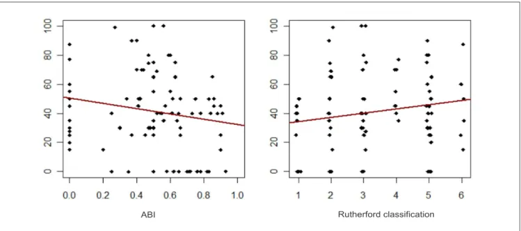
This study is characterized as a cros-sectional, prospective epidemiological one. We used univariate analysis to select potential predictors of severity of carotid artery stenosis by evaluating: the degree of carotid artery stenosis on a scale ranging from 0% (no stenosis) to 100% (occlusion), not accounting for the subgroups of stenosis degrees. For these calculations we employed the Mann-Whitney test and Spearman coefficient, used to evaluate the influence of ABI and classification of clinical severity (Rutherford classification).
We used the chi-square and Mann-Whitney tests and, where necessary, the Chi-Square was replaced by Fisher’s exact test to assess carotid stenosis. Patients were separated into two subgroups of stenosis degrees, one less than 50% (not significant stenosis) and another above 50% (significant stenosis). To select among the potential predictors in multiple regression, we used the backward algorithm. For the evaluation of the carotid treated in its original scale, without subdivision into groups of grades of stenosis, we employed the method of Quasi-Likelihood. We used logistic regression for the carotid evaluation in subgroups.
Since the objective of the study was to test the correlation between carotid stenosis and peripheral artery disease, and given a significance level of 0.05 for a two-tailed test with a medium effect size and a power of 80%, the sample should be composed of approximately 85 individuals (Figure 1).
RESULTS
RESULTS
RESULTS
RESULTS
RESULTS
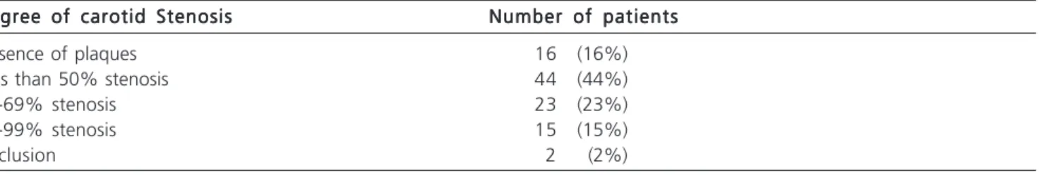
The prevalence of carotid stenosis was 84%, being significant in 40% of patients and severe in 17%. Absence of plaque in the carotid arteries was found in 16%. Two patients had unilateral internal carotid occlusion; one had a history of stroke two years before and the other was asymptomatic.
Regarding the classification of Rutherford, 50% of patients had grades 1, 2 or 3 (claudicating) and 50% of patients had grades 4, 5 or 6 (patients with rest pain or ulceration, i.e., critical ischemia). The ABI measurement showed an average of 0.58, with 50% of the patients with less than 0.56.
We note that 86% of the patients had hypertension, 66% exposure to smoke, 47% diabetes, 65% dyslipidemia, 24% coronary artery disease, 16% renal failure and 60% had family history of cardiovascular disease (Table 2). A history of vascular surgery was found in 48% of the patients, with a predominance of lower limb revascularization, performed in 34 patients (34%), and coronary artery bypass grafting (CABG) in ten (10% ).
Seven patients (7%) had a history of some prior ischemic cerebrovascular symptoms in the carotid territory,
two of them (2%) TIA and five patients (5%) stroke. In all these patients, this story had occurred more than two years before.
Patients were classified according to the arterial territory affecting the lower limbs, with findings of femoropopliteal disease in 63%, Infrapopliteal in 43% and aortoiliac in 26%.
After the diagnosis of carotid stenosis we evaluated the approach to to this finding. In 76% of patients observation was chosen, and 24% held some additional procedure. Of the latter, 15 were submitted only to more exams (new duplex-scan, angiography or magnetic resonance angiography) and nine patients underwent intervention in the carotid arteries, of which three were carotid endarterectomies and six were carotid angioplasties. All patients undergoing carotid endarterectomy or angioplasty had carotid stenosis higher or equal to 70% (severe stenosis). These patients had no complications postoperatively. Variables that significantly influenced the degree of stenosis of the carotid arteries showed a pd”0.25, being selected for multivariate analysis: prior symptomatic cerebrovascular disease, dyslipidemia, reduced motor activity, age, coronary artery disease, renal failure (Table 3) .
There was a negative correlation, although not significant (p = 0.073) between the ABI and the overall evaluation of carotid stenosis. The correlation between the assessment of carotid and the classification of Rutherford, on its turn, is positive, but also not significant (p = 0.110) (Figure 2).
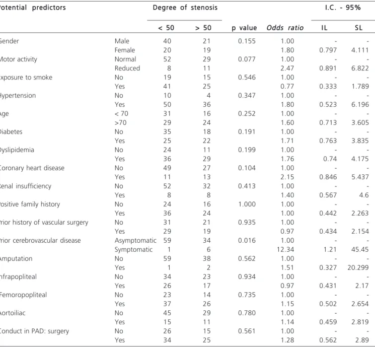
The following variables were selected as potential predictors of significant carotid stenosis (pd”0.25): reduced motor activity, history of previous symptomatic cerebrovascular disease, gender, age, diabetes, dyslipidemia and coronary artery disease (Table 4).
In multivariate analysis with the previously selected variables, only the variable previous symptomatic cerebrovascular disease remained in the final regression to globally explain the variation of carotid stenosis. The presence of prior symptomatic cerebrovascular disease increases in 30.98 the average value of carotid stenosis, with p = 0.0231.
In the assessment of carotid stenosis in groups of significant injuries and non-significant ones, only the variables prior symptomatic cerebrovascular disease and gender were retained in the final regression to explain the
Table 1 Table 1 Table 1 Table 1
Table 1 - Distribution of patients according to the degree of carotid artery stenosis.
Degree of carotid Stenosis Degree of carotid Stenosis Degree of carotid Stenosis Degree of carotid Stenosis
Degree of carotid Stenosis Number of patientsNumber of patientsNumber of patientsNumber of patientsNumber of patients
Absence of plaques 16 (16%)
Less than 50% stenosis 44 (44%)
50-69% stenosis 23 (23%)
70-99% stenosis 15 (15%)
Occlusion 2 (2%)
Figure 1 Figure 1 Figure 1 Figure 1
variation in evaluation of carotid stenosis. The results show that the chance of a female patient have an assessment of significant carotid stenosis is 2.28 (0.92 to 5.68) times the odds of male patients, but not statistically significant (p = 0.0766). The odds of a patient with prior symptomatic cerebrovascular disease present with a carotid stenosis considered significant is approximately 12.34 (1.47 to 125.0) times the chance of patients with asymptomatic disease, with a p value of 0.05.
DISCUSSION
DISCUSSION
DISCUSSION
DISCUSSION
DISCUSSION
This study sought to describe the clinical characteristics, risk factors and the association with carotid stenosis in a sample of patients with symptomatic peripheral
arterial disease treated by a reference service in Angiology and Vascular Surgery.
This is an unprecedented work in Brazil, since similar studies have not been found after research conducted in the SciELO and LILACS databases. The study of carotid artery disease and peripheral arterial disease are topics of great relevance, since stroke is currently the leading cause of death in Brazil and peripheral arterial disease is responsible for thousands of amputations 3-5.
Our results show a 84% prevalence of carotid stenosis in the sample, being significant in 40% of patients. Previous studies reported a prevalence of significant carotid stenosis ranging from 14.3% to 37.2%, but the samples of patients with peripheral arterial disease are different between the works, and in many of them there are only asymptomatic and claudicating patients 8,9,19,20. The higher
Table 2 Table 2 Table 2 Table 2
Table 2 - Distribution of the number of patients about the risk factors and associated diseases
V a r i a b l e s V a r i a b l e sV a r i a b l e s V a r i a b l e s
V a r i a b l e s N oN oN oN oN o Y e sY e sY e sY e sY e s
A F A FA F A F
A F A FA FA FA FA F
Hypertension 14 86
Exposure to smoke 34 66
Diabetes 53 47
Dyslipidemia 35 65
Coronary disease 76 24
Renal insufficiency 84 16
Family history of atherosclerosis 40 60
Prior history of vascular surgery 52 48
Previous cerebrovascular disease: asymptomatic 7 93
Amputation 97 3
AF: absolute frequency
Figure 2 Figure 2 Figure 2 Figure 2
prevalence of carotid stenosis found in our sample can be explained by the severity of the patients’ peripheral arterial disease, since half of the them had critical ischemia of the lower limbs, indicating greater arterial involvement.
As for the studied risk factors for atherosclerosis, the results are consistent with published data, indicating an association with hypertension, smoking, diabetes, dyslipidemia, coronary artery disease, positive family history and chronic renal failure 2. Dyslipidemia was statistically
significant for the presence of carotid stenosis in univariate analysis (p = 0.0497). None of these factors, however, was statistically significant for the severity of carotid stenosis after multivariate analysis and logistic regression. This may be explained by the fact that both factors have a high prevalence both in patients with peripheral arterial disease and in patients with carotid stenosis, indicating that these are potential markers of systemic atherosclerosis. The identification of modifiable risk factors configures an
important part of treatment. In all study patients, once risk factors and associated comorbidities were identified, medical therapy was initiated. The use of antiplatelet agents, statins, smoking cessation and controlling blood pressure and diabetes are essential to prevent stroke 21.
The average age of patients was 69.78 years, also indicating a direct relationship of arterial involvement with the elder population, in accordance to previously published work22. Distribution by age groups also revealed
a predominance of older patients, with 80% of individuals in the series with greater than or equal to 60 years of age. There was a predominance of male patients in our study, the proportion of about three men to two women, also compatible with the publications found in the literature, in which it is evident that atherosclerosis predominates in males 2. Our results show, however , an interesting finding
after multivariate analysis logistic regression. The females showed a trend (p = 0.0759) for significant carotid stenosis.
Table 3 Table 3 Table 3 Table 3
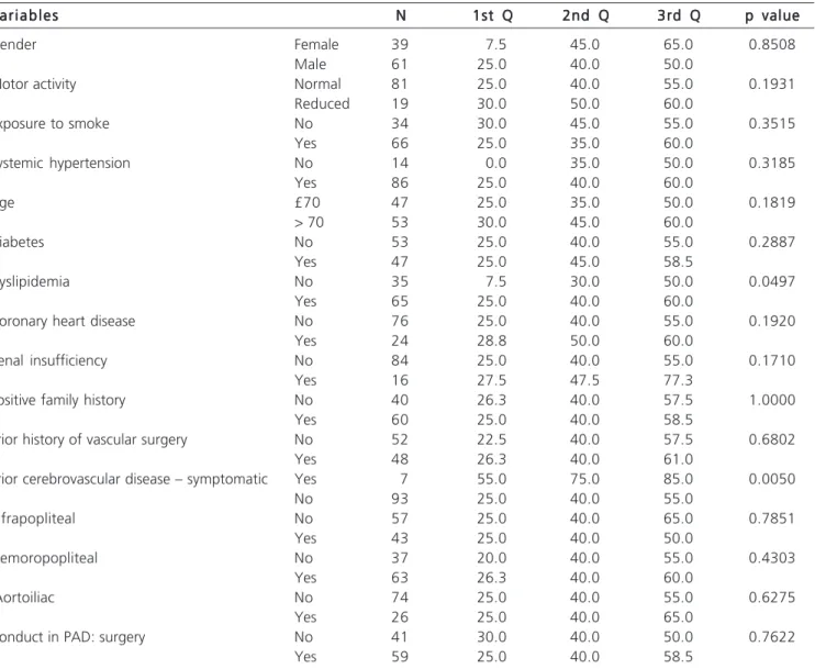
Table 3 - Descriptive measures and Mann-Whitney test for carotid stenosis and the variables studied.
V a r i a b l e s V a r i a b l e s V a r i a b l e s V a r i a b l e s
V a r i a b l e s NNNNN 1st Q1st Q1st Q1st Q1st Q 2nd Q2nd Q2nd Q2nd Q2nd Q 3rd Q3rd Q3rd Q3rd Q3rd Q p valuep valuep valuep valuep value
Gender Female 39 7.5 45.0 65.0 0.8508
Male 61 25.0 40.0 50.0
Motor activity Normal 81 25.0 40.0 55.0 0.1931
Reduced 19 30.0 50.0 60.0
Exposure to smoke No 34 30.0 45.0 55.0 0.3515
Yes 66 25.0 35.0 60.0
Systemic hypertension No 14 0.0 35.0 50.0 0.3185
Yes 86 25.0 40.0 60.0
Age £70 47 25.0 35.0 50.0 0.1819
> 70 53 30.0 45.0 60.0
Diabetes No 53 25.0 40.0 55.0 0.2887
Yes 47 25.0 45.0 58.5
Dyslipidemia No 35 7.5 30.0 50.0 0.0497
Yes 65 25.0 40.0 60.0
Coronary heart disease No 76 25.0 40.0 55.0 0.1920
Yes 24 28.8 50.0 60.0
Renal insufficiency No 84 25.0 40.0 55.0 0.1710
Yes 16 27.5 47.5 77.3
Positive family history No 40 26.3 40.0 57.5 1.0000
Yes 60 25.0 40.0 58.5
Prior history of vascular surgery No 52 22.5 40.0 57.5 0.6802
Yes 48 26.3 40.0 61.0
Prior cerebrovascular disease – symptomatic Yes 7 55.0 75.0 85.0 0.0050
No 93 25.0 40.0 55.0
Infrapopliteal No 57 25.0 40.0 65.0 0.7851
Yes 43 25.0 40.0 50.0
Femoropopliteal No 37 20.0 40.0 55.0 0.4303
Yes 63 26.3 40.0 60.0
Aortoiliac No 74 25.0 40.0 55.0 0.6275
Yes 26 25.0 40.0 65.0
Conduct in PAD: surgery No 41 30.0 40.0 50.0 0.7622
Yes 59 25.0 40.0 58.5
A previous publication has shown the female gender as a risk factor for the progression of carotid stenosis in patients with peripheral arterial disease 9. Other studies
have shown that the female sex is also an independent risk factor for significant carotid stenosis, but in a population of coronary patients in preoperative of surgical myocardial revascularization 23. The female gender
appears to be a risk factor for neurological complications after carotid angioplasty and restenosis after carotid endarterectomy 24-26.
The study of the variables ankle-brachial index (ABI) and classification of Rutherford showed a relationship between severity of peripheral artery disease and severity
of carotid stenosis. Despite the non-significant values found in the statistical analysis, there was a tendency towards lower measures of ABI and higher values in the classification of Rutherford, featuring a more severe peripheral arterial disease, and findings of more pronounced carotid stenosis. These findings are consistent with published data indicating that there is direct association between the severity of peripheral arterial disease and degree of carotid stenosis 19,27.
Patients were studied regarding the arterial territory of the affected lower limb. There was a predominance of the femoropopliteal segment, almost two thirds of patients, followed by the aortoiliac and infrapopliteal
Table 4 Table 4 Table 4 Table 4
Table 4 - Distribution of patients into two groups according to the degree of carotid stenosis and the variables studied.
Potential predictors Potential predictorsPotential predictors Potential predictors
Potential predictors Degree of stenosisDegree of stenosisDegree of stenosisDegree of stenosisDegree of stenosis I.C. - 95%I.C. - 95%I.C. - 95%I.C. - 95%I.C. - 95%
< 50 < 50 < 50 < 50
< 50 >>>> 50> 50 50 50 50 p valuep valuep valuep valuep value Odds ratioOdds ratioOdds ratioOdds ratioOdds ratio I LI LI LI LI L S LS LS LS LS L
Gender Male 40 21 0.155 1.00 -
-Female 20 19 1.80 0.797 4.111
Motor activity Normal 52 29 0.077 1.00 -
-Reduced 8 11 2.47 0.891 6.822
Exposure to smoke No 19 15 0.546 1.00 -
-Yes 41 25 0.77 0.333 1.789
Hypertension No 10 4 0.347 1.00 -
-Yes 50 36 1.80 0.523 6.196
Age < 70 31 16 0.252 1.00 -
->70 29 24 1.60 0.713 3.605
Diabetes No 35 18 0.191 1.00 -
-Yes 25 22 1.71 0.763 3.835
Dyslipidemia No 24 11 0.199 1.00 -
-Yes 36 29 1.76 0.74 4.175
Coronary heart disease No 49 27 0.104 1.00 -
-Yes 11 13 2.15 0.846 5.437
Renal insufficiency No 52 32 0.413 1.00 -
-Yes 8 8 1.40 0.567 4.6
Positive family history No 24 16 1.000 1.00 -
-Yes 36 24 1.00 0.442 2.263
Prior history of vascular surgery No 31 21 0.935 1.00 -
-Yes 29 19 0.97 0.434 2.154
Prior cerebrovascular disease Asymptomatic 59 34 0.016 1.00 -
-Symptomatic 1 6 12.34 1.21 45.45
Amputation No 59 38 0.562 1.00 -
-Yes 1 2 1.51 0.327 20.299
Infrapopliteal No 34 23 0.934 1.00 -
-Yes 26 17 0.97 0.431 2.17
Femoropopliteal No 23 14 0.735 1.00 -
-Yes 37 26 1.15 0.502 2.654
Aortoiliac No 45 29 0.780 1.00 -
-Yes 15 11 1.14 0.459 2.819
Conduct in PAD: surgery No 26 15 0.561 1.00 -
-Yes 34 25 1.28 0.562 2.89
territory. However, there was no statistically significant relationship between these variables and the involvement of the carotid arteries.
An important point to be emphasized concerns the approach to the patient after the duplex scan examination of the carotid. There was change in the conduct, which initially would be expectant without the prior diagnosis of carotid stenosis, in 24% of the patients. In these, the finding of stenosis was considered so relevant as to requested a more detailed study of the degree of stenosis. In 15% of patients, only additional exams were performed, without intervention. In 9%, carotid intervention was held according to the indication criteria established in large randomized studies 10,15,17,28. These
intervention patients showed carotid stenosis greater than 70%, and were indicated a treatment with level 1 scientific evidence for prevention of cerebrovascular accident. It should be noted that the required number of patients to be operated to prevent a single stroke accident in five years is high, about six endarterectomies in symptomatic patients and 17 in asymptomatic ones 29,30,
in services with low rates of postoperative neurological complications. Even for the non-operated patients we initiated the appropriate clinical treatment, currently considered the first option for treatment of asymptomatic patients by some authors 21. Both the diagnosis and the
indicated treatment would not have been carried if the
examination of the carotid arteries was not requested. Early diagnosis and treatment of carotid stenosis are of utmost importance for the prevention of stroke, as already emphasized 11.
The presence of prior neurological symptoms related to the carotid territory was also statistically significant for the presence of carotid stenosis after logistic regression and multivariate analysis. It should be emphasized, however, that the patients in this sample classified as having a history of neurological symptoms had presented with these at least more than two years before, ie, they had no recent history of neurological symptoms, which might not motivate the request of a carotid echo-color-doppler study. In the NASCET study 30 patients were considered symptomatic only if they
had neurological symptoms in the carotid territory in the past six months.
Patients with symptomatic peripheral arterial disease display a high prevalence of significant carotid stenosis (40%). There was a trend in the association between severity of peripheral artery disease and severity of carotid stenosis, although not significant. Previous history of neurological symptoms was statistically significant in explaining the severity of carotid stenosis. In conclusion, the study of the carotid arteries by duplex scan examination is of paramount importance in the evaluation of patients with symptomatic peripheral arterial disease and should be systematically indicated to such patients.
R E S U M O R E S U M O R E S U M O R E S U M O R E S U M O
Objetivo: Objetivo: Objetivo: Objetivo:
Objetivo: estudar estenose das artérias carótidas nos pacientes com doença arterial periférica sintomática. Métodos:Métodos:Métodos:Métodos:Métodos: avaliaram-se conavaliaram-secutivamente 100 portadores de doença arterial periférica sintomática, nos estágios de claudicação intermitente, dor em repouso ou lesão trófica. A estenose carotídea foi estudada pelo eco-color-doppler, sendo considerada significativa quando maior ou igual a 50%. A análise univariada foi utilizada para selecionar os potenciais preditores de estenose carotídea, levados posteriormente para análise multivariada. Resultados:Resultados:Resultados:Resultados:Resultados: a prevalência de estenose carotídea foi 84%, sendo significativa em 40% e acentuada em 17%. A idade variou de 43 a 89 anos (média de 69,78). Quanto ao sexo, 61% foram do sexo masculino e 39% do feminino. Metade dos pacientes da amostra era claudicante e metade tinha isquemia crítica. Quanto aos fatores de risco, 86% dos pacientes apresentaram hipertensão arterial sistêmica, 66% exposição ao fumo, 47% diabetes, 65% dislipidemia, 24% coronariopatia, 16% insuficiência renal e 60% história familiar positiva para doenças cardiovasculares. Em sete pacientes, havia história de alguma sintomatologia cérebro-vascular isquêmica no território carotídeo. A presença de sintomatologia cérebro-vascular mostrou-se estatisticamente significativa para influenciar o grau de estenose nas artérias carótidas (p=0,02 na avaliação global e p=0,05 nos subgrupos de estenoses significativas e não significativas). Conclusão:Conclusão:Conclusão:Conclusão: o estudo das artérias carótidas através do exame de duplex-Conclusão: scan é de suma importância na avaliação dos pacientes portadores de doença arterial periférica sintomática, devendo-se realizar o estudo de forma sistemática nos pacientes.
Descritores: Descritores: Descritores: Descritores:
Descritores: Artérias carótidas. Estenose das carótidas. Doença arterial periférica. Fatores de risco.
REFERENCES
REFERENCES
REFERENCES
REFERENCES
REFERENCES
1. Jotkowitz AB, Mark Clarfield A, Faust G, Wartman SA. Screening for carotid artery disease in the general public. Eur J Intern Med. 2005;16(1):34-6.
2. Alzamora MT, Forés R, Baena-Díez JM, Pera G, Toran P, Sorribes M, et al. The peripheral arterial disease study (PERART/ARTPER): prevalence and risk factors in the general population. BMC Public Health. 2010;10:38.
3. Lotufo PA, Goulart AC, Fernandes TG, Benseñor IM. A reappraisal of stroke mortality trends in Brazil (1979-2009). Int J Stroke. 2013;8(3):155-63.
4. Goulart AC, Bustos IR, Abe IM, Pereira AC, Fedeli LM, Benseñor IM, et al. A stepwise approach to stroke surveillance in Brazil: the EMMA (Estudo de Mortalidade e Morbidade do Acidente Vascular Cerebral) study. Int J Stroke. 2010;5(4):284-9.
6. Brasil. Ministério da Saúde. DATASUS. Sistema de Informações Hospitalares do SUS (SIH/SUS). Disponível em: http:// www2.datasus.gov.br/DATASUS/index.php.
7. Ankle Brachial Index Collaboration, Fowkes FG, Murray GD, Butcher I, Heald CL, Lee RJ, et al. Ankle brachial index combined with Framingham Risk Score to predict cardiovascular events and mortality: a meta-analysis. JAMA. 2008;300(2):197-208. 8. Brevetti G, Sirico G, Lanero S, De Maio JI, Laurenzano E, Giugliano
G. The prevalence of hypoechoic carotid plaques is greater in peripheral than in coronary artery disease and is related to the neutrophil count. J Vasc Surg. 2008;47(3):523-9.
9. Jahromi AS, Clase CM, Maggisano R, Bailey R, Safar HA, Cinà CS. Progression of internal carotid artery stenosis in patients with peripheral arterial occlusive disease. J Vasc Surg. 2009;50(2):292-8.
10. Jayasooriya G, Thapar A, Shalhoub J, Davies AH. Silent cerebral events in asymptomatic carotid stenosis. J Vasc Surg. 2011;54(1):227-36.
11. Weinberger J. Prevention of ischemic stroke. Curr Cardiol Rep. 2002;4(2):164-71.
12. North American Symptomatic Carotid Endarterectomy Trial Collaborators. Beneficial effect of carotid endarterectomy in symptomatic patients with high-grade carotid stenosis. N Engl J Med. 1991;325(7):445-53.
13. Randomised trial of endarterectomy for recently symptomatic carotid stenosis: final results of the MRC European Carotid Surgery Trial (ECST). Lancet. 1998;351(9113):1379-87.
14. Halliday A, Mansfield A, Marro J, Peto C, Peto R, Potter J, et al. Prevention of disabling and fatal strokes by successful carotid endarterectomy in patients without recent neurological symptoms: randomised controlled trial. Lancet. 2004;363(9420):1491-502. 15. Chambers BR, Donnan GA. Carotid endarterectomy for
asymptomatic carotid stenosis. Cochrane Database Syst Rev. 2005(4):CD001923.
16. Endarterectomy for asymtomatic carotid artery stenosis. Executive Committee for the Asymptomatic Carotid Atherosclerosis Study. JAMA. 1995;273:1421-8.
17. Halliday A, Harrison M, Hayter E, Kong X, Mansfield A, Marro J, et al. 10-year stroke prevention after successful carotid endarterectomy for asymptomatic stenosis (ACST-1): a multicentre randomised trial. Lancet. 2010;376(9746):1074-84.
18. Grant EG, Benson CB, Moneta GL, Alexandrov AV, Baker JD, Bluth EI, et al. Carotid artery stenosis: gray-scale and Doppler US diagnosis—Society of Radiologists in Ultrasound Consensus Conference. Radiology. 2003;229(2):340-6.
19. Goessens BM, Visseren FL, Algra A, Banga JD, van der Graaf Y, SMART Study Group. Screening for asymptomatic cardiovascular disease with noninvasive imaging in patients at high-risk and low-risk according to the European Guidelines on Cardiovascular Disease
Prevention: the SMART study. J Vasc Surg. 2006;43(3):525-32. 20. Mostaza JM, González-Juanatey JR, Castillo J, Lahoz C,
Fernández-Villaverde JM, Maestro-Saavedra FJ. Prevalence of carotid stenosis and silent myocardial ischemia in asymptomatic subjects with a low ankle-brachial index. J Vasc Surg. 2009;49(1):104-8. 21. Naylor AR. Time to rethink management strategies in asymptomatic
carotid artery disease. Nat Rev Cardiol. 2011;9(2):116-24. 22. Casella IB, Sotelo FJ, Yamazaki Y, Presti C, Vassoler A, Melo HA.
Comparison of common carotid artery intima-media thickness between Brazilian Euro-descendants and Afro-descendants with atherosclerosis risk factors. Clinics. 2009;64(7):657-64.
23. Shirani S, Boroumand MA, Abbasi SH, Maghsoodi N, Shakiba M, Karimi A, et al. Preoperative carotid artery screening in patients undergoing coronary artery bypass graft surgery. Arch Med Res. 2006;37(8):987-90.
24. Yun WS, Kwun WH, Suh BY. The early and mid-term results of carotid artery stenting in high-risk patients. J Korean Surg Soc. 2011;80(4):283-8.
25. Micari A, Stabile E, Cremonesi A, Vadalà G, Castriota F, Pernice V, et al. Carotid artery stenting in octogenarians using a proximal endovascular occlusion cerebral protection device: a multicenter registry. Catheter Cardiovasc Interv. 2010;76(1):9-15.
26. Crawford RS, Chung TK, Hodgman T, Pedraza JD, Corey M, Cambria RP. Restenosis after eversion vs patch closure carotid endarterectomy. J Vasc Surg. 2007;46(1):41-8.
27. Busch MA, Lutz K, Röhl JE, Neuner B, Masuhr F. Low ankle-brachial index predicts cardiovascular risk after acute ischemic stroke or transient ischemic attack. Stroke. 2009;40(12):3700-5.
28. Liapis CD, Bell PR, Mikhailidis D, Sivenius J, Nicolaides A, Fernandes e Fernandes J, et al. ESVS guidelines. Invasive treatment for carotid stenosis: indications, techniques. Eur J Vasc Endovasc Surg. 2009;37(4 Suppl):1-19.
29. Rothwell PM, Eliasziw M, Gutnikov SA, Warlow CP, Barnett HJ; Carotid Endarterectomy Trialists Collaboration. Endarterectomy for symptomatic carotid stenosis in relation to clinical subgroups and timing of surgery. Lancet. 2004;363(9413):915-24. 30. Inzitari D, Eliasziw M, Gates P, Sharpe BL, Chan RK, Meldrum HE,
et al. The causes and risk of stroke in patients with asymptomatic internal-carotid-artery stenosis. North American Symptomatic Carotid Endarterectomy Trial Collaborators. N Engl J Med. 2000;342(23):1693-700
Received on 15/10/2013
Accepted for publication 09/01/2014 Conflict of interest: none.
Source of funding: none.