Hypo m agne se m ia in critically ill
cance r patie nts: a pro spe ctive study
o f pre dictive facto rs
1Unidade de Terapia Intensiva, Departamentos de
2Análises Clínicas e Hemoterapia and 3Epidemiologia e Bioestatística,
Centro de Tratamento e Pesquisa, Hospital do Câncer, São Paulo, SP, Brasil D. Deheinzelin1,
E.M. Negri1, M.R. Tucci1,
M.Z. Salem1, V.M. da Cruz1,
R.M. O liveira2,
I.N. Nishimoto3
and C. Hoelz1
Abstract
Hypomagnesemia is the most common electrolyte disturbance seen upon admission to the intensive care unit (ICU). Reliable predictors of its occurrence are not described. The objective of this prospective study was to determine factors predictive of hypomagnesemia upon admission to the ICU. In a single tertiary cancer center,226 patients with different diagnoses upon entering were studied. Hypomagnese-mia was defined by serum levels <1.5 mg/dl. Demographic data, type of cancer, cause of admission, previous history of arrhythmia, cardio-vascular disease, renal failure, drug administration (particularly di-uretics, antiarrhythmics, chemotherapy and platinum compounds), previous nutrition intake and presence of hypovolemia were recorded for each patient. Blood was collected for determination of serum magnesium, potassium, sodium, calcium, phosphorus, blood urea nitrogen and creatinine levels. Upon admission, 103 (45.6%) patients had hypomagnesemia and 123 (54.4%) had normomagnesemia. A normal dietary habit prior to ICU admission was associated with normal Mg levels (P = 0.007) and higher average levels of serum Mg (P = 0.002). Postoperative patients (N = 182) had lower levels of serum Mg (0.60 ± 0.14 mmol/l compared with 0.66 ± 0.17 mmol/l, P = 0.006). A stepwise multiple linear regression disclosed that only normal dietary habits (OR = 0.45; CI = 0.26-0.79) and the fact of being a postoperative patient (OR = 2.42; CI = 1.17-4.98) were significantly correlated with serum Mg levels (overall model probability = 0.001). These findings should be used to identify patients at risk for such disturbance, even in other critically ill populations.
Co rre spo nde nce
D. Deheinzelin
Centro de Tratamento e Pesquisa Hospital do Câncer
Rua Prof. Antonio Prudente, 211 9º andar, UTI
01509-010 São Paulo, SP Brasil
Fax: + 55-11-3845-7457 E-mail: ddeheinz@ uol.com.br
Publication supported by FAPESP.
Received December 13, 1999 Accepted August 9, 2000
Ke y wo rds
·Magnesium
·Magnesium deficiency ·Intensive care unit ·Electrolytes ·Surgery ·Dietary habits ·Postoperative patient
Intro ductio n
Hypomagnesemia is a common finding in current medical practice, mainly in criti-cally ill and postoperative patients. Upon admission to the intensive care unit (ICU), the prevalence of this electrolyte disorder
morbidity of such patients. The etiology of magnesium deficiencies includes gastrointes-tinal and renal wasting, drug-induced loss, endocrine disorders, metabolic diseases, re-distribution of magnesium stores and other conditions (5).
The causes of hypomagnesemia found upon admission to an ICU have not been previously studied. The objective of the pre-sent study was to investigate prospectively the incidence of hypomagnesemia in criti-cally ill cancer patients and to determine predictive factors associated with such find-ing. Upon admission to the ICU, this popula-tion is expected to have electrolyte
distur-bances, not only because of the underlying disease, but also as a result of specific treat-ment regimens. The homogeneity of the popu-lation under study, admitted to the hospital with cancer as the primary cause, permitted analysis of the possible factors involved in this electrolyte disorder in these patients.
Me tho ds
Po pulatio n
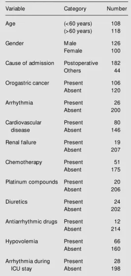
A total of 226 consecutive patients ad-mitted to the Critical Care Unit of the Hospi-tal do Câncer A.C. Camargo, São Paulo, SP, Brazil, were studied. The patient population consisted of 126 males (56%) and 100 fe-males (44%); 108 were under 60 years of age (48%) and 118 were 60 years old or over (52%). One hundred and eighty-two were postoperative patients (80%) and 44 were medically treated patients (20%). Popula-tion characteristics are detailed in Table 1. Most medical admissions were due to acute respiratory failure, acute renal failure, se-vere hypotension, sepsis, and cardiac ar-rhythmias. In all patients cancer was a pri-mary active diagnosis.
Study de sign
The Committee for Human Studies of the Cancer Hospital approved the following pro-tocol. Prior to admission to the ICU no pa-tient had a record of hypomagnesemia. Upon entering the ICU, demographic data (age, gender), type of cancer, cause of admission, previous history of arrhythmia, cardiovascu-lar disease or renal failure were recorded for each patient. Medications administered (par-ticularly diuretics, antiarrhythmics, chemo-therapy and previous use of platinum com-pounds) and types of nutritional support dur-ing the last 48 h before admission (normal habits, enteral support, parenteral support or fasting) were also recorded. On admission, special attention was paid to the presence of Table 1 - Population characteristics before
admis-sion and during permanence in the ICU.
Variable Category Number
Age (<60 years) 108
(>60 years) 118
Gender M ale 126
Female 100
Cause of admission Postoperative 182
Others 44
Orogastric cancer Present 106
Absent 120
Arrhythmia Present 26
Absent 200
Cardiovascular Present 80
disease Absent 146
Renal failure Present 19
Absent 207
Chemotherapy Present 51
Absent 175
Platinum compounds Present 20
Absent 206
Diuretics Present 24
Absent 202
Antiarrhythmic drugs Present 12
Absent 214
Hypovolemia Present 66
Absent 160
Arrhythmia during Present 28
hypovolemia (defined by skin turgor, mu-cosal dryness, oliguria or low central venous pressure when recorded).
Upon admission to the ICU, a blood sample was collected for serum magnesium determination by a calorimetric method us-ing chlorophosphanazo III (Cobas-Íntergra; Roche, Kaiser August, Switzerland). Potas-sium, sodium, calcium, phosphorus, blood urea nitrogen (BUN) and creatinine were also measured. Samples were run in dupli-cate.
When hypomagnesemia was found, mag-nesium sulfate was administered intrave-nously until normal levels were restored. After correction of magnesium levels, ar-rhythmia events were recorded until dis-charge from the ICU.
D e finitio ns
In our laboratory, normal magnesium concentration is 0.62 to 1.03 mmol/l, and hypomagnesemia is, therefore, defined as a serum magnesium level of 0.61 mmol/l or less; hypermagnesemia is defined as 1.04 mmol/l or more.
Statistical analysis
One-way ANOVA was used for analysis of continuous variables. For categorical vari-ables, serum magnesium levels were divided into low or normal levels and the chi-square test was used for comparison. Logistic gression was used to identify variables re-lated to hypomagnesemia. The STATA 6.0 software package was used for statistical analysis and the level of significance was set at 0.05.
Re sults
Upon admission to the ICU, 103 (45.6%) of the 226 patients studied had hypomagne-semia and 123 (54.4%) had normomagnese-mia. Hypermagnesemia was not detected in
any patient. Table 2 shows serum magne-sium levels and the levels of other electro-lytes obtained upon admission.
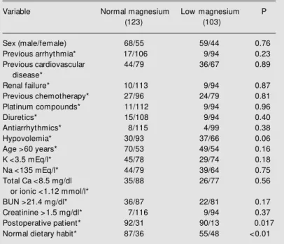
No correlation was found between mag-nesium concentration and age (P = 0.16), sex (P = 0.76), previous renal failure (P = 0.47), cardiovascular disease (P = 0.57),
chemo-Table 3 - Univariate analysis comparing patients w ith low and normal serum magnesium levels according to the different clinical variables studied (chi-square test).
* Yes/no. BUN, Blood urea nitrogen.
Variable Normal magnesium Low magnesium P
(123) (103)
Sex (male/female) 68/55 59/44 0.76
Previous arrhythmia* 17/106 9/94 0.23
Previous cardiovascular 44/79 36/67 0.89
disease*
Renal failure* 10/113 9/94 0.87
Previous chemotherapy* 27/96 24/79 0.81
Platinum compounds* 11/112 9/94 0.96
Diuretics* 15/108 9/94 0.40
Antiarrhythmics* 8/115 4/99 0.38
Hypovolemia* 30/93 37/66 0.06
Age >60 years* 70/53 49/54 0.16
K <3.5 mEq/l* 45/78 29/74 0.18
Na <135 mEq/l* 44/79 39/64 0.75
Total Ca <8.5 mg/dl 35/88 26/77 0.56
or ionic <1.12 mmol/l*
BUN >21.4 mg/dl* 36/87 22/81 0.17
Creatinine >1.5 mg/dl* 7/116 9/94 0.37
Postoperative patient* 92/31 90/13 0.017
Normal dietary habit* 87/36 55/48 <0.01
Table 2 - Serum electrolyte levels obtained upon admission to the intensive care unit, defined as low , normal or high.
Absolute (percentage). Definitions: M agnesium, low <0.61 mmol/l; potassium, low <3.5 mEq/l; sodium, low <135 mEq/l; calcium, low total Ca <8.5 mg/dl or ionic <1.12 mmol/l; phosphorus, low <2.5 mg/dl; creatinine, low <0.8 mg/dl and high >1.5 mg/dl; blood urea nitrogen (BUN), low <7.8 mg/dl and high >21.4 mg/dl.
Low Normal High
M agnesium 103 (45.6% ) 123 (54.4% )
Potassium 75 (33.0% ) 151 (67.0% )
Sodium 76 (33.6% ) 149 (66.4% )
Calcium 29 (12.8% ) 196 (87.2% )
Phosphorus 36 (16.5% ) 187 (83.5% )
Creatinine 122 (53.8% ) 88 (39.1% ) 16 (7.1% )
therapy (P = 0.80), use of platinum (P = 0.96), diuretics (P = 0.40) or antiarrhythmics (P = 0.38). The overall results are shown in Table 3.
There was no correlation between serum magnesium levels and serum potassium lev-els (r2 = 0.06, P = 0.34) or serum sodium levels (r2 = -0.04, P = 0.53). A significant, but weak correlation was found between serum magnesium levels and calcium levels (r2 = 0.295, P = 0.005), BUN levels (r2 = 0.208, P = 0.0016) and creatinine levels (r2 = 0.162, P = 0.01).
Serum magnesium levels varied accord-ing to previous nutritional support. A normal dietary intake prior to ICU admission was associated with normal magnesium levels (P = 0.007) and the average levels of serum magnesium in this group were higher than those of patients receiving other forms of nutritional support (P = 0.002). Because of this correlation, we decided to investigate if cancer itself influenced this finding. After selecting cancer cases affecting the gas-trointestinal tract and therefore impairing diet we found that, as expected, normal di-etary intake was more frequent in patients with no orogastric cancer (only 48% of pa-tients with orogastric cancer had a normal dietary intake compared to 78% of patients with other types of cancer; P<0.001). How-ever, orogastric cancer was not significantly more common in patients with low serum magnesium level (P = 0.65), nor related to absolute values of serum magnesium.
Postoperative patients also had lower lev-els of serum magnesium (0.60 ± 0.14 mmol/ l vs 0.66 ± 0.17 mmol/l, P = 0.006).
After administration of MgSO4 in order to normalize the magnesium balance, the incidence of cardiac arrhythmias in the en-tire group was 12.39% (28 of 226 patients) and was correlated only with previous car-diovascular disease (P<0.001, chi-square test). With regard to the incidence of ar-rhythmia, there was no difference between patients presenting with hypomagnesemia
or with normomagnesemia.
A stepwise multiple linear regression dis-closed that only normal dietary habit (OR = 0.45; CI = 0.26-0.79) and the fact of being a postoperative patient (OR = 2.42; CI = 1.17-4.98) were significantly correlated with se-rum magnesium levels (overall model prob-ability = 0.001).
D iscussio n
Magnesium is an essential element in many body functions. It plays a major role as a co-factor for adenosine triphosphatases and therefore is directly involved in impor-tant processes that require energy and also in protein synthesis mechanisms (6). In such a scenario, it is easy to predict the serious consequences brought about by magnesium deficiencies.
Magnesium is found in a large variety of foods, especially in green vegetables, nuts, grains, meat and seafood (6). In normal situ-ations the kidney and the small intestine control magnesium excretion and its reab-sorption, respectively, and serum levels are maintained within a normal range (5). Nev-ertheless, it is important to observe that se-rum levels of magnesium even remain nor-mal in the presence of intracellular magne-sium depletion and that low serum levels indicate severe magnesium deficiency (3).
Hypomagnesemia is a common finding in hospitalized patients (2) and it may basi-cally be related to drug-induced gastrointes-tinal or renal losses (diuretics, aminoglyco-sides, etc.), pancreatitis, chronic diarrhea and inadequate intake (7). In this prospec-tive study, we found that 46% of the patients admitted to an ICU in a tertiary cancer center presented hypomagnesemia. This finding agrees with previous reports showing a 61% incidence of hypomagnesemia in a postop-erative ICU (1) or an incidence ranging from 20 to 45.7% in patients admitted to a medical ICU (2-4,8).
to hypomagnesemia for patients in a critical care setting, such as cardiac arrhythmias, respiratory muscle weakness and seizures (4), the most intriguing finding is that an imbalance of this electrolyte is significantly associated with increased mortality (1,4). Therefore, understanding the causes of hy-pomagnesemia may be important to define and improve patient prognosis.
In our patients, logistic regression re-vealed that surgery and dietary regimen im-mediately before admission were the princi-pal causes of hypomagnesemia. The lack of correlation observed in our study with previ-ously described factors associated with hy-pomagnesemia such as use of diuretics, aminoglycosides or platinum compounds should be interpreted with caution. One pos-sibility is that awareness of such side effect leads the physicians in charge to supplement magnesium when administering such drugs (9). Another possible explanation for our results is that, due to the relatively low num-ber of patients with previous use of such drugs, a beta error occurred (10).
During surgery, magnesium loss has been ascribed to administration of magnesium-free fluids and to serum catecholamine lev-els (11,12). In fact, after hip surgery magne-sium concentrations not only decreased sig-nificantly, but also led to worsening of car-diac arrhythmia (13).
Association of dietary habits with low serum magnesium levels has been described in ambulatory patients. Furthermore, low
dietary magnesium was often associated with atherosclerosis, cardiovascular diseases, dia-betes (14), wheezing and airway hyperreac-tivity (15). We did not record the dietary habits of our patients for a period over 48 h prior to admission. However, the significant correlation between different dietary regi-mens and low magnesium levels suggests that disturbances in the homeostasis of this electrolyte may occur very rapidly and that patients at risk should be closely monitored. Another explanation for this finding is that free access to a normal diet is only one indicator of better health conditions. How-ever, there is no correlation betweenseverity of illness scores and serum magnesium level, suggesting that in this case the dietary habit is not a simple marker of overall health conditions (1,4).
We conclude that the incidence of hypo-magnesemia in critically ill cancer patients is high. Basically, it is related to metabolic characteristics and not to any type of associ-ated pathologies or previous treatment regi-mens. It is also directly related to the type of dietary intake, magnesium levels being best maintained with a free oral diet. Surgery is a major determinant of hypomagnesemia and such patients should be closely evaluated.
The incidence of complications was similar for non-magnesium deficient patients and for those who continued to be deficient after magnesium replacement. Therefore, the above findings should be used to identify patients at risk for such deficiency.
Re fe re nce s
1. Chernow B, Bamberger BSS, Stoiko M , Vadnais RNM , M ills RNS, Hoellerich V & Warshaw AL (1995). Hypomagnesemia in patients in postoperative intensive care.
Chest, 2: 391-397.
2. Reinhart RA & Desbiens NA (1985). Hypo-magnesemia in patients entering the ICU.
Critical Care M edicine, 13: 506-507. 3. Guérin C, Cousin C, M ignot F, M anchon
M & Fournier G (1996). Serum and eryth-rocyte magnesium in critically ill patients.
Intensive Care M edicine, 22: 724-727. 4. Rubeiz GJ, Thill-Baharozian M , Hardie D &
Carlson RW (1993). Association of hypo-magnesemia and mortality in acutely ill medical patients. Critical Care M edicine, 21: 203-209.
5. Al-Ghamdi SM G, Cameron EC & Sutton RAL (1994). M agnesium def iciency: pathophysiologic and clinical overview .
American Journal of Kidney Diseases, 24: 737-752.
6. M cLean RM (1994). M agnesium and its therapeutic uses: a review . American Journal of M edicine, 96: 63-76.
7. Arnold A, Tovey J, M angat P, Penny W & Jacobs S (1995). M agnesium deficiency in critically ill patients. Anaesthesia, 50: 203-205.
9. Shah GM & Kirschenbaum M A (1991). Renal magnesium w asting associated w ith therapeutic agents. M ineral and Elec-trolyte M etabolism, 17: 58-64.
10. Altman DG (1997). Practical Statistics for M edical Students. Chapter 8: Principles of Statistical Analysis. Chapman & Hall, London, UK, 152-178.
11. Hamill-Ruth RJ & M acGory R (1996). M ag-nesium repletion and its effects on potas-sium homeostasis in critically ill adults: results of a double-blind, randomized, con-trolled trial. Critical Care M edicine, 24: 38-45.
12. Kasaoka S, Tsuruta R, Nakashim a K, Soejima Y, M iura T, Sadamitsu D, Tateishi A & M aekaw a T (1996). Effect of intrave-nous magnesium sulfate on cardiac ar-rhythmias in critically ill patients w ith low serum ionised magnesium. Japanese Cir-culation Journal, 60: 871-875.
13. Zuccalà G, Pahor M , Lattanzio F, Vagnoni S, Rodolà F, De Sole P, Cittadini A, Cocchi A & Bernabei R (1997). Detection of ar-rhythmogenic cellular magnesium deple-tion in hip surgery patients. British Jour-nal of Anaesthesia, 79: 776-781. 14. M a J, Folsom AR, M elnick SL, Eckfeldt
JH, Sharrett AR, Nabulsi AA, Hutchinson RG & M etcalf PA (1995). Associations of serum and dietary magnesium w ith car-diovascular disease, hypertension, diabe-tes, insulin, and carotid arterial w all thick-ness: the ARIC study. Atherosclerosis Risk in Communities Study. Journal of Clinical Epidemiology, 48: 927-940. 15. Britton J, Pavord I, Richards K, W