The role of reelin in the development
and evolution of the cerebral cortex
1Developmental Genetics Unit, GEDE 7382,
University of Louvain Medical School, Brussels, Belgium
2Developmental Neurobiology Unit 2853,
University of Namur Medical School, Namur, Belgium F. Tissir1,
C. Lambert de Rouvroit2
and A.M. Goffinet1
Abstract
Reelin is an extracellular matrix protein that is defective in reeler mutant mice and plays a key role in the organization of architectonic patterns, particularly in the cerebral cortex. In mammals, a reelin signal is activated when reelin, secreted by Cajal-Retzius neurons, binds to receptors of the lipoprotein receptor family on the surface of cortical plate cells, and triggers Dab1 phosphorylation. As reelin is a key component of cortical development in mammals, comparative embryological studies of reelin expression were carried out during cortical development in non-mammalian amniotes (turtles, squamates, birds and crocodiles) in order to assess the putative role of reelin during cortical evolution. The data show that reelin is present in the cortical marginal zone in all amniotes, and suggest that reelin has been implicated in the evolution of the radial organization of the cortical plate in the synapsid lineage leading from stem amniotes to mammals, as well as in the lineage leading to squamates, thus providing an example of homoplastic evolution (evolutionary convergence). The mechanisms by which reelin instructs radial cortical organization in these two lineages seem different: in the synapsid lineage, a drastic amplification of reelin production occurred in Cajal-Retzius cells, whereas in squamates, in addition to reelin-secreting cells in the marginal zone, a second layer of reelin-producing cells developed in the subcortex. Altogether, our results suggest that the reelin-signaling pathway has played a significant role in shaping the evolution of cortical development.
Correspondence
A.M. Goffinet
Developmental Genetics Unit GEDE 7382
University of Louvain Medical School Avenue Mounier, 73
B1200 Brussels Belgium
Fax: +32-2-764-7430
E-mail: [email protected]
Presented at the IV International UNESCO Symposium on “What the Developing Cerebral Cortex Tells About the Adult Cortex (and Vice Versa)”, Rio de Janeiro, RJ, Brazil, December 3-7, 2001.
Received July 12, 2002 Accepted September 18, 2002
Key words ·Reeler ·Dab1 ·VLDLR ·ApoER2
Introduction
The role of the reelin-signaling pathway is well established, and there is some evi-dence that this pathway also played a key role during the evolution of the cerebral cor-tex from stem amniotes to mammals, provid-ing an interestprovid-ing example of an Evo-Devo approach of cortical development (1,2). The arguments for this view will be introduced in
two parts. First, we will summarize our cur-rent knowledge of the reelin-signaling path-way and its role during normal cortical de-velopment, and we will then overview com-parative embryological data that support a role of reelin during cortical evolution.
Reelin and cortical development
compared in normal mouse and reeler (reelin-deficient) mice, the most evident difference is at the level of the cortical plate, which is dense and radially organized in the normal mouse, but really disordered in reeler mice (3,4). Normal cortical development begins with a neuroepithelium, which lasts until about E11 in the mouse and is followed by a very transient stage, called preplate, when the first postmitotic neurons migrate to the periphery of the telencephalon and form a loose, horizontal network.
The next stage is the appearance of the cortical plate, which occurs in mice at E13-E14, and in man around the 7th or 8th gesta-tional week. In normal mice and all mam-mals as far as we know, neurons are gener-ated close to the ventricle. Most glutamater-gic cells migrate radially from the ventricu-lar zone, along radial cells, whereas most GABAergic neurons are generated in the ganglionic eminences, the primordium of the striatum, and gain access to the cortex by tangential migration (5,6). At the end of migration, cortical neurons form a dense, radially organized structure named cortical plate. The cortical plate is very dense in normal animals and this compact cell layer splits the preplate into two components. Some preplate derivatives settle externally in the marginal zone, whereas others settle below the cortical plate and form the sub(cortical) plate. In reeler mice and mice homozygous for mutations in members of the reelin path-way, neurons are generated at the normal time, at the normal place and in normal numbers. They first migrate normally, but something goes wrong at the end of migra-tion. The neurons of the reeler mouse corti-cal plate differentiate normally, they even connect normally. However, instead of as-suming a radial architectonic organization, they develop an abnormal, distorted orienta-tion.
The gene that is affected in reeler mice was named reelin. It encodes a large protein of about 400 to 450 kDa. The protein has a
signal peptide. It is an extracellular protein, with an N-terminal region similar to another extracellular protein named spondin. This is followed by a region that does not resemble anything else, and then by eight reelin repeats. Each of these reelin repeats is quite large, 300-350 amino acids, and has an EGF motif at the center. The protein ends with a highly basic C terminus. There is no transmem-brane region, so this is a purely extracellular protein that probably is incorporated into the extracellular matrix, although at this stage we do not know with what components in the matrix it interacts.
As the morphological defect in reeler mice consists of a disorganization of the cortical plate, it was surprising when the gene was cloned to find out that it was not expressed in the cells that are abnormal in mutated animals. Instead, reelin is found, both using immunohistochemistry and in situ
hybridization, in early marginal zone neu-rons that correspond to the Cajal-Retzius cells. A frequently made oversimplification should be noted: the marginal zone contains at that stage at least two types of neurons, namely subpial, large reelin-positive Cajal-Retzius neurons that extend their axons in the marginal zone, and other large, reelin-negative neurons located deeper in the mar-ginal zone that probably pioneer the early corticofugal projections and will not be dis-cussed further here (7).
at a later stage, around birth in rodents, and that they are different from Cajal-Retzius cells but probably belong to the contingent of GABAergic interneurons of the cortex.
Cajal-Retzius cells are among the earliest generated cortical neurons. They have fea-tures typical of neuronal cells, such as an axonal arbor and a dendritic tree, but they also have some atypical features, both mor-phological and physiological. For example, their axon potentials are relatively slower than those generated by other cortical cells. Morphologically, a unique characteristic is that Cajal-Retzius neurons come into direct contact with the basal lamina. Most cortical neurons never touch the basal lamina di-rectly but are kept separated from it by glial end-feet. Cajal-Retzius neurons are an ex-ception, as they send processes that insinu-ate between glial end-feet and adhere to the basal lamina. In humans, Cajal-Retzius neu-rons reach a highly elaborate organization and extend numerous radial branches to the pial surface. It is reasonable to suggest that these radial extensions reflect the focal ad-hesion to the basal lamina, and that this adhesion progressively gives rise to radial cytoplasmic extensions when the cell is pro-gressively displaced during development from its immediate subpial location to a deeper level in the marginal zone.
In contrast to the preplate derivatives that form the early horizontal and loose network, the cells in the cortical plate are initially oblique but not horizontal, and then become rapidly organized in a strictly radial shape, with formation of the dense cortical plate that covers basically all cortical areas. Most cortical plate cells are bipolar, with a den-dritic pole that ramifies towards the mar-ginal zone and an inner pole from which the axon emerges radially and then, as it reaches the subplate and the border of the intermedi-ate zone, turns at a right angle and runs tangentially in the future white matter en route towards the thalamus. Of course, at later developmental stages, corticofugal
axons become directed at other subcortical targets. As discussed in more detail by others (5), it is most probable that the early cortico-fugal axons serve to guide later cortical pro-jections as well as early afferent fibers from the thalamus, and that they also play a role in the guidance of migrating GABAergic cells from the medial ganglionic eminence to the cortex. With the appearance of a dense corti-cal plate, the early horizontal derivatives of the preplate become divided into two contin-gents. Some are displaced outward and settle in the marginal zone, whereas others are displaced at a deeper level and settle in the subplate.
As the cortical plate thickens with further cell migration, newly arriving cells pass through previously established layers of older neurons. As a consequence, the cortical rib-bon is formed following an inside to outside gradient, with older cortical plate neurons being found in cortical layer VI and progres-sively younger neurons in layers V, IV, III and II.
matura-tion of the cortical plate proceeds from out-side to inout-side, instead of the inout-side-outout-side gradient that occurs in the normal cortical plate.
In case of defective reelin signaling, as mentioned above, the preplate is not split by the condensation of the cortical plate. As a consequence, things develop as if the radial orientation was not perceived by cells when they reach the inner aspect of the cortical plate, and young cells fail to migrate past previously established layers but instead settle at progressively deeper levels. Thus, in reelin-deficient animals, cortical development pro-ceeds from outside to inside and in adult animals the order of layers II to VI is grossly inverted. In addition, there is a wide dispersion of cortical layers that are clearly not sharply defined as they are in normal mammals.
Other mutations with an intermediate phenotype are worth mentioning. This phe-notype occurs, for example, if the cyclin-dependent kinase 5 (Cdk5) gene or its acti-vator co-factor p35 is mutated, or even better if both activator co-factors p35 and p39 are mutated (9,10). In these animals, early de-velopment is almost normal, including preplate splitting and formation of an early cortical plate that is quite well organized radially. However, the next generation of neurons cannot migrate through this early cortical plate. It looks as if neurons were lacking migratory capacity when the dis-tance they need to traverse increases. The mature phenotype is thus the following. There is a marginal zone, then a minute radial cortical plate, then a subplate, and finally a large stream of ectopic neurons that failed to achieve migration and settled between ven-tricular zones and the subplate. In this case, the gradient of cortical maturation is also inverted, directed from outside to inside, even though the phenotype is different from that in reeler-like brains. This group of mal-formations shows that preplate splitting and the establishment of an inside to outside gradient are not necessarily linked.
After this summary of the cortical pheno-type in various mutants, we will briefly in-troduce the other known members of the reelin-signaling pathway. Although cortical plate neurons do not produce any reelin (at least at early stages), they are the main tar-gets of reelin. Reelin acts on target cells by binding to largely redundant receptors that are members of the LDL receptor family, namely the very-low-density lipoprotein ceptor (VLDLR), and apolipoprotein E re-ceptor type 2 (ApoER2) (11,12). Single knockout mice for these two genes have a phenotype that is almost, although not en-tirely, normal. However, inactivation of both genes yields a reeler-like phenotype. The names are thus misleading because, although they can bind other proteins besides reelin and notably ApoE, obviously reelin is their most relevant functional ligand. VLDLR and ApoER2 are characterized by the presence of seven or eight so-called LA repeats that are responsible for binding reelin. Like all the members of the lipoprotein receptor fam-ily they have a very short cytoplasmic region that cannot transmit a signal. The cytoplas-mic tail contains an NPxY sequence that is a docking site for the phosphotyrosine-bind-ing protein domain of an adapter, named disabled-1 or Dab1. Dab1 can be considered an extension of the cytoplasmic region of the receptors. Mice that are deficient in Dab1, by inactivation, by homologous recombina-tion or by spontaneous mutarecombina-tion as in scram-bler or yotari mutant mice, have exactly a reeler-like phenotype (13-15). Thus, the same phenotype is produced by inactivation of reelin, Dab1, and both VLDLR and ApoER2. Reelin binds to receptors directly, and this induces tyrosine phosphorylation of Dab1. The other partners of the signaling pathway are still unknown. How this reelin signal is translated in terms of neuronal phenotype similarly remains mysterious.
as the tip of migrating cortical plate cells touches reelin, a signal generated by VLDLR/ ApoER2 and Dab1 phosphorylation is re-sponsible for their radial organization. The mechanistic details are unknown.
Reelin and cortical evolution
Since reelin is critical to the normal de-velopment of the cortex, it could be that the modulation of reelin expression was im-portant during the evolution of the cerebral cortex, particularly in the synapsid lineage that led from stem amniotes to mammals (16). In order to assess this idea, we exam-ined the development of the embryonic cor-tical plate and studied reelin expression
us-ing in situ hybridization and
immunohis-tochemistry in selected representatives of the main different phyla, i.e., turtles (anapsid lineage; 17), lizards (diapsid, lepidosaurian lineage; 18), crocodiles (diapsid, archosau-rian lineage), and chick (19).
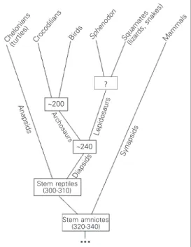
All living reptiles, birds and mammals are thought to have evolved from a common ancestor, the stem amniote (Figure 1). In other words, there is general consensus that the amniote egg evolved only once and that amniotes are monophyletic. All living rep-tiles and birds have a simple, mostly uni-laminar cortex. The questions that we wanted to address are: i) How is this simple cortex organized at the embryonic stage of cortical plate formation? Is the cortical plate strictly radial and laminated as in normal mamma-lian embryos, or is it poorly defined as in reeler mutant mouse embryos? ii) What is the direction of the maturation gradient in this thin cortex? iii) Does the pattern of reelin expression differ from phylum to phy-lum and from mammals? As a corollary, do reptiles and birds have cells homologous to Cajal-Retzius cells?
Before we consider the development of the cortex, it is useful to present a simple overview of the evolution of the different phyla (Figure 1). Stem amniotes (formerly
often named stem reptiles) are thought to have evolved during the carboniferous. From this common ancestor, several lineages evolved, four of which led to the present reptiles, birds and mammals. The first lin-eage, the synapsids, separated early and evolved into the present marsupials and eu-therian mammals. Another line, the anapsids, seems also to have separated early and gave rise to chelonians (turtles). The third branch, the lepidosaurs and eosuchians, contains the ancestors of the Rhynchocephalia, of which
Spenodon punctatus (New Zealands
tua-tara) is the only living representative, and the squamates (lizards and snakes). The fourth lineage, the archosaurs, evolved into dino-saurs and modern crocodilians (via theco-donts) and to birds (via saurischian dino-saurs). This summarized cladogram is sche-matically illustrated in Figure 1.
Organization of the cortical plate
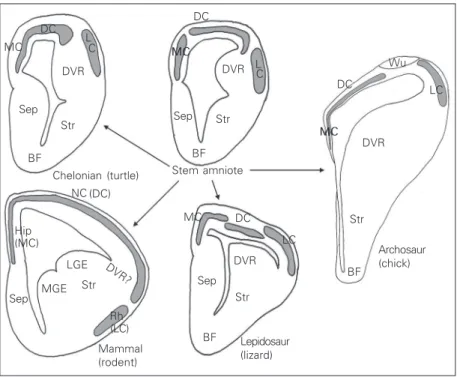
The anatomy of the embryonic telen-cephalon of reptiles is different from that of mammals (Figure 2). First, as mentioned above, the cortical plate is mainly composed of a layer that is a few cells thick. The main
Lepidosaurs
Synapsids
Diapsids
Mammals Squamates
(lizards, snakes) Sphenodon
Birds Crocodilians Chelonians
(turtles)
Stem amniotes (320-340) Stem reptiles
(300-310) Anapsids
Archosaurs
?
~200
~240
components of the cortex are the medial cortex, which is homologous to the mamma-lian hippocampal formation, the dorsal cor-tex (with some subdivisions in some spe-cies), homologous to the neocortex, and the lateral cortex, homologous to the pyriform cortex. In addition, reptiles and birds have a huge telencephalic compartment located ven-trally to the lateral ventricle and dorsally to the striatum, named the dorsal ventricular ridge (DVR, formerly named hyperstriatum in birds). There has been (and still is) contro-versy about the mammalian structure that is homologous to the DVR, but the current trend is that the DVR regressed during mam-malian evolution and is mainly represented by the claustrum. In this description, we focus on the cortical plate itself, and particu-larly the medial and dorsal cortex. Although stem amniotes are extinct and the organization of their embryonic brain will remain unknown, there is significant evidence that chelonians are, among living species, those most closely
related to stem amniotes and the present dis-cussion will accept that premise.
In all species, early cortical development proceeds by following a common theme: neurons are generated in ventricular zones and migrate outward to form a cortex (16). The formation of a cortical plate is preceded by an early horizontal network or preplate. On the other hand, as soon as the cortical plate appears, important species differences are noted in terms of its architectonic organ-ization. In turtle embryos (Figure 3), the cortical plate is loosely ordered, with poorly defined inner and outer borders. Although the cortical plate contains predominantly ra-dially oriented cells, cortical plate neurons are not densely packed and their radial shape is highly variable. This rudimentary cortical plate organization stands in sharp contrast to that of squamates, particularly lacertilian liz-ards (Figure 3), in which the cortical plate is clearly defined, sharply separated from the marginal zone and the subplate, and popu-lated with strictly radial neurons, in a pattern that is somewhat reminiscent of the mamma-lian one. The organization of the cortical plate in other reptiles can be described as intermediate between the rudimentary type found in chelonians and the elaborate type of lizards and mammals. For example, croco-diles have a well-populated cortical plate, but its borders are not clearly visible. The cortical plate of birds is similarly rather poorly defined and diminutive. In Sphenodon, there is an interesting sectorial variation: the me-dial cortex contains an elaborate cortical plate, similar to that of lizards, whereas the architectonics of the cortical plate in the dorsal cortex is more rudimentary, reminis-cent of that of turtles. This large species difference in terms of embryonic cortical plate architectonics indicates that this com-plex structure is the product of an evolution-ary process that occurred differently in dif-ferent phyla, and that the elaborate pattern presently found in lizards and mammals re-flects homoplasy (as opposed to a
homolo-DC L
C MC
DVR
Sep Str
BF
(DC) NC
BF Str Sep
DVR MC
DC
DC
Wu
MC DVR
LC
Str
BF
Archosaur (chick) LC
MC
DVR
Sep Str
BF LGE
Str
Rh (LC) MGE
Sep
DVR? (MC)
Hip
Chelonian (turtle)
Mammal (rodent)
Lepidosaur (lizard) DC Stem amniote
L C
gous feature), the product of evolutionary convergence. If evolution bothers to build similar structures repeatedly, it usually means that these features are beneficial from an evolutionary standpoint (20). And indeed the radial organization of the embryonic cor-tical plate is required for the acquisition of a laminate cortex, as clearly demonstrated from observation in reeler-like phenotypes.
In summary, if turtles are considered to be related to stem amniotes, comparative studies of the embryonic cortical plate sug-gest that the radial cortical plate architecton-ics evolved differently in the various lin-eages to reach its more elaborate status in lizards and mammals. With respect to radial architectonic organization alone, the normal mouse and reeler cortical plate are reminis-cent of the cortical plate of lizards and turtles, respectively.
The gradient of cortical plate maturation
In all mammals, marsupials included, the cortical plate develops following the inside-outside rule that we have outlined above. When the architectonics of the cortical plate is defective, as in reeler and similar mutant mice, this gradient is grossly inverted, di-rected from outside to inside. An obvious question concerns the histogenetic gradient in the reptilian cortical plate. Several years ago, we examined this question in turtle and lizard embryos by using in ovo injection of tritiated thymidine (21). Thymidine availa-bility is sufficient to label all or at least most neurons that are generated after the injec-tion, as demonstrated by the profuse labeling of some continuously proliferating epider-mal or mesoderepider-mal cell types. The neurons that are not labeled are thus those that were generated prior to thymidine injection. Us-ing this method, we could show that the maturation of the cortical plate is directed from outside to inside both in turtles and in lizards. Other species were not studied, but it
is known from the literature that the gradient is also outside-inside in the chick cortical plate (22). There was a relative exception to the rule in that in the lizard medial cortex, no gradient could be defined and all neurons in that compact layer were either labeled or not labeled. A first conclusion from these obser-vations is that the cortical plate of stem mammals most probably developed follow-ing an outside-inside gradient, and that the
V P V
I A
P I A
P I A
P I A V
V
Turtle DC Lizard DC
Turtle MC Lizard MC
inside-out development of the mammalian cortical plate represents an evolutionary ac-quisition. As maturation can be directed from outside to inside in cases when the cortical plate is radially organized (as in lizards), a second conclusion is that radial cortical plate organization, although necessary for the ac-quisition of the inside to outside maturation in mammals, is not sufficient. In other words, in addition to radial neuronal orientation and differentiation, other factors are needed for maturation to proceed from inside to out-side. Among necessary factors are probably a larger number of neurons leading to a thicker cortical plate, and the capacity of immature neurons to migrate over large dis-tances. This idea receives strong support from observations in mice deficient in the Cdk5 and in its p35 and p39 activators (9,10). In these mice, a thin, early cortical plate develops with an almost normal radial or-ganization, but later migrating cells cannot achieve migration over longer distances and settle at different levels in the intermediate zone, so that the maturation gradient of the cortex is still directed from outside to inside. Another example is the Tbr1 mouse muta-tion in which the cortical plate is reasonably well organized, yet maturation is from out-side to inout-side (23).
From comparative data and observations in mice with reeler-like and Cdk5-like phe-notypes, it is tempting to propose that the cortical plate was probably poorly organized radially in stem amniotes, and that the capac-ity to differentiate radially evolved sepa-rately in the lineages leading to mammals and squamates. In mammals with functional control mechanisms - such as the reelin, Cdk5 and other less welldefined pathways -the increase in -the thickness of -the cortex led to the inside-out maturation.
Comparative studies on the expression of reelin in the telencephalon
The drastic differences in cortical plate
organization among different phyla, together with the observation that the gradient of cortical plate maturation is directed from outside to inside in all non-mammalian spe-cies and in reeler mice, prompted us to com-pare reelin expression between representa-tives of all amniote lineages. Reelin expres-sion was studied in embryos from a turtle
(Emys orbicularis), from lizards (several
Lacerta species) and from chicks and
croco-diles (2,17-19). It was not possible to obtain an embryonic brain from Sphenodon
punc-tatus because of strict conservation
single reelin-positive cell was detected in the dense cortical plate in medial and dorsal pallial areas, which contained Dab1-posi-tive neurons. In the lateral cortex, reelin- and Dab1-positive neurons were somewhat mixed, although reelin expression seemed more diffuse than Dab1 expression, which was restricted to the dorsal aspect. At this level, some neurons may co-express reelin and Dab1, but this should be confirmed us-ing both markers simultaneously. The pat-tern in lizards is unique, with heavy reelin expression in the marginal zone and subcor-tex, and Dab1 expression in the dense corti-cal plate. The reelin expression level in the marginal zone and subcortex is comparable to that in mitral cells. One may perhaps imagine that reelin defines the upper and lower borders of the dense Dab1-positive cortical plate by preventing neuronal somata from migrating in reelin-rich zones. In the crocodilian embryonic telencephalon (Fig-ure 6), fut(Fig-ure cortical areas are less defined than in lizards, but clearly all of them contain reelin-positive cells in their marginal zone. In contrast to turtles, very few reelin-posi-tive cells are dispersed within the cortex. Unlike lizards, crocodiles lack reelin expres-sion in the subcortex. The level of reelin expression in marginal zone neurons is lower than or equivalent to that in the olfactory bulb. Not surprisingly, the chick telencepha-lon is rather similar to that of crocodiles (Figure 7). Cortical areas are, however, re-duced in birds compared to crocodilians and even compared to turtles, suggesting that crocodiles are more closely related to stem archosaurs. In parallel, reelin-positive neu-rons are dispersed in the chick marginal zone as in crocodiles, but they are less abundant and prominent than in the crocodilian cor-tex. It should be added that the DVR contains some reelin- and Dab1-positive cells in all reptiles studied, which, however, have not been studied in detail since the architectonic patterns of the DVR are poorly defined in most species, except lizards and Sphenodon.
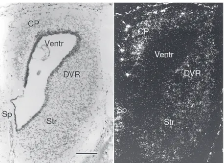
Figure 4. Reelin mRNA expression in the turtle embryonic cortex. Bright and dark field view of a coronal section through a turtle embryo (Emys orbicularis). In situ hybridization with a
[33P]-labeled, species-specific reelin riboprobe. The poor laminar organization of the cortical
plate (CP) in all cortical fields is evident. Reelin-positive cells are heavily labeled in the marginal zone and less intensely labeled cells are dispersed in the cortical plate. The strong darkfield signal of melanophores (arrows) is not related to probe hybridization. DVR: dorsal ventricular ridge; Sp: septum; Str: striatum; Ventr: ventricle. Adapted from Ref. 17. Magnifi-cation bar: 200 µm.
Figure 5. Reelin mRNA expression in the lizard embryonic cortex. Bright and dark field view
of a coronal section through a lizard embryo (Lacerta viridis). In situ hybridization with a
[33P]-labeled, species-specific reelin riboprobe. The elaborate radial organization of the
Conclusion: A synthetic model
Our comparative studies still contain many gaps. It would be useful to study in more detail the expression patterns of Dab1 and of the reelin receptors VLDLR and ApoER2 to confirm the expression canvas in a few other species, and particularly to ana-lyze Sphenodon embryos. In spite of these caveats, it is reasonable to propose the fol-lowing model of reelin expression and its role in cortical evolution (2). First, reelin is most probably expressed in the embryonic brain in all vertebrates, with a common pat-tern that includes (with some anecdotal ex-ceptions) expression in the retina, spinal cord, reticular formation, olfactory mitral cells, cerebellar granule cells, thalamic reticular nuclei, septal nuclei and some scattered cells in the telencephalon. In stem amniotes, this common pattern of reelin expression prob-ably included high expression in marginal zone neurons and lower expression in neu-rons scattered in a loosely defined cortical plate. In this loose cortical plate, most neu-rons expressed Dab1 and maturation pro-ceeded from outside to inside; this pattern apparently changed little during evolution of the anapsid lineage and is still found in modern turtles. During evolution of the archosaur lineage, an increase in the number of reelin-positive neurons occurred in the marginal zone, but fewer neurons expressed reelin in the cortical plate. The cortical plate increases in thickness and neuronal content but remains poorly organized from an archi-tectonic standpoint, and maturation still pro-ceeds from outside to inside; this pattern is found in modern crocodilians and, to a lesser extent, in birds. A different pattern evolved in the lepidosaurian lineage, in which reelin expression is amplified not only in neurons in the marginal zone, but also in the subcor-tex, whereas Dab1 expression is confined to the reelin-negative cortical plate. Further-more, in this lineage, the pattern evolved differently in various cortical areas and in
Figure 6. Reelin mRNA expression in the embryonic cortex of crocodiles. Bright and dark
field view of a coronal section through a crocodile embryo. In situ hybridization with a [33
P]-labeled, species-specific reelin riboprobe. The cortical plate (CP) has an intermediate level of radial organization and is reelin negative, whereas the marginal zone contains reelin-positive neurons (arrows). Ventr: ventricle; Str: striatum; DVR: dorsal ventricular ridge; Sp: septum. Magnification bar: 500 µm.
Figure 7. Reelin mRNA expression in the chick embryonic cortex. Bright and dark field view
of a coronal section through a chick embryo. In situ hybridization with a [33P]-labeled,
parallel to the evolution of architectonic lami-nation: it is the most evident in the medial and dorsal cortex and less marked in lateral cortical fields, and intermediate in DVR areas. Cortical plate maturation proceeds from outside to inside in all areas, except in the medial cortex in which no gradient is perceived. This pattern is found to different extents in lizards, snakes and probably
Sphe-nodon. In the three lineages above (turtles,
lizards, crocodiles and birds), and thus prob-ably in stem amniotes, the level of reelin expression in marginal zone cells was not heavily amplified and remained comparable to that observed in olfactory mitral cells. A different situation is found in mammals. Evo-lution of the synapsid lineage from stem
amniotes is characterized by two key fea-tures. First, at an unknown stage, a spectacu-lar amplification of reelin expression occurs in marginal zone cells, whereas the early cortical plate becomes reelin negative. Sec-ond, again at an unknown stage of cortical evolution, the outside to inside gradient of cortical plate maturation changes and be-comes directed from inside to the outside. Mutations in the reelin pathway and other genes (Cdk5, p35) show that the evolution of reelin expression is necessary but not suffi-cient for inside-outside maturation of the cortex. Clearly, a lot of work remains to be done in order to better define the molecular determinants of cortical evolution.
References
1. Raff RA (1996). The Shape of Life. Chi-cago University Press, ChiChi-cago, IL, USA. 2. Bar I, Lambert de Rouvroit C & Goffinet
AM (2000). The evolution of cortical de-velopment. An hypothesis based on the role of the Reelin signalling pathway. Trends in Neurosciences, 23: 633-638. 3. Lambert de Rouvroit C & Goffinet AM
(1998). The reeler mouse as a model of brain development. Advances in Ana-tomy, Embryology and Cell Biology, 150: 1-108.
4. Rice DS & Curran T (2001). Role of the reelin signaling pathway in central ner-vous system development. Annual Re-view of Neuroscience, 24: 1005-1039. 5. Parnavelas JG (2000). The origin and
mi-gration of cortical neurones: new vistas. Trends in Neurosciences, 23: 126-131. 6. Marin O & Rubenstein JL (2001). A long,
remarkable journey: tangential migration in the telencephalon. Nature Reviews. Neuroscience, 2: 780-790.
7. Meyer G, Soria JM, Martinez-Galan JR, Martin-Clemente B & Fairen A (1998). Dif-ferent origins and developmental histo-ries of transient neurons in the marginal zone of the fetal and neonatal rat cortex. Journal of Comparative Neurology, 397: 439-518.
8. Meyer G, Schaaps JP, Moreau L & Goffinet AM (2000). Embryonic and early fetal development of the human
neocor-tex. Journal of Neuroscience, 20:
1858-1868.
9. Ohshima T, Ward JM, Huh CG, Longenecker G, Veeranna Pant HC, Brady RO, Martin LJ & Kulkarni AB (1996). Tar-geted disruption of the cyclin-dependent kinase 5 gene results in abnormal cortico-genesis, neuronal pathology and perinatal death. Proceedings of the National Acad-emy of Sciences, USA, 93: 11173-11178. 10. Ko J, Humbert S, Bronson RT, Takahashi S, Kulkarni AB, Li E & Tsai LH (2001). p35 and p39 are essential for cyclin-depend-ent kinase 5 function during
neurodevel-opment. Journal of Neuroscience, 21:
6758-6767.
11. Trommssdorf M, Gotthardt M, Hiesberger T, Shelton J, Stockinger W, Nimpf J, Ham-mer RE, Richardon JA & Herz J (1999). Reeler/disabled-like disruption of neuronal migration in knockout mice lacking the VLDL receptor and ApoE receptor 2. Cell, 97: 689-701.
12. Hiesberger T, Trommssdorf M, Howell BW, Goffinet AM, Mumby MC, Cooper JA & Herz J (1999). Direct binding of Reelin to VLDL receptor and ApoE recep-tor 2 induces tyrosine phosphorylation of disabled-1 and modulates tau phosphory-lation. Neuron, 24: 481-489.
13. Howell BW, Hawkes R, Soriano P & Coo-per JA (1997). Neuronal position in the developing brain is regulated by mouse
disabled-1. Nature, 389: 733-737. 14. Ware ML, Fox JW, Davis NM, Lambert de
Rouvroit C, Russo C, Chua Jr SC, Goffinet AM & Walsh CA (1997). Aberrant splicing of a mouse disabled homolog, mDab1, in the scrambler mouse. Neuron, 19: 239-249.
15. Howell BW, Herrick TM, Hildebrand JD, Zhang Y & Cooper JA (2000). Dab1 ty-rosine phosphorylation sites relay posi-tional signals during mouse brain devel-opment. Current Biology, 10: 877-885. 16. Goffinet AM (1983). The embryonic
de-velopment of the cortical plate in reptiles: a comparative study in Emys orbicularis and Lacerta agilis. Journal of Comparative Neurology, 215: 437-452.
17. Bernier B, Bar I, Pieau C, Lambert de Rouvroit C & Goffinet AM (1999). Reelin mRNA expression during embryonic brain development in the turtle. Journal of Comparative Neurology, 413: 463-479. 18. Goffinet AM, Bar I, Bernier B, Trujillo C,
Raynaud A & Meyer G (1999). Reelin ex-pression during embryonic brain develop-ment in lacertilian lizards. Journal of Com-parative Neurology, 414: 533-550. 19. Bernier B, Bar I, D’Arcangelo G, Curran T
& Goffinet AM (2000). Reelin mRNA ex-pression during embryonic brain develop-ment in the chick. Journal of Comparative Neurology, 422: 448-463.
tel-encephalon in non-mammals. Annual Re-view of Neuroscience, 4: 301-350. 21. Goffinet AM, Daumerie Ch, Langerwerf B
& Pieau C (1986). Neurogenesis in
reptil-ian cortical structures: 3H thymidine
au-toradiographic analysis. Journal of Com-parative Neurology, 243: 106-116.
22. Tsai HM, Garber GB & Larramendi LMH (1981). Thymidine autoradiographic analy-sis of telencephalic histogeneanaly-sis in the chick embryo: I. Neuronal birthdates of telencephalic compartments in situ. Jour-nal of Comparative Neurology, 198: 275-292.