Cop
yright
© ABE&M t
odos os dir
eit
os r
eser
vados
.
1 Santa Casa de Belo Horizonte, Belo Horizonte, MG, Brazil
Correspondence to: Pedro Weslley Rosário Instituto de Ensino e Pesquisa da Santa Casa de Belo Horizonte Rua Domingos Vieira, 590
30150-240 – Belo Horizonte, MG, Brazil [email protected]
Received on Oct/25/2012 Accepted on Dec/12/2012
Usefulness of preoperative
serum calcitonin in patients with
nodular thyroid disease without
suspicious history or cytology for
medullary thyroid carcinoma
Utilidade da calcitonina sérica pré-operatória em pacientes com doença nodular tireoidiana sem história ou citologia suspeitas para carcinoma medular de tireoide
Pedro Weslley Rosário1, Gustavo Cancela Penna1, Kamilla Brandão1, Bárbara Érika Souza1
ABSTRACT
Objective: To evaluate the usefulness of preoperative serum calcitonin (sCT) in patients with nodular disease without suspicion of medullary thyroid carcinoma (MTC) in history or cytology. Patients and methods: sCT was measured before thyroidectomy in 494 patients with nodular disease who had no family history of MTC or multiple endocrine neoplasia type 2, and no cyto-logical suspicion of MTC. Results: Basal sCT was < 10 ng/mL in 482 patients and none of them had MTC. One patient with basal sCT > 100 pg/mL had MTC. Among the 11 patients with basal sCT between 10 and 100 pg/mL, MTC was diagnosed in only one. The two patients with MTC were submitted to total thyroidectomy, combined with elective lymph node dissection indicated exclusively based on hypercalcitoninemia, and sCT was undetectable after six months. Con-clusions: Preoperative sCT is useful for the detection of sporadic MTC in patients with nodular disease, even in the absence of suspicious history or cytology. Arq Bras Endocrinol Metab. 2013;57(4):312-6
Keywords
Calcitonin; medullary carcinoma; nodular disease
RESUMO
Objetivo: Avaliar a utilidade da calcitonina sérica (sCT) pré-operatória em pacientes com doen-ça nodular sem suspeita de carcinoma medular de tireoide (CMT) pela história e citologia. Pacientes e métodos: Antes da tireoidectomia, sCT foi dosada em 494 pacientes com doença nodular, sem história familiar de CMT ou neoplasia endócrina múltipla tipo 2 e sem citologia suspeita para CMT. Resultados: sCT basal foi < 10 ng/ml em 482 pacientes e nenhum possuía CMT. Um paciente com sCT basal > 100 pg/ml realmente possuía CMT. Dos 11 pacientes com sCT basal entre 10 e 100 pg/ml, CMT foi diagnosticado em apenas um. Os dois pacientes com CMT foram submetidos à tireoidectomia total com dissecção eletiva de linfonodos, indicada exclusivamente pela hipercalcitoninemia, e após seis meses apresentaram sCT indetectável. Conclusões: Em pacientes com doença nodular, mesmo sem história ou citologia suspeitas, a sCT pré-operatória é útil para detecção do CMT esporádico. Arq Bras Endocrinol Metab. 2013;57(4):312-6
Descritores
Cop
yright
© ABE&M t
odos os dir
eit
os r
eser
vados
.
INTRODUCTION
T
here is consensus regarding the usefulness of se-rum calcitonin (sCT) in patients with nodular di-sease and family history of medullary thyroid carcinoma (MTC) or multiple endocrine neoplasia type 2 (MEN-2) (1-3), or with suspicious cytology for MTC (4). Controversy exists in cases without suspicious history or cytology, with some authors defending routine me-asurement of sCT (1), whereas others consider the evi-dence to be insuficient for this recommendation (2,5). In the case of patients with an indication for thy-roidectomy, irrespective of sCT, the inding of hyper-calcitoninemia modiies the extent of the procedure (to total thyroidectomy with elective dissection of cervical lymph nodes). This approach enables effective treat-ment of MTC already in the irst intervention, without the need for surgical reintervention, which might be necessary when histology reveals this tumor and the irst surgery was less extensive (4).However, the cost of sCT measurement (basal and after stimulation in some cases), which needs to be per-formed in hundreds of patients in order to detect one case of MTC exclusively by this method, and the risk of false-positive results are limitations of routine preopera-tive sCT measurement in patients without suspicious history or cytology.
The different positions on this topic in current guidelines (1-3,5) clearly demonstrate that more stu-dies are desirable. The objective of the present investi-gation was to evaluate the usefulness of sCT in patients with nodular thyroid disease and indication for surgical treatment, and who had no suspicion of MTC in his-tory or cytology.
PATIENTS AND METHODS
Design
Prospective study
Patients
First, patients with nodular thyroid disease and an in-dication for surgery were evaluated. In these cases, the decision for surgery was made before sCT measurement and, therefore, was not inluenced by this result. The following patients were excluded: (i) those with a family history of MTC or MEN-2; (ii) those with suspicious cytology or diagnosis of MTC; (iii) and those with known kidney failure, hyperparathyroidism, chronic atrophic gastritis, neuroendocrine tumor, or pulmonary
carcino-ma. Only patients whose indication for surgery was in ac-cordance with current guidelines were included (1-3,5).
Measurement of serum calcitonin and management of the patients
For the measurement of sCT performed 30 days or less before thyroidectomy, patients were asked not to con-sume alcoholic beverages for at least 1 week, and not to use proton pump inhibitors for at least 4 weeks. At the time of measurement, none of the patients had bacterial infections or hypercalcemia.
Patients with basal sCT > 10 pg/mL underwent a calcium stimulation test [rapid venous infusion of 2.5 mg calcium/kg in the form of 10% calcium gluconate (10 ml/min)]. Serum calcitonin was measured before and 2, 5 and 10 min after the infusion of calcium. Pa-tients with stimulated sCT > 100 pg/mL were sub-mitted to total thyroidectomy combined with elective dissection of the cervical lymph nodes (indicated ex-clusively based on the inding of hypercalcitoninemia).
The study was approved by the local Research Ethics Committee.
Assay
Serum calcitonin was measured by an immunochemilu-minescent assay (Immulite, Diagnostic Products Cor-poration, Los Angeles, CA, USA). The sensitivity of the assay was 2 pg/mL.
Fine-needle aspiration biopsy
Fine-needle aspiration biopsy (FNAB) as performed with a 22-gauge needle and a 5- or 10-mL syringe and guided by ultrasound. The smears were analyzed by pa-thologists experienced in thyroid pathology.
RESULTS
Cop
yright
© ABE&M t
odos os dir
eit
os r
eser
vados
.
T1aN0M0 and T1bN1aM0, respectively. Basal sCT was undetectable in the two patients 6 months after the initial surgery.
DISCUSSION
This study included only patients without a suspicion of MTC in history and cytology. In fact, there is con-sensus regarding the usefulness of sCT in patients with family history of MTC or MEN-2 (1-3), or those with suspicious cytology for this cancer (4). Cytology re-sult of MTC may be interpreted as papillary carcino-ma, indeterminate, or even benign (6-8). In addition, in subjects who are at low clinical risk for malignancy, only nodules ≥ 1 cm are submitted to FNAB (5), and an eventual microcarcinoma associated with macrono-dules may therefore not be detected. These arguments favor the measurement of sCT even in patients who al-ready underwent FNAB and whose cytology result is not suggestive of MTC.
We evaluated only patients with surgical indication, irrespective of sCT. In these cases, the inding of hy-percalcitoninemia modiies the extent of the procedure, enabling adequate treatment of possible MTC already in the irst intervention. In fact, two cases of MTC di-agnosed by preoperative sCT were submitted to total thyroidectomy and elective cervical lymph node dis-section exclusively based on the inding of hypercalci-toninemia, which led to complete biochemical remis-sion. Speciically in this situation (measurement of sCT
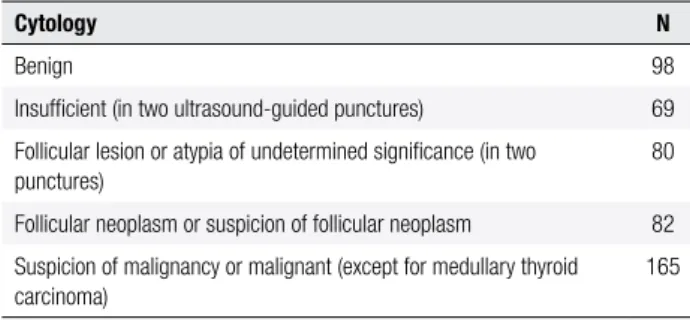
Table 1. Results of cytology in the patients studied
Cytology N
Benign 98
Insuficient (in two ultrasound-guided punctures) 69
Follicular lesion or atypia of undetermined signiicance (in two punctures)
80
Follicular neoplasm or suspicion of follicular neoplasm 82
Suspicion of malignancy or malignant (except for medullary thyroid carcinoma)
165
Basal sCT was < 10 ng/mL in 482 patients. MTC was not detected in any of these cases. Serum calcitonin was > 100 pg/mL in only one patient and histology conirmed MTC in this case. Finally, 11 patients had basal sCT > 10 ng/mL, but < 100 pg/mL. Histology revealed MTC in one of these patients (the only case with basal sCT > 50 pg/mL). Ten patients with basal sCT > 10 ng/mL (< 50 pg/mL) did not have MTC, and only one of these patients presented stimulated CT > 100 pg/mL. Three patients were submitted to par-tial thyroidectomy. In these cases, MTC in the remnant gland was ruled out by the absence of nodules in post-operative ultrasound scans and a reduction of basal CT to levels < 5 ng/mL six months after surgery. Data of the patients with basal sCT > 10 pg/mL are shown in table 2.
The two patients with MTC were submitted to total thyroidectomy and elective dissection of the cer-vical lymph nodes. Micrometastases were detected in two lymph nodes of one of these patients. The tumors measured 7 and 12 mm, and the initial stages were
Table 2. Data of patients with basal serum calcitonin > 10 pg/mL
Gender Age (years) Cytology sectionFrozen Basal sCT (pg/mL) Stimulated sCT (pg/mL) Surgery Histology
F 78 Benign NA 20 42 TT CG
F 50 Benign NA 25 56 TT CG
M 75 Insuficient NA 12 38 PT CG
F 16 Follicular lesion of undetermined signiicance NA 18 45 PT PTC
M 50 Atypia of undetermined signiicance Benign 32 75 TT HT
F 46 Follicular neoplasm NA 112 887 TT + ND MTC
F 62 Follicular neoplasm NA 23 40 PT FA
F 53 Suspicion of PTC Malignancy 56 216 TT + ND MTC
F 72 Suspicion of PTC Malignancy 21 40 TT PTC
F 29 PTC NA 16 31 TT PTC
M 42 PTC NA 30 108 TT + ND PTC + CCH
F 18 PTC NA 25 43 TT PTC
Cop
yright
© ABE&M t
odos os dir
eit
os r
eser
vados
.
in patients with surgical indication), the frequency of MTC was 0.4% in the present study. Two other series reported a frequency of 0.5% (9) and 1.37% (10).
Another matter of discussion are the cutoff values of sCT. Whereas sporadic MTC is very unlikely in the presence of basal sCT < 10 pg/mL (9,10), serum con-centration > 100 pg/mL has an excellent positive pre-dictive value (PPV) for this tumor in the absence of any apparent cause (e.g., chronic renal failure, use of pro-ton pump inhibitors, other known secretory tumors) (6,8-21), and stimulation tests are not necessary in these cases. Despite the traditional cutoff value of 100 pg/mL, at least in women, PPV of 100% was reported in many series for basal sCT > 60 pg/mL (6,8-10,12-14,17-21). In agreement with this inding, in the pres-ent study, after known causes of hypercalcitoninemia were excluded, none of the 492 patients without MTC had basal sCT > 40 pg/mL.
In patients in whom a stimulation test is indicated (intermediate basal sCT levels), stimulated sCT < 100 pg/mL also renders sporadic MTC unlikely. In con-trast, PPV of stimulated sCT > 100 pg/mL is contro-versial. Although some studies reported PPV of 100% (8,10,20), many series found a value of only 25% (21), 20% (22), and 0/13 (9) in patients with elevated basal sCT, but < 100 pg/mL, who converted to levels > 100 pg/mL after stimulation. As a consequence, different cutoffs of stimulated sCT have been proposed in the literature (11-13,15,19), and this value remains unde-ined.
In countries such as Brazil where pentagastrin is not readily available, although its importation is pos-sible, calcium can be used for stimulation tests. In ad-dition to being a known stimulus of CT secretion and showing an excellent correlation with post-pentagastrin peak, calcium is better tolerated (13,23). In the present study, all 11 patients with basal sCT between 10 and 100 pg/mL received venous infusion of calcium for the stimulation test. Two of these patients presented stimu-lated sCT > 100 pg/mL, one had MTC and the other had C-cell hyperplasia.
Finally, the strengths of the present study are its prospective design; the fact that cytology was available for all patients; the stimulation test using calcium, and the fact of being the largest Brazilian series evaluating measurement of sCT as a screening method for spo-radic MTC.
We conclude that preoperative measurement of sCT is useful for the detection of sporadic MTC in patients
with nodular thyroid disease, even those without suspi-cious history or cytology for MTC, enabling adequate surgical treatment of this cancer already in the irst in-tervention. In cases in which basal sCT does not eluci-date the case, the calcium stimulation test seems to be of value for the identiication of patients with MTC.
Disclosure: no potential conlict of interest relevant to this article was reported.
REFERENCES
1. Pacini F, Schlumberger M, Dralle H, Elisei R, Smit J, Wiersinga W. European Consensus for the management of patients with di-fferentiated thyroid carcinoma of the follicular epithelium. Eur J Endocrinol. 2006;154:787-803.
2. Camargo R, Corigliano S, Friguglietti C, Gauna A, Harach R, Mu-nizaga F, et al. Latin American Thyroid Society recommendations for the management of thyroid nodules. Arq Bras Endocrinol Me-tab. 2009;53:1167-75.
3. Gharib H, Papini E, Paschke R, Duick DS, Valcavi R, Hegedus L, et al. American Association of Clinical Endocrinologists, Associazio-ne Medici Endocrinologi, and European Thyroid Association me-dical guidelines for clinical practice for the diagnosis and mana-gement of thyroid nodules. Endocr Pract. 2010;16(Suppl 1):1-43. 4. Kloos RT, Eng C, Evans DB, Francis GL, Gagel RF, Gharib H, et al.
Medullary thyroid cancer: management guidelines of the Ameri-can Thyroid Association. Thyroid. 2009;19:565-612.
5. Cooper DS, Doherty GM, Haugen BR, Kloos RT, Lee SL, Mandel SJ, et al. Revised American Thyroid Association management guidelines for patients with thyroid nodules and differentiated thyroid cancer. Thyroid. 2009;19:1167-214.
6. Henry JF, Denizot A, Puccini M, Gramatica L, Kvachenyuk A, Conte Devolx B, et al. Latent subclinical medullary thyroid carcinoma: diagnosis and treatment. World J Surgery. 1998;22:752-6. 7. Bugalho MJ, Santos JR, Sobrinho L. Preoperative diagnosis of
medullary thyroid carcinoma: ine needle aspiration cytology as compared with serum calcitonin measurement. J Surg Oncol. 2005;91:56-60.
8. Elisei R, Bottici V, Luchetti F, Di Coscio G, Romei C, Grasso L, et al. Impact of routine measurement of serum calcitonin on the diag-nosis and outcome of medullary thyroid cancer: experience in 10,864 patients with nodular thyroid disorders. J Clin Endocrinol Metab. 2004;89:163-8.
9. Chambon G, Alovisetti C, Idoux-Louche C, Reynaud C, Rodier M, Guedj AM, et al. The Use of preoperative routine measurement of basal serum thyrocalcitonin in candidates for thyroidectomy due to nodular thyroid disordes: results from 2733 consecutive patients. J Clin Endocrinol Metab. 2011;96:75-81.
10. Niccoli P, Wion-Barbot N, Caron P, Henry JF, De Micco E, SaintAn-dre JP, et al. Interest of routine measurement of serum calcitonin: study in a large series of thyroidectomized patients. J Clin Endo-crinol Metab. 1997;82:338-41.
11. Costante G, Meringolo D, Durante C, Bianchi D, Nocera M, Tumi-no S, et al. Predictive value of serum calcitonin levels for preo-perative diagnosis of medullary thyroid carcinoma in a cohort of 5817 consecutive patients with thyroid nodules. J Clin Endocrinol Metab. 2007;92:450-5.
Cop
yright
© ABE&M t
odos os dir
eit
os r
eser
vados
.
13. Colombo C, Verga U, Mian C, Ferrero S, Perrino M, Vicentini L, et al. Comparison of calcium and pentagastrin tests for the diagno-sis and follow-up of medullary thyroid cancer. J Clin Endocrinol Metab. 2012;97:905-13.
14. Vierhapper H, Niederle B, Bieglmayer C, Kaserer K, Baumgart-ner-Parzer S. Early diagnosis and curative therapy of medullary thyroid carcinoma by routine measurement of serum calcitoninin patients with thyroid disorders. Thyroid. 2005;15:1267-72. 15. Iacobone M, Niccoli-Sire P, Sebag F, DeMicco C, Henry JF. Can
sporadic Medullary thyroid carcinoma be biochemically predic-ted? Prospective analysis of 66 operated patients with elevated serum calcitonin levels. World J Surg. 2002;26:886-90.
16. Gibelin H, Essique D, Jones C, Levillain P, Maréchaud R, Kraimps JL. Increased calcitonin level in thyroid nodules without medulla-ry carcinoma. Br J Surg. 2005;92:574-8.
17. Papi G, Corsello SM, Cioni K, Pizzini AM, Corrado S, Carapezzi C, et al. Value of routine measurement of serum calcitonin concen-trations in patients with nodular thyroid disease: A multicenter study. J Endocrinol Invest. 2006;29:427-37.
18. Kaserer K, Scheuba C, Neuhold N, Weinhäusel A, Vierhapper H, Haas OA, et al. C-cell hyperplasia and medullary thyroid
carcino-ma in patients routinely screened for serumcalcitonin. Am J Surg Pathol. 1998;22:722-8.
19. Machens A, Hoffmann F, Sekulla C, Dralle H. Importance of gender-specific calcitonin thresholds in screening for occult sporadic me-dullary thyroid cancer. Endocr Relat Cancer. 2009;16:1291-8. 20. Pacini F, Fontanelli M, Fugazzola L, Elisei R, Romei C, Di Coscio G, et
al. Routine measurement of serum calcitonin in nodular thyroid dise-ases allows the preoperative diagnosis of unsuspected sporadic me-dullary thyroid carcinoma. J Clin Endocrinol Metab. 1994;78:826-9. 21. Herrmann BL, Schmid KW, Goerges R, Kemen M, Mann K.
Calci-tonin screening and pentagastrin testing: predictive value for the diagnosis of medullary carcinoma in nodular thyroid disease. Eur J Endocrinol. 2010;162:1141-5.
22. Rink T, Truong PN, Schroth HJ, Diener J, Zimny M, Grunwald F. Calculation and validation of a plasma calcitonin limit for early detection of medullary thyroid carcinoma in nodular thyroid di-sease. Thyroid. 2009;19:327-32.