Protein 3-nitrotyrosine formation during
Trypanosoma cruzi
infection in mice
1Departamento de Bioquimica and Center for Free Radical and Biomedical Research, 2Departamento de Reumatología, 3Departamento de Anatomía Patológica,
Hospital de Clínicas, 4Departamento de Medicina, Facultad de Medicina, Universidad de la República, Montevideo, Uruguay
M. Naviliat1,2, G. Gualco3, A. Cayota4 and R. Radi1
Abstract
Nitric oxide (•••••NO) is a diffusible messenger implicated in
Trypanoso-ma cruzi resistance. Excess production of •••••NO and oxidants leads to
the generation of nitrogen dioxide (•••••NO
2), a strong nitrating agent.
Tyrosine nitration is a post-translational modification resulting from the addition of a nitro (-NO2) group to the ortho-position of tyrosine
residues. Detection of protein 3-nitrotyrosine is regarded as a marker of nitro-oxidative stress and is observed in inflammatory processes. The formation and role of nitrating species in the control and myocar-diopathy of T. cruzi infection remain to be studied. We investigated the levels of •NO and protein 3-nitrotyrosine in the plasma of C3H and
BALB/c mice and pharmacologically modulated their production during the acute phase of T. cruzi infection. We also looked for protein 3-nitrotyrosine in the hearts of infected animals. Our results demon-strated that C3H animals produced higher amounts of •NO than
BALB/c mice, but their generation of peroxynitrite was not propor-tionally enhanced and they had higher parasitemias. While NG
-nitro-arginine methyl ester treatment abolished •NO production and
drasti-cally augmented the parasitism, mercaptoethylguanidine and guanido-ethyl disulfide, at doses that moderately reduced the •NO and
3-nitrotyrosine levels, paradoxically diminished the parasitemia in both strains. Nitrated proteins were also demonstrated in myocardial cells of infected mice. These data suggest that the control of T. cruzi
infection depends not only on the capacity to produce •NO, but also on
its metabolic fate, including the generation of nitrating species that may constitute an important element in parasite resistance and collat-eral myocardial damage.
Correspondence
R. Radi
Departamento de Bioquímica Facultad de Medicina Universidad de la República Avda. General Flores, 2125 Montevideo 11800 Uruguay
E-mail: [email protected]
Research supported by CSIC, Universidad de la República and the Howard Hughes Medical Institute.
Received January 4, 2005 Accepted August 30, 2005
Key words
•Peroxynitrite •Nitric oxide •Nitrotyrosine •Free radicals •Trypanosoma cruzi
•Chagas’ disease
Introduction
Chagas’ disease is a parasitic condition caused by Trypanosoma cruzi that affects 16 to 18 million people in Central and South America and that can lead to impairment of heart function. During the acute phase of the
the anti-parasitic defense, mainly due to the production of nitric oxide (•NO) (1).
• •• •
•NO is a diffusible messenger produced by
the reaction of L-arginine with different iso-forms of nitric oxide synthases (NOS). •NO
has been implicated in different physiological functions, including vasodilation, inhibition of platelet aggregation, neurotransmission, and immune regulation (2), among others. In in-flammatory conditions, large amounts of •NO
can be produced after the induction of induc-ible NOS (iNOS) in different cell types, nota-bly macrophages. During T. cruzi infection,
•NO production, measured by its end products
nitrite (NO2-) plus nitrate (NO3-) (NOx-) in
serum or NO2- in splenocyte culture
superna-tants, rises early after the infection and re-mains high throughout the acute phase (3,4). Most of the current data indicate the necessity for •NO to control the infection. The
produc-tion of •NO correlates with the resistance to T.
cruzi infection in C57BL/6 mice (3), and the
use of different iNOS inhibitors dramatically increases parasitemia and mortality (5). In-deed, interferon-γ (INF-γ) receptor or iNOS knock-out mice are extremely susceptible to
T. cruzi infection (6-8). However, in contrast,
a recent publication by Cummings and Tarleton (9) showed that iNOS knock-out mice are not more susceptible than wild type to T. cruzi infection.
Macrophage infection with T. cruzi may also induce the generation of reactive oxy-gen species (ROS) (10,11), particularly su-peroxide (O2•-) from the NADPH
oxidase-dependent respiratory burst (12). ROS have been implicated early in T. cruzi killing. The J774 macrophage cell clone C3C, unable to produce O2•- and hydrogen peroxide (H2O2),
cannot control parasite growth when infected with T. cruzi (13).
Tyrosine nitration is a post-translational modification resulting from the addition of a nitro (-NO2) group to the ortho-position of
tyrosine residues. Two principal reaction pathways lead to 3-nitrotyrosine formation, both centered on the formation of nitrogen
dioxide (•NO
2) (14), a strong nitrating agent,
namely, the peroxynitrite- and hemeperoxi-dase-dependent mechanisms. The formation of 3-nitrotyrosine is regarded as a marker of nitro-oxidative stress and is observed in in-flammatory processes under excess produc-tion of •NO and oxidants.
3-Nitrotyrosine-containing proteins have been described in different human and animal diseases, par-ticularly in various forms of myocarditis (for a review, see Ref. 15).
Peroxynitrite, myeloperoxidase and eosin-ophil peroxidase are cytotoxic for T. cruzi. Previous work from our laboratory has shown that macrophage-derived peroxynitrite is able to kill T. cruzi epimastigotes and to impair their replication and motility as well as their energetic, calcium and trypanothione metabo-lism (16,17). In addition, peroxynitrite causes the nitration of T. cruzi proteins (18). Neutro-phils are able to kill intracellular amastigotes by a mechanism mediated by myeloperoxi-dase and H2O2 (19). Also, eosinophil
peroxi-dase-coated T. cruzi trypomastigotes became sensitized to macrophage destruction in the presence of H2O2 (20).
On the basis of this evidence, we hypoth-esize that nitrating molecules are formed during T. cruzi infection as part of the anti-parasitic mechanisms, and also contribute to collateral host tissue damage. To test this hypothesis we estimated inflammatory cell-derived •NO and nitrotyrosine production in
the plasma of two strains of mice with differ-ent susceptibility to T. cruzi infection (11) and pharmacologically modulated its pro-duction during the acute phase of the dis-ease. We also searched for protein 3-nitroty-rosine in the hearts of infected animals.
Material and Methods
Chemicals
Animals
Eight- to 12-week-old BALB/c and C3H mice were obtained from the animal house of Instituto de Higiene (Facultad de Medi-cina, Universidad de la República, Montevi-deo, Uruguay) and breed and maintained as recommended (21).
Parasites and experimental infections
The Tulahuen-2 strain of T. cruzi was used in all studies. The animals were in-fected intraperitoneally with sublethal doses of blood-derived trypomastigote forms, i.e., 500 and 100 live parasites for BALB/c and C3H mice, respectively. Parasitemia was measured in 8 µL of blood obtained from the tail vein as described (22).
Pharmacological intervention
To modulate •NO production we used
the NOS inhibitors NG-nitro-L-arginine
meth-yl ester (L-NAME) and guanido-ethmeth-yl disul-fide (GED) as well as mercaptoethylguani-dine (MEG), an iNOS inhibitor and peroxy-nitrite scavenger (23) that also reduces neu-trophil infiltration (24). Mice received a daily intraperitoneal dose of 50 mg/kg L-NAME, 10 mg/kg MEG or 7 mg/kg GED diluted in PBS or vehicle from the day of infection and throughout the experimental period. Similar L-NAME doses have been shown to inhibit •NO production during T. cruzi
infec-tion in mice (3,5). Also, 10 mg/kg MEG inhibited •NO formation and nitrotyrosine
immunoreactivity in a rat model of lung inflammation (24). GED (7 mg/kg) repre-sents the quantity that provides the same molar amount of guanidine residues as 10 mg/kg MEG. Clinically healthy non-infected mice receiving L-NAME, MEG or GED were used as controls. MEG and GED were kindly provided by Dr. Csaba Szabó (Inoteck Phar-maceuticals Corporation, Beverly, MA, USA).
Blood collection
Blood was collected on day 17 post-infection (pi) from L-NAME-treated mice and on day 25 pi from MEG- and GED-treated mice, when parasitemia started to decline. The hearts were removed, washed in PBS and fixed in 10% formalin (v/v) in PBS. Blood leaking from the heart was col-lected into heparinized tubes and centrifuged and then submitted to five cycles of freezing and thawing in order to eliminate parasites. Finally, plasma samples were maintained at -20ºC until use.
Measurement of •••••NO
The production of •NO was estimated by
measuring NOx- in plasma by the Griess
method (25) adapted to work with small volumes of sample. Briefly, 10 µL of the sample or standard dilutions of NO3- were
incubated on an ELISA plate (NUNC, Roskilde, Denmark) in the presence of 0.1 U/mL Aspergillus sp nitrate reductase, 5 µM flavin adenine dinucleotide (and 30 µM NADPH for 15 min at 37ºC in a final volume of 100 µL. Next, excess NADPH was oxi-dized with 100 U/mL lactate dehydrogenase and 0.3 mM sodium pyruvate for 5 min at 37ºC and then for 15 min at 4ºC, in a final volume of 200 µL. Diazotization was per-formed using 10 mM sulfanilamide and 0.6 M HCl. In order to eliminate the precipitated proteins, the samples were centrifuged at 3000 g for 15 min at 4ºC. The supernatant was then transferred to a new ELISA plate, 10 mM N-(1-naphthyl)-ethylenediamine was added, and absorbance was measured at 548 nm in a microplate reader (Labsystems Multiskan MS, Vantaa, Finland).
Measurement of nitrated plasma proteins
in 100 mM phosphate buffer, pH 7.4, was nitrated with 1 mM peroxynitrite added as a single bolus. The nitrotyrosine content of the nitrated BSA was calculated spectropho-tometrically as the difference of the absorb-ance at pH 6 and 11, at 420 nm (ε = 4400 M-1
cm-1) (27). ELISA plates (NUNC) were
coated with dilutions of nitrated BSA or plasma samples diluted 1/100 in carbonate buffer (50 mM, pH 9) overnight at 4ºC and then blocked with 1% gelatin diluted in PBS for 1 h at 37ºC. After washing, an anti-nitrotyrosine polyclonal antibody (Ab) gen-erated in our laboratory (28) diluted to 250 ng/mL in PBS-0.05% Tween 20 was incu-bated for 1 h at 37ºC and detected using a goat anti-rabbit peroxidase-conjugated im-munoglobulin polyclonal antibody (Amer-sham-Pharmacia, Arlington Height, IL, USA). The plates were then developed using
o-phenylenediamine and absorbance was
read at 452 nm using a microplate reader. The results are reported as pmol/mg plasma proteins.
Immunohistochemistry
Hearts were probed with anti-nitroty-rosine and anti-myeloperoxidase polyclonal Ab. After fixing, the specimens were cut into 5-µm sections on silanized microscope slides. The anti-nitrotyrosine polyclonal Ab (5 ng/ mL) was diluted in Tris-saline (50 mM Tris, 150 mM NaCl, pH 7.4), incubated over-night, and then developed with a secondary
antibody coupled to biotin using a streptavi-din-peroxidase kit and diaminobenzidine as chromogen. Histological sections were coun-terstained with hemalum. Diaminobenzidine tetrahydrochloride staining was enhanced by adding 0.03% NiCl2 (w/v) to the solution.
Controls were performed with a pre-immune rabbit serum as the first antibody. The tech-nical control was omission of the primary Ab. The specificity of immunostaining was always assessed by competition with soluble 5 mM 3-nitrotyrosine. For anti-myeloper-oxidase staining we used a rabbit anti-hu-man myeloperoxidase polyclonal Ab (RDI Research Diagnosis Inc., Flanders, NY, USA) diluted in Tris-saline containing goat serum, 1/8 (v/v). A preliminary study on mouse bone marrow performed in order to confirm the cross-reaction was positive.
Statistical analysis
Five to eight animals were included in each experimental group. All experiments were repeated three times. The results were compared by the Mann-Whitney-Wilcoxon test and a P value ≤0.05 was considered to be significant.
Results
Parasitemia and mortality
Previous studies have shown that, when infected with T. cruzi, BALB/c and C3H
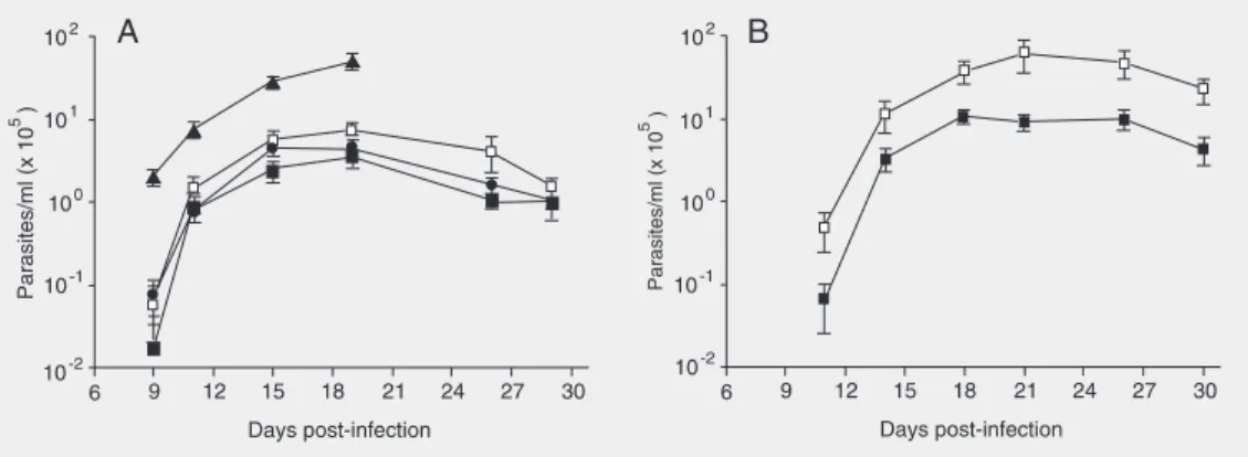
Figure 1. Parasitemia of Trypa-nosoma cruzi-infected BALB/c (A) and C3H/c mice (B). Mice were infected with sublethal doses of Tulahuen-2 trypomas-tigotes and treated with mercap-toethylguanidine (filled squares), guanido-ethyl disulfide (circles) or NG-nitro-L-arginine methyl
mice display different degrees of macro-phage activation as indicated by different
amounts of ROS and TNF-α production
(11,29). Thus, since in the present study we evaluated the formation of •NO and
nitroty-rosine as a marker of nitro-oxidative stress during T. cruzi infection, we chose to work with the C3H and BALB/c strains. The ani-mals were infected with a sublethal dose of parasites and treated with the NOS inhibi-tors L-NAME or GED or with MEG, the latter an iNOS inhibitor and peroxynitrite scavenger (23). L-NAME is a general NOS inhibitor which has already been used to inhibit •NO production during T. cruzi
infec-tion (3,4,30). In turn, MEG and GED are guanidino compounds which are more spe-cific for the inhibition of iNOS (31). In addition, due to its free thiol moiety, MEG reacts with peroxynitrite and its decomposi-tion products, carbonate (CO3•-) and •NO2
radicals, and inhibits the nitration and oxida-tion reacoxida-tions of peroxynitrite (23). MEG also displays different anti-inflammatory ef-fects, including the inhibition of neutrophil migration and thus can inhibit the two main pathways of 3-nitrotyrosine formation (24, 32).
The parasitemias of control and L-NAME-, MEG- or GED-treated BALB/c and C3H mice are shown in Figure 1A and B, respectively. Untreated C3H mice developed a parasitemia about one order of magnitude higher than untreated BALB/c animals. L-NAME-treated BALB/c mice had a signifi-cantly higher parasitemia (P < 0.05) through-out the infection than control animals. Un-expectedly, MEG-treated BALB/c and C3H mice developed a significantly lower parasi-temia (P < 0.05, from day 15 to 25 pi, i.e., 4-and 10-fold less for MEG than control at day 20 for BALB/c and C3H, respectively) than non-treated animals. GED-treated BALB/c mice also showed a trend towards a de-creased parasitemia which, however, was not statistically significant (P = 0.07). While L-NAME-treated BALB/c mice showed
100% mortality at day 19 pi, all the MEG-and GED-treated animals were alive at the end of the acute phase of the infection (Fig-ure 2). Similarly, mortality of MEG-treated C3H mice was null at 25 days pi (data not shown). A direct lethal effect of MEG and GED on the parasite was ruled out because none of these compounds at a concentration up to 3 mM affected the growth of T. cruzi epimastigotes in culture (data not shown).
Production of •••••NO
•NO production during the immune
re-sponse was evaluated by measuring the NOx
-levels in plasma of control and infected, treated and untreated animals, at day 25 pi.
T. cruzi infection led to an elevation of NOx
-in both stra-ins of mice (Table 1). However, the infected C3H mice produced higher lev-els of •NO (similar to published data, 33)
than infected BALB/c animals. In turn, while L-NAME produced a profound inhibition of
•NO synthesis, MEG and GED induced only
a partial inhibition. Thus, there was no direct correlation between •NO levels (Table 1)
and parasitemia (Figure 1), indicating that
•NO is not the only molecule implicated in
the control of the parasite growth, and that some •NO-derived intermediate may
partici-pate in T. cruzi clearance.
Detection of nitrated plasma proteins
We measured the levels of nitrated plasma proteins in MEG- and GED-treated mice by
Figure 2. Mortality of BALB/c mice infected with sublethal doses of Tulahuen-2 trypomas-tigotes and treated with different iNOS inhibitors or a peroxyni-trite scavenger or not treated (open squares). Mercaptoeth-ylguanidine (filled squares), guanido-ethyl disulfide (circles), NG-nitro-L-arginine methyl ester
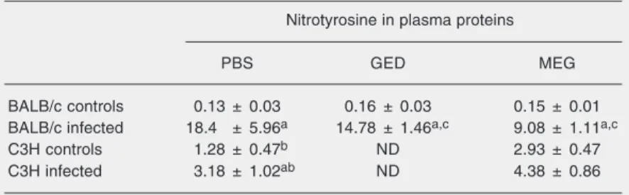
ELISA as an index of the systemic formation of nitrating species. Table 2 shows that in-fected BALB/c mice had significantly higher levels of nitrated plasma proteins than un-infected mice (>7-fold), while MEG or GED treatment of infected animals significantly decreased the amount of protein 3-nitroty-rosine in plasma. Uninfected C3H mice had significantly higher basal levels of nitrated serum proteins than BALB/c mice and, when infected with T. cruzi, showed a moderate increase (~1.6-fold) in nitrated proteins com-pared to controls. Thus, in spite of the
capac-ity of C3H mice to produce much higher amounts of •NO than BALB/c mice (Table
1), in infected animals the levels of nitrated proteins were ~2-fold lower than those of BALB/c animals. In BALB/c mice, a signifi-cant decrease of nitrated serum proteins was observed when the animals were treated with MEG.
Protein 3-nitrotyrosine in chagasic myocardiopathy
3-Nitrotyrosine in heart proteins was evaluated by immunohistochemistry. Pro-tein 3-nitrotyrosine residues were not de-tected in hearts of infected animals until day 25 pi (data not shown). At this time, hem-alum-eosin staining of the hearts indicated scarce inflammatory infiltrate and fibrosis. No polymorphonuclear cells were observed. Immunohistochemistry with a polyclonal anti-nitrotyrosine Ab showed diffuse myo-cardial immunoreactivity in infected ani-mals (Figure 3D). The immunochemical la-beling was stronger in the inflammatory ar-eas. Intense labeling was observed in myo-cardial cells, with a membranous and granu-lar cytoplasmic pattern (Figure 4). This study could not be performed in L-NAME-treated mice since all the animals died before pro-tein 3-nitrotyrosine was detectable in the hearts. In MEG- and GED-treated mice, a trend towards less intense immunostaining was observed (data not shown), although the technique was not sensitive enough for quan-titative purposes. In non-infected animals, no immunoreactivity against nitrotyrosine was observed (Figure 3A). Controls were performed to substantiate the specificity of the immunostaining, including specimens incubated with pre-immune serum (Figure 3B) and the co-incubation of the first Ab with 5 mM nitrotyrosine, which completely blocked the reactivity (Figure 3C). Anti-myeloperoxidase immunostaining showed no reactivity, consistent with the lack of neutrophil infiltration in this model.
Table 1.NOx- levels in plasma of Trypanosoma cruzi-infected and treated mice and
controls.
NOx- (µM)
PBS GED MEG L-NAME
BALB/c controls 50.8 ± 8.3 60 ± 12 56.8 ± 11.5 47 ± 6.3 BALB/c infected 246.6 ± 14.3a,b 166.8 ± 28.4a,c 186.8 ± 42.1a,c 90 ± 8.7a,c
C3H controls 71.8 ± 8 ND 49 ± 5c ND
C3H infected 434.7 ± 63.4a,b ND 251 ± 41.2a,c ND
Data are reported in µM as means ± SEM for 5 to 8 mice in each group. NOx- levels
were measured as described in Material and Methods at day 25 post-infection. PBS = phosphate-buffered saline; GED = guanido-ethyl disulfide; MEG = mercaptoethylguani-dine; L-NAME = NG-nitro-L-arginine methyl ester; ND = not determined. Significantly
different values (P < 0.05) between: a) infected and non-infected mice of the same strain; b) non-treated BALB/c and C3H mice; c) treated and non-treated animals of the same strain (Mann-Whitney-Wilcoxon test).
Table 2. Nitrotyrosine in plasma proteins of Trypanosoma cruzi-infected and treated mice and controls.
Nitrotyrosine in plasma proteins
PBS GED MEG
BALB/c controls 0.13 ± 0.03 0.16 ± 0.03 0.15 ± 0.01 BALB/c infected 18.4 ± 5.96a 14.78 ± 1.46a,c 9.08 ± 1.11a,c
C3H controls 1.28 ± 0.47b ND 2.93 ± 0.47
C3H infected 3.18 ± 1.02ab ND 4.38 ± 0.86
Protein nitrotyrosine is reported in pmol/mg protein as means ± SEM for 5 to 8 mice in each group. Nitrotyrosine levels were measured as described in Material and Methods at day 25 post-infection. PBS = phosphate-buffered saline; GED = guanido-ethyl disulfide; MEG = mercaptoethylguanidine; L-NAME = NG-nitro-L-arginine methyl
Discussion
Over the last few years, abundant evi-dence has been obtained for a role of •NO in
the host defense against T. cruzi infection as well as in other infectious and inflammatory diseases. It is now thought that many of the toxic effects of •NO are due to its conversion
to secondary •NO-derived oxidants
includ-ing peroxynitrite (14).
In the present study, we investigated the production of 3-nitrotyrosine as an index of nitro-oxidative stress in order to determine if there is a link between 3-nitrotyrosine for-mation and host defense and/or pathology. Our results demonstrate that in C3H ani-mals, despite a higher •NO synthesis than in
BALB/c mice, the generation of 3-nitroty-rosine was not proportionally enhanced and the infection was poorly controlled. L-NAME treatment effectively reduced •NO
synthe-sis, inducing high parasitism and mortality. As expected, MEG- and GED-treated mice produced lower levels of •NO and
3-nitroty-rosine than untreated animals, but surpris-ingly a lower extent of parasitism was ob-served. Thus, in untreated animals, the anti-parasitic mechanisms seem to depend not only on the individual capacity to produce
•NO, but also on the way its metabolism is
directed, i.e., the ability to generate •
NO-derived oxidants constitutes an important element in parasite resistance. In agreement with this idea, the results of Cardoni et al. (11) demonstrated that, when infected with
T. cruzi, BALB/c mice produced higher
lev-els of ROS (O2•-, H2O2) than C3H mice.
These results could not be reproduced with MEG and GED treatments since the inhibi-tion of •NO and 3-nitrotyrosine formation
occurred with a decreased parasitemia. In addition to its capacity to scavenge peroxy-nitrite, MEG has been shown to reduce IL-1ß and TNF-α and to prevent Iκ-B degrada-tion (32). On the basis of these data, we reasoned that the results obtained with phar-macological intervention of NOS should be
Figure 3. Immunohistochemical staining for protein nitrotyrosine residues in heart of BALB/c mice. The immunostaining was carried out as described in Ma-terial and Methods. A, Non-in-fected animal; B, C and D, in-fected animals. B, Control with pre-immune serum; C, the anti-nitrotyrosine Ab binding was blocked with 5 mM nitrotyrosine;
D, slide incubated with anti-ni-trotyrosine Ab. A diffuse immu-nostaining (brown color) is ob-served in infected mice (D), but not in non-infected mice (A), nei-ther in control (B, C). Magnifica-tion 2X.
Figure 4. Immunohistochemical staining for protein nitrotyrosine residues in heart of an infected BALB/c mice. Diffuse immunostaining is observed in the myocardium (brown color), with stronger labeling in inflammatory areas, particularly around inflammatory cells (asterisk). Exceptionally, an amastigote nest was found in this slide (thin arrow), (magnifica-tion 10X). Hemalum stain nuclei in dark violet. Insert: NiCl2-intensified immunostaining. A
strong immunostaining is observed in a myocardial cell membrane (thick arrow) and in the cytoplasm, where the immunostaining shows a clear granular pattern (arrowhead). Magnifi-cation, 40X.
interpreted with caution since a drug (i.e., MEG) can modulate other processes in addi-tion to the inflammatory response. There are no data on the effects of L-NAME and GED on the inflammatory response.
infection. It has been proved that mice treated with the •NO inhibitors L-NAME (5) or
NG-monomethyl-arginine (NMMA) (3,4) and
iNOS knock-out mice (6) are very suscep-tible to the infection. However, it is impor-tant to note that •NO induces
immunosup-pression by impairing T cell function. Early works have suggested its participation in the immunosuppression developed during in-fectious diseases characterized by strong macrophage activation, including T. brucei (34) and Salmonella typhimurium (35).
Dur-ing T. cruzi infection, it has been shown that
the inhibition of the proliferative response of T lymphocytes of infected mice could be restored by the addition of L-NMMA (30). In addition, spleen cells from T. cruzi -in-fected INF-γ knockout mice, that are unable to produce •NO, have a normal proliferative
response (7). Thus, •NO cannot be regarded
as a simple parasitized molecule since it displays multiple functions in the homeosta-sis of immune response.
We demonstrated anti-nitrotyrosine stain-ing in hearts of infected mice. While the diffuse immunostaining suggests the forma-tion of nitrotyrosine by myocardial cells, the concomitant induction of iNOS (36) and the presence of a stronger labeling in inflamma-tory areas indicate that these cells also par-ticipate in the generation of nitrating agents. A partial inhibition of iNOS and the scav-enging of peroxynitrite resulted in a lower formation of 3-nitrotyrosine in chagasic myocardium. There is abundant evidence that peroxynitrite participates in different cardiac diseases, including autoimmune myocarditis, heart failure and cardiac al-lograft rejection (15) secondary to the nitra-tion and inhibinitra-tion of myofibrillar creatine kinase and the reduction of cell contractility. Thus, it can be postulated that peroxynitrite can mediate myocardial dysfunction during Chagas’ disease. Another interesting point to investigate in chagasic myocardiopathy is the role of peroxynitrite in the loss of periph-eral autonomic neurons. It has been
demon-strated that T. cruzi-infected rats treated with NMMA have a decreased loss of peripheral autonomic neurons in heart and colon than untreated animals in spite of an increased parasitism in these tissues (37). A question that rises from these results is whether the neuronal damage is due directly to the inhi-bition of •NO or to peroxynitrite production.
The nitrating species in our experimental model cannot be identified with certainty. However, we can speculate that peroxyni-trite is responsible for most of the nitration observed. The anti-parasite response is de-veloped mainly in lymphoid organs where the synthesis of peroxynitrite has been de-scribed (8) and in the myocardium where the anti-nitrotyrosine staining co-localizes with anti-iNOS labeling (36). In the present study, the reduced participation of polynuclear cells in the inflammatory infiltrate of the myocar-dium, even during the early stages of the infection, and the negativity of the immuno-staining against myeloperoxidase (data not shown) allow us to suggest the idea that, in our experimental model, peroxynitrite is the main nitrating species.
The precise role of these nitrogen and ROS during T. cruzi infection in vivo re-mains to be elucidated. In the particular case of peroxynitrite we can speculate, in the light of published data, that it could have a direct toxic effect on the parasite and a role in the regulation of the immune response. Work from our group has demonstrated that peroxynitrite impairs the antioxidant defenses (18) as well as the energy and calcium me-tabolism of cultured epimastigotes and re-duces their motility and replication rates (38). In agreement with this idea, Linares et al. (39) showed that peroxynitrite is the mole-cule responsible for the resistance of C57BL/ 6 mice to L. amazonensis infection. In addi-tion, by affecting tyrosine phosphorylation pathways, peroxynitrite is able to modulate the activation and proliferation of T lympho-cytes (28).
that the precise role of •NO during T. cruzi
infection needs to be reconsidered. In gen-eral, these data suggest that while •NO is
necessary for parasite clearance, when effi-ciently synergizing with other ROS (14), high •NO levels can inhibit the anti-parasite
response and produce tissue injury through the formation of peroxynitrite or other ni-trating molecule. An efficient control of the infection requires •NO levels high enough
for a lytic effect, probably due to the genera-tion of peroxynitrite or other nitrating
mole-cule, but insufficient to promote the sup-pression of the immune response and tissue injury. Further work is necessary to under-stand the respective role of •NO and
peroxy-nitrite in the pathophysiology of Chagas’ disease1.
Acknowledgments
We thank Dr. Otto Pritsch for a critical reading of the manuscript.
References
1. Silva JS, Machado FS & Martins GA (2003). The role of nitric oxide in the pathogenesis of Chagas’ disease. Frontiers in Bioscience, 8: 314-325.
2. Mariotto S, Menegazzi M & Suzuki H (2004). Biochemical aspects of nitric oxide. Current Pharmaceutical Design, 10: 1627-1645. 3. Vespa GNR, Cunha FQ & Silva JS (1994). Nitric oxide is involved in
control of Trypanosoma cruzi-induced parasitemia and directly kills the parasite in vitro. Infection and Immunity, 62: 5177-5182. 4. Petray P, Rottenberg ME, Grinstein S et al. (1994). Release of nitric
oxide during the experimental infection with Trypanosoma cruzi. Parasite Immunology, 16: 193-199.
5. Petray P, Castanos-Velez E, Grinstein S et al. (1995). Role of nitric oxide in resistance and histopathology during experimental infection with Trypanosoma cruzi.Immunology Letters, 47: 121-126. 6. Hölscher C, Köhler G, Müller U et al. (1998). Defective nitric oxide
effector functions lead to extreme susceptibility of Trypanosoma cruzi-infected mice deficient in gamma interferon receptor or induc-ible nitric oxide synthase. Infection and Immunity, 66: 1208-1215. 7. Martins GA, Vieira LQ, Cunha FQ et al. (1999). Gamma interferon
modulates CD95 (Fas) and CD95 ligand (Fas-L) expression and nitric oxide-induced apoptosis during the acute phase of Trypanoso-ma cruzi infection: a possible role in immune response control.
Infection and Immunity, 67: 3864-3871.
8. Rottemberg ME, Castanos-Velez E, de Mesquita R et al. (1996). Intracellular co-localization of Trypanosoma cruzi and inducible ni-tric oxide synthase (iNOS): evidence for dual pathway of iNOS induction. European Journal of Immunology, 26: 3203-3213. 9. Cummings KL & Tarleton RL (2004). Inducible nitric oxide synthase
is not essential for control of Trypanosoma cruzi infection in mice.
Infection and Immunity, 72: 4081-4089.
10. Cardoni R, Rottemberg M & Segura E (1990). Increased production of reactive oxygen species by cells from mice acutely infected with
Trypanosoma cruzi. Cellular Immunology, 128: 11-21.
11. Cardoni R, Antunez MI, Morales C et al. (1997). Release of reactive oxygen species by phagocytic cells in response to live parasites in mice infected with Trypanosoma cruzi. American Journal of Tropical Medicine and Hygiene, 56: 329-334.
12. Docampo R, Casellas AM, Madeira ED et al. (1983). Oxygen-de-rived radicals from Trypanosoma cruzi-stimulated human neutro-phils. FEBS Letters, 155: 25-30.
13. Tanaka Y, Tanowitz H & Bloom BR (1983). Growth of Trypanosoma cruzi in a cloned macrophage cell line and in a variant defective in oxygen metabolism. Infection and Immunity, 41: 1322-1331. 14. Radi R (2004). Nitric oxide, oxidants, and protein tyrosine nitration.
Proceedings of the National Academy of Sciences, USA, 101: 4003-4008.
15. Turko IV & Murad F (2002). Protein nitration in cardiovascular dis-eases. Pharmacological Reviews, 54: 619-634.
16. Rubbo H, Radi R, Trujillo M et al. (1994). Nitric oxide regulation of superoxide and peroxynitrite-dependent lipid peroxidation. Forma-tion of novel nitrogen-containing oxidized lipid derivatives. Journal of Biological Chemistry, 269: 26066-26075.
17. Alvarez MN, Piacenza L, Irigoin F et al. (2004). Macrophage-derived peroxynitrite diffusion and toxicity to Trypanosoma cruzi. Archives of Biochemistry and Biophysics, 432: 222-232.
18. Thomson L, Denicola A & Radi R (2003). The trypanothione-thiol system in Trypanosoma cruzi as a key antioxidant mechanism against peroxynitrite-mediated cytotoxicity. Archives of Biochemis-try and Biophysics, 412: 55-64.
19. Villalta F & Kierszenbaum F (1983). Role of polymorphonuclear cells in Chagas’ disease. I. Uptake and mechanism of destruction of intracellular (amastigote) forms of Trypanosoma cruzi by human neutrophils. Journal of Immunology, 131: 1504-1510.
20. Nogueira NM, Klebanoff SJ & Cohn ZA (1982). T. cruzi: sensitization to macrophage killing by eosinophil peroxidase. Journal of Immunol-ogy, 128: 1705-1708.
1Moreover, the genetically modified NADPH oxidase knock-out mice (40) can help to define the role of macrophage-derived
21. Consejo DCdlUdlR (2000). Ordenanza sobre uso de animales de experimentación, docencia e investigación universitaria. Diario Oficial, Montevideo, Paraguay, 64-68.
22. Brener Z (1962). Therapeutic activity and criterion of cure in mice experimentally infected with Trypanosoma cruzi. Revista do Insti-tuto de Medicina Tropical de São Paulo, 4: 396-398.
23. Szabo C, Ferrer-Sueta G, Zingarelli B et al. (1997). Mercaptoeth-ylguanidine and guanidine inhibitors of nitric-oxide synthase react with peroxynitrite and protect against peroxynitrite-induced oxida-tive damage. Journal of Biological Chemistry, 272: 9030-9036. 24. Cuzzocrea S, Zingarelli B, Hake P et al. (1998). Antiinflammatory
effects of mercaptoethylguanidine, a combined inhibitor of nitric oxide synthase and peroxynitrite scavenger, in carrageenan-induced mod-els of inflammation. Free Radical Biology and Medicine, 24: 450-459. 25. Schmidt HHHW & Kelm M (1996). Determination of nitrite and nitrate by the Griess reaction. In: Feelisch M & StamLer JS (Editors),
Methods in Nitric Oxide Research. John Wiley & Sons Ltd., New York.
26. Radi R, Peluffo G, Alvarez MN et al. (2001). Unraveling peroxynitrite formation in biological systems. Free Radical Biology and Medicine, 30: 463-488.
27. Crow JP & Ischiropoulos H (1996). Detection and quantitation of nitrotyrosine residues in proteins: in vivo marker of peroxynitrite.
Methods in Enzymology, 269: 185-194.
28. Brito C, Naviliat M, Tiscornia AC et al. (1999). Peroxynitrite inhibits T lymphocyte activation and proliferation by promoting impairment of tyrosine phosphorylation and peroxynitrite-driven apoptotic death.
Journal of Immunology, 162: 3356-3366.
29. Russo M, Starobinas N, Ribeiro-Dos-Santos R et al. (1989). Sus-ceptible mice present higher macrophage activation than resistant mice during infections with myotropic strains of Trypanosoma cruzi.
Parasite Immunology, 11: 385-395.
30. Abrahamsohn IA & Coffman RL (1995). Cytokine and nitric oxide regulation of the immunosuppression in Trypanosoma cruzi infec-tion. Journal of Immunology, 155: 3955-3963.
31. Hasan K, Heesen BJ, Corbett JA et al. (1993). Inhibition of nitric oxide by guanidines. European Journal of Pharmacology, 249: 101-106.
32. Lancel S, Tissier S, Mordon S et al. (2004). Peroxynitrite decompo-sition catalysts prevent myocardial dysfunction and inflammation in endotoxemic rats. Journal of the American College of Cardiology, 43: 2348-2358.
33. Shedlofsky SI, Tosheva RT & Snawdwe JA (2000). Depression of constitutive murine cytochromes P450 by staphylococcal entero-toxin B. Biochemical Pharmacology, 59: 1295-1303.
34. Stenberg J & McGuigan F (1992). Nitric oxide mediates suppression of T cell responses in murine Trypanosoma brucei infection. Euro-pean Journal of Immunology, 22: 2741-2744.
35. Eisenstein TK, Huang D, Meissler JJ et al. (1994). Macrophage nitric oxide mediates immuno-suppression in infectious inflamma-tion. Immunobiology, 191: 493-502.
36. Chandrasekar B, Melby PC, Troyer DA et al. (2000). Differential regulation of nitric oxide synthase isoforms in experimental acute chagasic cardiomyopathy. Clinical and Experimental Immunology, 121: 112-119.
37. Garcia SB, Paula JS, Giovannetti GS et al. (1999). Nitric oxide is involved in the lesions of the peripheral autonomic neurons ob-served in the acute phase of experimental Trypanosoma cruzi infec-tion. Experimental Parasitology, 93: 191-197.
38. Rubbo H, Denicola A & Radi R (1994). Peroxynitrite inactivates thiol-containing enzymes of Trypanosoma cruzi energetic metabo-lism and inhibits cell respiration. Archives of Biochemistry and Bio-physics, 308: 96-102.
39. Linares E, Giorgio S, Mortara RA et al. (2001). Role of peroxynitrite in macrophage microbicidal mechanisms in vivo revealed by protein nitration and hydroxylation. Free Radical Biology and Medicine, 30: 1234-1242.