Airway Inflammation Requires Patency and Foxp3
+
Treg
Cells
Laura E. Layland1,2., Kathrin Straubinger1., Manuel Ritter1
, Eva Loffredo-Verde1, Holger Garn3, Tim Sparwasser4, Clarissa Prazeres da Costa1*
1Institute of Medical Microbiology, Immunology and Hygiene (MIH), Technische Universita¨t Mu¨nchen, Munich, Germany, 2Institute of Medical Microbiology, Immunology and Parasitology (IMMIP), University Clinic Bonn, Bonn, Germany,3Institute of Laboratory Medicine and Pathobiochemistry, Medical Faculty, Philipps-University Marburg, Marburg, Germany,4Institut fu¨r Infektionsimmunologie TWINCORE - Zentrum fu¨r Experimentelle und Klinische Infektionsforschung GmbH, Hannover, Germany
Abstract
The continual rise of asthma in industrialised countries stands in strong contrast to the situation in developing lands. According to the modified Hygiene Hypothesis, helminths play a major role in suppressing bystander immune responses to allergens, and both epidemiological and experimental studies suggest that the tropical parasitic trematodeSchistosoma mansonielicits such effects. The focus of this study was to investigate which developmental stages of schistosome infection confer suppression of allergic airway inflammation (AAI) using ovalbumin (OVA) as a model allergen. Moreover, we assessed the functional role and localization of infection-induced CD4+Foxp3+regulatory T cells (Treg) in mediating such suppressive
effects. Therefore, AAI was elicited using OVA/adjuvant sensitizations with subsequent OVA aerosolic challenge and was induced during various stages of infection, as well as after successful anti-helminthic treatment with praziquantel. The role of Treg was determined by specifically depleting Treg in a genetically modified mouse model (DEREG) during schistosome infection. Alterations in AAI were determined by cell infiltration levels into the bronchial system, OVA-specific IgE and Th2 type responses, airway hyper-sensitivity and lung pathology. Our results demonstrate that schistosome infection leads to a suppression of OVA-induced AAI when mice are challenged during the patent phase of infection: production of eggs by fecund female worms. Moreover, this ameliorating effect does not persist after anti-helminthic treatment, and depletion of Treg reverts suppression, resulting in aggravated AAI responses. This is most likely due to a delayed reconstitution of Treg in infected-depleted animals which have strong ongoing immune responses. In summary, we conclude that schistosome-mediated suppression of AAI requires the presence of viable eggs and infection-driven Treg cells. These data provide evidence that helminth derived products could be incorporated into treatment strategies that specifically target suppression of immune responses in AAI by inducing Treg cells.
Citation:Layland LE, Straubinger K, Ritter M, Loffredo-Verde E, Garn H, et al. (2013)Schistosoma mansoni-Mediated Suppression of Allergic Airway Inflammation Requires Patency and Foxp3+Treg Cells. PLoS Negl Trop Dis 7(8): e2379. doi:10.1371/journal.pntd.0002379
Editor:Edward Mitre, Uniformed Services University of the Health Sciences, United States of America
ReceivedDecember 12, 2012;AcceptedJuly 5, 2013;PublishedAugust 15, 2013
Copyright:ß2013 Layland et al. This is an open-access article distributed under the terms of the Creative Commons Attribution License, which permits unrestricted use, distribution, and reproduction in any medium, provided the original author and source are credited.
Funding:This work was supported by grants awarded by the German Research Council which were incorporated into the research consortiums SFB TR22 (KS and CPdC) and SFB 704 (LEL). We hereby declare that the funders had no role in study design, data collection and analysis, decision to publish or preparation of the manuscript.
Competing Interests:The authors have declared that no competing interests exist.
* E-mail: [email protected]
.These authors contributed equally to this work.
Introduction
Over the last century and in strong contrast to third world countries, Western populations have shown a consistent rise in autoimmune disorders (e.g. Crohn’s disease) and allergic conditions such as asthma [1]. Indeed, allergic asthma is the most common disease in industrialized countries, with a prevalence of up to 29% [2]. It is a potentially life-threatening illness since severe allergic responses within the respiratory airways elicit inflammation and swelling that results in varying breathing difficulties, e.g. dyspnea, tight chest, cough or bronchospasm [3]. Further clinical features of bronchial asthma include inflammatory cell recruitment and impaired lung function. Moreover, several defined immune-response characteristics can be assessed including levels of IgE specific for the allergic agent and the development of Th2 responses upon challenge with the allergen.
independent epidemiological surveys, studying the influence of geohelminth infections on allergy prevalence, the ‘‘Parasites in Asthma Study Group’’ concluded that protective effects are dependent on the worm species, age, state of infection (chronic versus acute) and parasite burden [5]. Consequently, despite their Th2 inducing capacity, helminth infections are now incorporated into the ‘‘Expanded Hygiene Hypothesis’’.
Interestingly, the blood flukeSchistosoma mansoniwas one of the parasites found to have a protective effect. More than 250 million people in 74 tropical and subtropical countries are chronically infected with this trematode, which has life-stages that pass through both the skin and lung of the definite host. During the course of the disease an immune homeostasis eventually evolves that is supported by long-lasting immunomodulatory mechanisms and potentially deviates other responses. Worm development, pathology and immune responses, including the switch from Th1 to Th2 upon egg expulsion, are parallel to those seen in man and studies in mice have shown the ability of schistosomes to decrease autoimmune and allergic diseases [6–10]. These manipulative strategies are directed through immune cell populations such as Foxp3+
regulatory T cells (Treg) or B regulatory cells [11]. Treg are essential for controlling unwarranted responses to ‘‘self-antigens’’ [12] and during schistosomiasis this cell population increases within the CD4+
T cell compartment in a homeostatic fashion. Moreover, Foxp3+
Treg maintain granuloma develop-ment, the main cause of morbidity and develop a unique genetic signature [13,14]. Using murine models of allergic airway inflammation (AAI), Treg in general have been shown to control overt allergic responses [15,16] and appear to be required in mediating protection elicited via schistosome infection [17–19]. Here we evaluate in detail which life-cycle stage of the worm confers protection and assess the capacity of Foxp3+Treg induced
during infection to suppress allergic airway disease by depletion Foxp3+
Treg cells in the molecularly defined DEREG (Depletion in Regulatory T cell) mouse model [20].
Methods
Ethics statement
This animal study was conducted in accordance with an application to perform in vivo experiments (license number AZ. 55.2.1.54-2532-147-08) and was approved by the local govern-ment authorities Bezirksregierung Oberbayern. Animals were housed at the Institute of Medical Microbiology, Immunology and Hygiene (MIH), Technische Universita¨t Mu¨nchen, Germany, in accordance with the German Tierschutzgesetz (German animal protection laws) and the EU guidelines 86/809.
Infection experiments, Treg depletion and parasitological assessment
Wildtype BALB/c female mice (6–8 weeks old) were purchased from Harlan (Borchen Germany). DEREG C57BL/6 mice were bred in house at the MIH. Infections with a Brazilian strain ofS. mansoni were instigated with the injection of 90 cercariae per mouse and were performed as depicted in Figure 1A–E. Praziquantel (PZQ) was obtained from Bayer Healthcare, Leverkusen, Germany and was administered orally at a dose of 100 mg/kg body weight over 5 consecutive days during the 6th week of infection (Figure 1D).S. mansoni-infection was confirmed through visible granuloma development in liver sections and egg burden in the liver following KOH digestion following standard techniques [21]. In the PZQ experiments, Masson’s stained liver sections were also used to determine the percentage of viable eggs. In Figure 1E, gfp+
Foxp3+
Treg in C57BL/6 DEREG mice were depleted by i.p. application of 1mg diphtheria toxin (DT) purchased from Merck KGaA, Darmstadt, Germany and was dissolved in endotoxin-free PBS (PAA Laboratories GmbH, Linz, Austria) [20].
Ovalbumin-induced allergic airway inflammation model On the days depicted in Figure 1, intraperitoneal (i.p.) sensitizations were performed using 10mg of grade VI ovalbumin (OVA), (Sigma, Deisenhofen, Germany) emulsified in 1.5 mg alum (aluminium hydroxide (Al[OH]3)) (Sigma). Subcutaneous injections of OVA (10mg) (Figure 1A) were administered without
alum at the back of the neck. Control groups were injected with PBS. Mice were challenged over three consecutive days with 10mg
of grade V OVA using the Pari-Master (PARI GmbH) aerosolic nebulizer [16]. AAI parameters were analyzed 72 hours after the last challenge. Since age has been shown to influence the intensity of developing AAI in mice [22], we limited possible age-bias by ensuring that all groups of mice were age-matched at the onset of the experiment. Moreover, they were housed for the entire experimental period under the same conditions.
Airway Hyperresponsiveness (AHR)
Following challenge, airway responsiveness to methacholine (MCh) (Sigma) was determined in individual mice using the Flexivent system (SCIREQ, Montreal, QC, Canada). Following anaesthesia with Ketanest (Inresa Arzneimittel GmbH, Freiburg, Germany) and Rompun (Bayer Health Care, Leverkusen, Germany) mice were paralyzed with Esmeron (N.V. Organon, Oss, Netherlands). The trachea was then intubated with a 1.2 mm tracheal cannula and the lungs mechanically ventilated at a respiratory frequency of 150 breaths per min, a tidal volume of 10 ml/kg and a positive end-expiratory pressure of 3 ml H2O. After exposing mice to aerosolized PBS to retrieve the baseline value, bronchoconstriction was induced by increasing the concen-tration of MCh. Resistance was recorded over 1 minute intervals using a standardized inhalation maneuver (SnapShot-150) [23]. Author Summary
Infections with schistosomes, such as S. mansoni, S. japonicum and S. haematobium, are considered a major public health concern. Morbidity arises through granulo-matous responses to eggs that become trapped in infected tissues. Interestingly, schistosomes belong to the group of helminths that have been shown to reduce allergy or autoimmunity. Indeed, the evidence provided by epidemiological surveys and experimental animal models has been so overwhelming that such helminths are now included in the Hygiene Hypothesis. However, since helminths provoke immunological responses that are similar to those seen in allergy (increased eosinophilia and IgE) it is suggested that additional mechanisms dampen such allergic responses. Helminth-induced regu-latory T cells (Treg) are considered a component of these modulatory networks. Using an allergic airway inflamma-tion model, we have elucidated that schistosome-mediat-ed protection requires patency, that is, active egg production from fecund female worms. In addition, protection was shown to be mediated by infection-induced Treg. Interestingly, in endemic countries it is usually individuals with strong patent infections that show reduced allergic prevalence. Thus, further research into the immunomodulatory capacity of schistosome-egg derived factors may elucidate novel drug candidates or enhance treatment strategies to reduce allergic responses on the cellular level.
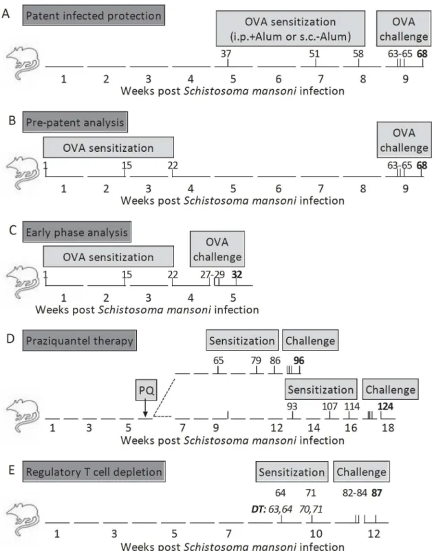
Figure 1. Experimental models for examiningS. mansoni-mediated suppression of AAI.APatent Infected Protection (PiP). Groups of BALB/c female mice were infected withS. mansoni. Inf/OVA and OVA groups of mice were then sensitised with OVA either i.p. with Alum or s.c. without adjuvant at the indicated timepoints. Challenge occurred over three consecutive days in the 9th week of infection and analysis 5 days thereafter. B Prepatent (PP). Sensitizations were started in Inf/OVA and OVA groups during the first weeks of infection, before eggs are released from fecund females. Challenge and analysis were performed as in A. CEarly Phase (EP). AAI was performed entirely over the first 5 weeks of infection. D Praziquantel therapy. During the 6th week of infection, mice were orally treated with PZQ for 5 consecutive days. AAI was then begun after either 2 weeks (upper track) or 6 weeks (lower track). EDepletion of Treg. Groups of C57BL/6 DEREG mice were infected withS. mansoni. In some groups, Treg were depleted using DT injections prior to OVA sensitization at the indicated timepoints. Challenge and analysis occurred in the 12th week of infection.
Bronchoalveolar lavage and cell differentiation analysis To obtain bronchoalveolar lavage (BAL) cells, the lungs of individual mice were rinsed with 1 ml of PBS containing proteinase inhibitor cocktail tablets (Roche Diagnostics Mann-heim, Germany). The resulting fluid was weighed and centrifuged at 230 g for 5 minutes at 4uC. Cells were then re-suspended in PBS containing 2% FCS and then centrifuged (400 rpm for 5 minutes) onto glass slides using the Shandon Cytospin 3 Centrifuge (Thermo Scientific, Hamburg, Germany). After overnight drying slides were stained using the Diff-Quick staining kit (Medion Diagnostics, Langen, Germany). Cell differentiation was determined microscopically.
Detection of OVA-specific IgE
OVA-specific IgE levels were measured in the sera of individual mice. In brief, 96-well ELISA plates (Nunc , Langenselbold, Germany) were coated overnight (4uC) with 1mg per well of OVA grade V diluted in aqua dest. containing
NaHCO3 and Na2CO3. After washing and blocking in 50 mM Tris solution containing 3% BSA, sera was diluted in blocking buffer (1:200 to 1:100,000 dilutions) and a standard of mousea
-OVA IgE antibody (Biozol, Eching, Germany) was applied in two-fold serial dilutions and incubated overnight (4uC). Subse-quently, plates were washed and a-mouse IgE biotinylated
detection antibody (Biozol) was applied and incubated for 2 h (RT). After further washing streptavidin-horseradish peroxidase (HRP) conjugate (R&D Systems GmbH, Wiesbaden, Germany) was added and plates were incubated for 30 min (RT) in the dark. After a final washing step, TMB substrate (BD, Heidelberg, Germany) was applied and reactions were stopped with 2MH2SO4. Finally, ODs were determined at 450 nm using the Sunrise ELISA microplate reader (Tecan, Crailsheim, Germany). The concentration of the samples was then calculated according to the standard curve.
Lung pathology and immunohistochemistry
Paraffin embedded sections (3mm) from the left lungs of individual mice were stained with PAS (Periodic acid-Schiff). Lung tissue inflammation was microscopically determined by the degree of cell infiltration around the basal membrane of bronchi or vessels, which was graded on a scale from 1 to 4. In brief, a value of 1 was assigned for occasional cuffing with inflammatory cells, a value of 2 was assigned for a thin layer (two to three cells thick) of inflammatory cells, a value of 3 was assigned when bronchi or vessels were surrounded by a thick layer of four to five inflammatory cells and a value of 4 was assigned when bronchi or vessels were surrounded by a layer of more than five inflammatory cells. Per lung section, a mean inflammation score was determined. Immunohistochemistry of CD3+
Foxp3+
Treg was performed as previously described [14]. In brief, lung sections were deparaffinized and heated in sodium citrate buffer (pH6.0) for 2 min at high pressure. After washing and blocking, slides were incubated with a goat polyclonal antibody (Ab) against the C terminus of the Foxp3 protein (ab2481, dilution 1:50; Abcam Limited, Cambridge, U.K.). Sections were then exposed to a biotin-conjugated rabbit anti-goat and the EnVision peroxidase kit (Dako-Cytomation). After a further incubation with the second Ab against CD3 (clone UCHT1, 1:25; DakoCytomation) cells were visualized using the alkaline phosphatase–anti-alkaline phospha-tase method [24]. In a blind fashion, Foxp3+
T cells were quantified in individual lung sections within a 1 mm2area using a Zeiss microscope (Axio Lab.A1, Oberkochen, Germany) at 206
magnification.
Flow cytometry staining of Treg Intracellular staining of Foxp3+
CD4+
T cell populations was performed on erythrocyte-depleted mediastinal lymphnode cells (medLN). Prior to staining, Fc receptors were blocked using anti-CD16/CD32 (eBiosciences, Frankfurt, Germany). Thereafter, cells were stained with APC-conjugated anti-CD4 mAb (eBios-ciences). Intracellular Foxp3 levels were detected using the PE-conjugated anti-Foxp3 mAb staining kit according to the manufacture’s instruction (eBiosciences). Fluorescence was ana-lyzed using a flow cytometer and software from BDbiosciences (Heidelberg, Germany). The presence or absence of egfp+
cells in DEREG mice was also detected by flow cytometry in peripheral blood samples from individual mice [20].
In vitrore-stimulation
Erythrocyte-depleted cell suspensions (26105) from individual lung lymph node (LLN) or individual spleens were re-stimulatedin vitrofor 72 hours with 10mg/ml OVA (Grade VI) or with 20mg/
ml soluble egg antigen (SEA) prepared from schistosomal eggs as previously described [13]. Cytokine content in the culture supernatant was then determined using ELISA in accordance with the manufacturer’s instructions (eBiosciences).
Statistical analysis
Statistical differences were analyzed using GraphPad Prism 5 software (San Diego, CA, USA). Parametrically distributed data were analyzed using unpaired t-tests or one-way ANOVA.
Results
Patent infections ofS. mansonireduce allergic airway inflammation
OVA upon challenge did not present any signs of inflammation or cell infiltration (Figures 2A–I and S1C and D). Airway resistance was also measured in the PiP investigation studies and as with the other measured parameters, Inf/OVA mice had resistance levels that were comparable to the control groups (Figure S1E). With regards to immunological responses, OVA-specific Th2 (Figures S2A and B) and regulatory (Figure S2C) recall responses of draining lymph node cells were significantly dampened in Inf/ OVA groups and this was regardless to the sensitisation route. Interestingly, OVA-specific IgE levels in the sera were elevated in Inf/OVA mice that were sensitised i.p. but not s.c. (Figure S2D). Next, we assessed whether protection occurred when infected mice received sensitization rounds within the first 21 days of infection (prepatency) but were challenged during patency (Figure 2B - PP: prepatent). Interestingly, although leucocyte
and eosinophil infiltration was still significantly dampened (Figures 2B and E respectively), within this experimental scenario, no protective influence on lung inflammation could be observed in the Inf/OVA group (Figure 2H). Finally, experiments were performed in which AAI was induced and assessed during prepatency i.e. the first five weeks of infection (Figure 2C - EP: early phase). Here,S. mansoniinfection elicited no protection since cellular infiltration and lung inflammation scores were equal to OVA groups (Figures 2C, F and I). Parasitological assessment of the S. mansoni groups, in all infection scenarios, showed no differences between granuloma size or egg burden in the liver (data not shown). Eggs were not detected in the EP experiments since at week 4–5, adult female worms are not yet fecund.
In another set of experiments we investigated the development of AAI upon curing a patentS. mansoniinfection (Figure 1D). In Figure 2. Suppression of AAI inS. mansoni-infected mice requires patency.Groups of BALB/c female mice were infected withS. mansoni. Left PiP experiments: After the 5th week of infection, OVA sensitizations were administered either i.p. with alum or s.c. in the absence of adjuvant (Figure 1A). Middle PP experiments: sensitizations were given during the first weeks of infection (before patency) but challenge was performed at the same time-point of infection as in PiP investigations (Figure 1B). Right EP studies: both sensitizations and challenge were performed before the onset of patency (Figure 1C). Upon AAI analysis, individual mice were assessed for leucocyte numbers (A–C) and eosinophil levels (D–F). Lung sections were also scored for inflammation (G–I). In i.p.+Alum experiments displayed in A, D and G, bars show mean+SD of individual mice from 5 independent infection/allergy experiments (PBS, n = 21; OVA, n = 34; Inf/OVA, n = 29 and Inf., n = 28). In s.c.-Alum experiments OVA, n = 8 and Inf/OVA, n = 7. In PP experiments B, E and H, bars show mean+SD of individual mice from 2 independent infection/allergy experiments (PBS, n = 2; OVA, n = 14; Inf/OVA, n = 13 and Inf., n = 10). In EP experiments C, F and I, bars show mean+SD of individual mice from 1 or 3 similar independent infection/allergy experiments (PBS, n = 2; OVA, n = 10; Inf/OVA, n = 10 and Inf., n = 4). Asterisks show statistical differences (ANOVA or Student’s t test) between the groups indicated by the brackets (*p,0.05, **p,0.01, ***p,0.001).
brief, groups of infected mice were treated with praziquantel (PZQ) during the 6th week of infection. After a further 2 or 6 weeks, OVA sensitization commenced, thus, analysis of AAI was performed after either 12 or 18 weeks post infection (or 6 and 10 weeks post PZQ-therapy). Interestingly, infected PZQ-treated groups of mice (Inf/OVA/PQ) analysed after 12 weeks but not 18 weeks post-infection were still significantly protected from cellular infiltration (Figures S3A–D) but not inflammation (Figures S3E and F). As shown in Figure S3G, egg counts in the liver were reduced after 10 weeks of PZQ therapy. This was also confirmed in the amount of released eggs in the stool since none were detected on the days of analysis (data not shown). Moreover, upon assessment of individual liver sections, there was a significant reduction in viable eggs in the liver after 10 weeks of PZQ treatment (Figure S3H).
Absence of Foxp3+Treg in lung sections of Inf/OVA mice As mentioned above, previous studies have shown that Treg play a role in helminth-mediated immunomodulation during AAI. However, questions pertaining to their redistribution into the lymphatics or relevant organ tissues remain unanswered. Thus, we analysed the levels and distribution of Foxp3+
T cells in the draining lymph nodes (LLN) of mice from the PiP experiments (Figure 2A). Figure 3A shows that the absolute cell counts in OVA treated mice, regardless of infection, is higher than in PBS groups. No significant differences could be observed between OVA and Inf/OVA groups. With regards to the percentage of Foxp3+T
cells in the CD4+
T cell compartment, these regulatory cells were elevated in Inf/OVA mice but not in OVA groups alone (c.fOVA and Inf/OVA in Figure 3B). Interestingly, Treg numbers were high in the Inf. group as well. Within the lung however, numerous CD3+
Foxp3+
T cells could be identified in sections from OVA mice (Figure 3C) but not Inf/OVA groups (Figure 3D). These sections were comparable to those sampled from the PBS groups (Figure 3E). These impressions were verified through further quantification of the Treg numbers in the lung sections of individual mice. As shown in Figure 3F, CD3+Foxp3+Treg were
significantly higher in OVA but not Inf/OVA group.
Lack of Treg during sensitization impedesS. mansoni-mediated suppression of AAI
To address whetherS. mansoni-mediated protection of AAI was mediated by Treg we infected DEREG mice, which allow the specific depletion of egfp+
Foxp3+
cells at specified time-points through the administration of DT [20]. Initially we performed such depletion experiments in BALB/c DEREG mice but due to their higher sensitivity to DT application and schistosome infection, Treg depletion during AAI development resulted in high mortality rates (over 90%, data not shown). Therefore, we continued the experiments with DEREG mice on a C57BL/6 background. As depicted in Figure 1E, Treg were depleted in groups of infected and non-infected mice during sensitization. As observed before within the PiP studies described above, Inf/OVA groups had significantly lower levels of leucocytes and eosinophils when compared to OVA groups (c.f.bars 1 and 4 in Figures 4A and B). However, depletion of Treg in the infected group (Inf/ OVADT) prevented the previously observed suppressive effects (c.f. bar 3 with bars 1 and 4 in Figures 4A and B) and levels of cellular infiltration even equalled those of the OVADT groups. In association with the current literature [16], non-infected OVA groups, depleted of Treg, also showed increased leucocyte and eosinophil infiltration(c.f.bars 1 and 2 in Figures 4A and B). With regards to inflammation score depletion of Treg abrogated the suppressive effect of schistosome infection (Figure 4C c.f. bars 3
and 4 and Figures 4E and G). Moreover, Treg depletion led to enhanced lung inflammation when compared with the OVA group (Figures 4Cc.fbar 1 with 2 and 3 and Figures 4D,4F and 4G). Immunologically, both Treg depleted groups showed significantly higher levels of OVA-specific IgE in the sera (Figure 4H) and OVA-specific IL-5 upon recall of splenocytes cells (Figure 4I). Interestingly, schistosome-specific responses in the infected groups were also significantly increased in the Inf/ OVADTgroups (Figure 4J). Moreover, these mice showed lower egg burden in the liver (Figure 4K). These data correlate to our earlier studies in which we depleted Tregs inS. mansoniinfected mice by administering an anti-CD25 antibody. There, we also observed elevated schistosome-specific Th2 responses and strongly decreased egg numbers in the livers of Treg-depleted S. mansoni infected animals [13]. During these experiments, we controlled for the depletion of Treg by observing levels of CD4+
egfp+
T cells in peripheral blood. Figure S4A shows a representative comparison of CD4+
egfp+
T cells in infected (left) and non-infected (right) mice on the day before depletion and 3 days thereafter. As shown in the bottom panel of flow cytometry images, no egfp+
cells are visible after depletion. Levels were measured again following asthma induction (Figure S4B). Here, the percentage of Foxp3+ Treg in infected-depleted mice was
approximately 50% lower than in non-infected depleted mice (4.7% vs 8.24%). This was observed in all the experimental mice (Figure S4C) and indicates that the re-establishment of Treg in infected mice is slower than in non-infected controls and provides a possible explanation as to why infected mice are no longer suppressed from AAI development.
Discussion
Another factor which has to be stringently addressed is age, since it is known that age effects the development of AAI in mice [22]. To ensure that our findings were not due to an age-bias, all mice were age–matched at the onset of experiments and housed under the same conditions.
Evidently, S. mansoni-infected mice had reduced AAI when sensitized and challenged during patent infection and moreover, Inf/OVA mice still had dampened responses when challenge but not sensitization occurred in the patent phase. However, no protection was observed in Inf/OVA mice when both sensitization Figure 3. Increased Foxp3+Treg frequency in the draining lymph nodes of Inf/OVA mice is not reflected in lung sections.A) Total cell counts and B) the number of CD4+
Foxp3+
T cells in the draining lymph nodes of individual mice on the day of analysis in PiP studies. Symbols depict the levels in each mouse and data shows values from 5 independent infection studies (PBS, n = 21; OVA, n = 34; Inf/OVA, n = 29 and Inf., n = 28). CD3+Foxp3+T cells in the lung sections of C) OVA mice, D) Inf/OVA mice and E) control groups (PBS) were determined by immunohistochemical staining. Arrows depict positive cells. F) Quantification of CD3+
Foxp3+
T cells in individual lung sections was determined within a 1 mm2area. Asterisks show statistical differences (ANOVA) between the groups indicated by the brackets (*p,0.05, **p,0.01, ***p,0.001).
and challenge were performed in the prepatent phase (EP studies) or six weeks after praziquantel treatment. Taken together these results clearly highlight the requirement of patency for infection-mediated AAI suppression. Interestingly, our anti-helminthic treatment experiments revealed that some level of protection against AAI remained when sensitization commenced two weeks after treatment. At this time-point although many tissue-residing eggs are still viable, most of the worms have died and only stunted, non-egg producing worms are left over [31,32]. Complete abrogation of the protective effects was only observed in those experiments analyzed on the 18th week of infection, when sensitization began 6 weeks after PZQ treatment. Here it is considered that the proportion of viable eggs has strongly decreased and our findings here coincide with these data [13]. In association, a human study demonstrated that schistosome-infected individuals displayed increased levels of HDM-specific IgE and low responses to skin prick tests [33]. However, following anti-helminthic treatment these patients presented increased HDM skin reactivity [34–36] indicating that in order to maintain a suppressive milieu an ongoing patent infection is required. The patently-induced protection was also independent of the OVA-application route and the presence of alum. OVA-specific IgE levels in infected mice were increased when OVA was injected i.p. with alum, which has been reported to drive the induction of the disease independently from mast-cell mediated early phase
reactions [37,38]. OVA-specific IgE levels remained unaltered when alum was applied subcutaneously which indicates that the suppression of AAI occurs independently of IgE levels. Thus, we conclude that the suppressive effects mediated by schistosome infection are not due to defective OVA priming which supports earlier findings [30].
Concerning the possible underlying mechanisms that lead to dampened allergic responses in schistosome infected animals, we and others have shown previously that Foxp3+
Treg expand homeostatically during the course of infection and this phenom-enon already starts when the first SEA-specific Th1 responses can be detected around the 5th week of infection [13]. The first hint that Treg might be involved in schistosome-mediated protection of AAI came from the finding that when compared to OVA groups, Foxp3+
Treg were significantly elevated in the LLN of Inf/OVA and Inf alone groups of mice. However, whereas numerous CD3+Foxp3+Treg were present in the lung tissues of OVA groups
very few were detectable in Inf/OVA mice. Therefore, we consider that during schistosome infection, regulation of lung infiltrate upon challenge occurs at the draining LLN and not within the lung tissue itself. It will be interesting to decipher how Treg in Inf/OVA mice are retained in the LLN and whether these are indeed infection-induced Foxp3+
T cells. In our previous studies we showed that infection-induced Treg but not Treg from naive mice suppress SEA-specific CD4+
T cell effector responses Figure 4. Depletion of Treg during sensitization reverts schistosome-mediated dampening of AAI.As depicted in Figure 1F, groups of C57BL/6 DEREG mice were infected withS. mansoni. On the 9thand 10thweek of infection mice were sensitized i.p. with OVA in the presence of alum. Prior to sensitization, Treg were depleted by the administration of DT. During the 12th week of infection, mice were challenged and assessed for the number of leucocytes (A) and eosinophils (B) in the BAL. Levels of inflammation were also scored using lung sections stained with PAS (C–G). OVA-specific IgE levels were measured in the sera of individual mice via ELISA (H). OVA-OVA-specific responses were measured following stimulation of spleen cells (26105) with 10mg/ml OVA (I). In addition, schistosome-specific responses were evaluated upon re-stimulation of spleen cells with SEA (25mg/ ml) (J). After 72 hours, IL-5 levels were determined in the culture supernatant by ELISA (I and J). Egg burden was measured in livers of individually infected mice (K). Bars show data from one of two similar experiments (OVA, n = 7; OVADT, n = 7; Inf/OVADT, n = 5; Inf/OVA n = 8; PBSDTn = 3). Asterisks show statistical differences (ANOVA or Student’s t test) between the groups indicated by the brackets (*p,0.05, p,0.01, ***p,0.001).
doi:10.1371/journal.pntd.0002379.g004
and develop a unique gene expression profile that includes upregulation of granzyme B and anti-inflammatory molecules such as SLPI (secretory leucocyte peptidase inhibitor) [14]. This change of phenotype could account for their selective suppressive activity since it was shown, for example, that activated Treg cells can actually kill B cells in a granzyme-B-dependent manner [39]. It is now discussed that Treg adapt their mode of immune suppression according to the altered requirements found under inflammatory conditions in comparison to those in the steady-state [12]. The role of these molecules in Treg-mediated suppression of allergiesin vivo will be an interesting field of future research.
To assess whether Treg played a role in the development of AAI during infection we employed the DEREG mouse model which allows depletion of Treg at the experimenter’s desired time-points [20,40]. This model ensures that all Foxp3+
T cells are depleted regardless of their CD25-coexpression and also circumvents the criticized application of anti-CD25 antibodies in which recently activated effector T cells are targeted as well [20,41,42]. BALB/c mice have a higher morbidity due to schistosome infection and additional DT application further increased mortality rates. Thus, we changed to the DEREG mice on a C57BL/6 background, which tolerated our experimental procedure [20]. Interestingly, Treg depletion during OVA-sensitization in C57BL/6 DEREG mice resulted in a loss of protection inS. mansoni infected mice. Furthermore, cellular infiltration levels and pathology scores were higher in Inf/OVADTgroups when compared to control OVA groups. These findings confirm other studies that have shown the requirement for Treg in AAI [16] and that Foxp3+
T cells down-modulate airway eosinophilia but not AHR and IgE levels in a model of SIT-induced tolerance [43]. Indeed, OVA-specific IgE levels were enhanced in both depleted groups indicating a role of Treg in initiating Th2 responses. In addition, schistosome-specific immune responses were elevated in Inf/OVADT mice and alongside the strongly reduced egg burden this confirms our previous findings in schistosome-infected mice treated with anti-CD25 antibody [13]. In addition, we noted a delayed reconsti-tution of expanding Treg in Inf/OVADT mice compared to OVADT mice after Treg depletion. Thus, the removal of Treg during a chronic infection, in contrast to the steady-state or immunization situation, probably gives rise to more pronounced and quicker Th responses against schistosome antigens. This might favor immune reactions against OVA antigens and eventually results in decelerated or unbalanced Treg reconstitution.
Collectively, by comparative experiments using different proto-cols for AAI our data support recent findings that schistosome infection can indeed suppress allergic airway responses and that this suppression requires patency and the continuous release of eggs. Furthermore, this suppression is partly mediated by expanding Foxp3+Treg cells within the draining lymph nodes of
the lung. We therefore propose the concept that patent infection inhibits the effector phase of AAI through schistosome egg-derived factors and not worm or schistosomula antigens. An in-depth analysis of egg components and their potential to drive Treg expansion or induction could therefore have potential therapeutic value.
Supporting Information
Figure S1 Lung pathology and airway resistance in PiP
investigations. Groups of BALB/c mice were infected with S. mansoni. During the 6th to 8th week of infection, mice were thrice sensitised i.p. with OVA and Alum. Following aerosolic OVA challenge lung sections from individual mice were assessed for their level of inflammation. Representative PAS stained lung
sections from OVA, Inf/OVA, PBS control and infected alone groups of mice are depicted in A–D respectively and in E airway resistance from groups of mice in PiP investigations. Symbols show mean+SD of each group of mice (n = 12/group). Asterisks show significant differences between the groups indicated by the brackets (**p,0.01).
(TIF)
Figure S2 S. mansoni infected mice present suppressed OVA-specific Th responses and IgE levels upon the development of AAI. Groups of BALB/c mice were infected withS. mansoniaccording to the PiP investigation protocol. During the 6th to 8th week of infection, mice were thrice sensitised either i.p. with OVA and Alum or s.c. with OVA alone. Following challenge, erythrocyte-depleted LLN cells (26105per well) were re-stimulatedin vitrowith OVA (10mg/ml) for 72 hours. Culture supernatants were then screened for their content of A) IL-5, B) IL-13 or C) IL-10 by ELISA. D) OVA-specific IgE levels were measured in the sera of individual mice. Bars depict mean+SD. Asterisks show statistical differences (Student’s t test) between the groups indicated by the brackets (*p,0.05, **p,0.01).
(TIF)
Figure S3 Worm elimination through praziquantel therapy
reverts protection against AAI. Groups of BALB/c mice were infected with S. mansoni. During the 6th week of infection, schistosomes were killed by the oral administration of praziquantel (100 mg/kg body weight) over 5 consecutive days. i.p. OVA sensitizations commenced after either 2 or 6 weeks post PZQ treatment (Figure 1D). Groups of non-infected OVA mice were maintained under the same conditions throughout the experiment. A–F shows the changes in AAI parameters in mice analyzed after 6 (A, C and E) or 10 (B, D and F) post PZQ therapy. Graphs show leucocyte infiltration (A and B), eosinophil number (C and D) and inflammation score (E and F) in lungs of individual mice. G shows egg counts in the liver following digestion with KOH. H depicts number of viable eggs in individual livers following assessment with Masson’s stained liver sections. Bars show mean6SD from one of two experiments containing 6–8 mice per group. Asterisks show statistical differences (ANOVA) between the groups indicated by the brackets (*p,0.05, **p,0.01).
(TIF)
Figure S4 Effective Treg depletion but reduced recovery of Foxp3+
T cells inS. mansoniinfected DEREG mice upon asthma induction. In A) the efficacy of Treg depletion was controlled by analyzing the percentage of cells in peripheral blood by flow cytometry (see Figure 1E). In brief, prior to depletion (d50, upper panel) and 3 days after DT injections (d53, lower panel) the percentage of CD4+
egfp+
T cells was observed in S. mansoni infected (left) and naive DEREG mice (right). B) Upon asthma induction, the percentage of Treg was observed again in peripheral blood (d74). Representative dot plot on the left depicts the levels of CD4+
egfp+
T cells in a Inf/OVADTmouse and the right image those observed in a OVADTmouse. C) Bars represent the mean+SEM of CD4+
egfp+
T cells on d74 recovered from 4–5 mice per group. Percentages were calculated by flow cytometry. Asterisks show statistical differences (Student’s t test) between the groups indicated by the brackets (**p,0.01).
(TIF)
Acknowledgments
Author Contributions
Conceived and designed the experiments: CPdC LEL KS. Performed the experiments: KS LEL MR ELV. Analyzed the data: CPdC LEL KS HG.
Contributed reagents/materials/analysis tools: HG TS. Wrote the paper: CPdC LEL KS.
References
1. Masoli M, Fabian D, Holt S, Beasley R (2004) The global burden of asthma: executive summary of the GINA Dissemination Committee report. Allergy 59: 469–478.
2. Beasley R, Crane J, Lai CK, Pearce N (2000) Prevalence and etiology of asthma. J Allergy Clin Immunol 105: S466–472.
3. Barnes PJ (2008) Immunology of asthma and chronic obstructive pulmonary disease. Nat Rev Immunol 8: 183–192.
4. Carvalho EM, Bastos LS, Araujo MI (2006) Worms and allergy. Parasite Immunol 28: 525–534.
5. Leonardi-Bee J, Pritchard D, Britton J (2006) Asthma and current intestinal parasite infection: systematic review and meta-analysis. Am J Respir Crit Care Med 174: 514–523.
6. Zaccone P, Burton O, Miller N, Jones FM, Dunne DW, et al. (2009) Schistosoma mansoni egg antigens induce Treg that participate in diabetes prevention in NOD mice. Eur J Immunol 39: 1098–1107.
7. Smits HH, Hammad H, van Nimwegen M, Soullie T, Willart MA, et al. (2007) Protective effect of Schistosoma mansoni infection on allergic airway inflammation depends on the intensity and chronicity of infection. J Allergy Clin Immunol 120: 932–940.
8. Song X, Shen J, Wen H, Zhong Z, Luo Q, et al. (2011) Impact of Schistosoma japonicum infection on collagen-induced arthritis in DBA/1 mice: a murine model of human rheumatoid arthritis. PLoS One 6: e23453.
9. Correale J, Farez M (2009) Helminth antigens modulate immune responses in cells from multiple sclerosis patients through TLR2-dependent mechanisms. J Immunol 183: 5999–6012.
10. Medeiros M, Jr., Figueiredo JP, Almeida MC, Matos MA, Araujo MI, et al. (2003) Schistosoma mansoni infection is associated with a reduced course of asthma. J Allergy Clin Immunol 111: 947–951.
11. Lloyd CM, Hawrylowicz CM (2009) Regulatory T cells in asthma. Immunity 31: 438–449.
12. Yamaguchi T, Wing JB, Sakaguchi S (2011) Two modes of immune suppression by Foxp3(+) regulatory T cells under inflammatory or non-inflammatory conditions. Semin Immunol 23: 424–430.
13. Layland LE, Rad R, Wagner H, da Costa CU (2007) Immunopathology in schistosomiasis is controlled by antigen-specific regulatory T cells primed in the presence of TLR2. Eur J Immunol 37: 2174–2184.
14. Layland LE, Mages J, Loddenkemper C, Hoerauf A, Wagner H, et al. (2010) Pronounced phenotype in activated regulatory T cells during a chronic helminth infection. J Immunol 184: 713–724.
15. Kearley J, Robinson DS, Lloyd CM (2008) CD4+CD25+regulatory T cells reverse established allergic airway inflammation and prevent airway remodeling. J Allergy Clin Immunol 122: 617–624 e616.
16. Baru AM, Hartl A, Lahl K, Krishnaswamy JK, Fehrenbach H, et al. (2010) Selective depletion of Foxp3+ Treg during sensitization phase aggravates experimental allergic airway inflammation. Eur J Immunol 40: 2259–2266. 17. van der Vlugt LE, Labuda LA, Ozir-Fazalalikhan A, Lievers E, Gloudemans
AK, et al. (2012) Schistosomes induce regulatory features in human and mouse CD1d(hi) B cells: inhibition of allergic inflammation by IL-10 and regulatory T cells. PLoS One 7: e30883.
18. Amu S, Saunders SP, Kronenberg M, Mangan NE, Atzberger A, et al. (2010) Regulatory B cells prevent and reverse allergic airway inflammation via FoxP3-positive T regulatory cells in a murine model. J Allergy Clin Immunol 125: 1114–1124 e1118.
19. Pacifico LG, Marinho FA, Fonseca CT, Barsante MM, Pinho V, et al. (2009) Schistosoma mansoni antigens modulate experimental allergic asthma in a murine model: a major role for CD4+CD25+Foxp3+T cells independent of interleukin-10. Infect Immun 77: 98–107.
20. Lahl K, Loddenkemper C, Drouin C, Freyer J, Arnason J, et al. (2007) Selective depletion of Foxp3+regulatory T cells induces a scurfy-like disease. J Exp Med 204: 57–63.
21. Layland LE, Wagner H, da Costa CU (2005) Lack of antigen-specific Th1 response alters granuloma formation and composition in Schistosoma mansoni-infected MyD88-/- mice. Eur J Immunol 35: 3248–3257.
22. Busse PJ, Zhang TF, Srivastava K, Schofield B, Li XM (2007) Effect of ageing on pulmonary inflammation, airway hyperresponsiveness and T and B cell
responses in antigen-sensitized and -challenged mice. Clinical and Experimental Allergy 37: 1392–1403.
23. Conrad ML, Yildirim AO, Sonar SS, Kilic A, Sudowe S, et al. (2009) Comparison of adjuvant and adjuvant-free murine experimental asthma models. Clin Exp Allergy 39: 1246–1254.
24. Hsu SM, Raine L, Fanger H (1981) The use of antiavidin antibody and avidin-biotin-peroxidase complex in immunoperoxidase technics. Am J Clin Pathol 75: 816–821.
25. Wilson RA, Coulson PS (2009) Immune effector mechanisms against schistosomiasis: looking for a chink in the parasite’s armour. Trends Parasitol 25: 423–431.
26. Schulte S, Sukhova GK, Libby P (2008) Genetically programmed biases in Th1 and Th2 immune responses modulate atherogenesis. American Journal of Pathology 172: 1500–1508.
27. Lohoff M, Gessner A, Bogdan C, Rollinghoff M (1998) Experimental murine leishmaniasis and the Th1/Th2 cell concept. Tokai J Exp Clin Med 23: 347– 350.
28. Baumgart M, Tompkins F, Leng J, Hesse M (2006) Naturally occurring CD4+Foxp3+regulatory T cells are an essential, IL-10-independent part of the immunoregulatory network in Schistosoma mansoni egg-induced inflammation. J Immunol 176: 5374–5387.
29. Kane CM, Cervi L, Sun J, McKee AS, Masek KS, et al. (2004) Helminth antigens modulate TLR-initiated dendritic cell activation. J Immunol 173: 7454–7461.
30. Mangan NE, van Rooijen N, McKenzie AN, Fallon PG (2006) Helminth-modified pulmonary immune response protects mice from allergen-induced airway hyperresponsiveness. J Immunol 176: 138–147.
31. Cioli D, Botros SS, Wheatcroft-Francklow K, Mbaye A, Southgate V, et al. (2004) Determination of ED50 values for praziquantel in praziquantel-resistant and -susceptible Schistosoma mansoni isolates. Int J Parasitol 34: 979–987. 32. Wilson MS, Cheever AW, White SD, Thompson RW, Wynn TA (2011) IL-10
Blocks the Development of Resistance to Re-Infection with Schistosoma mansoni. Plos Pathogens 7: e1002171.
33. van den Biggelaar AH, van Ree R, Rodrigues LC, Lell B, Deelder AM, et al. (2000) Decreased atopy in children infected with Schistosoma haematobium: a role for parasite-induced interleukin-10. Lancet 356: 1723–1727.
34. van den Biggelaar AH, Lopuhaa C, van Ree R, van der Zee JS, Jans J, et al. (2001) The prevalence of parasite infestation and house dust mite sensitization in Gabonese schoolchildren. Int Arch Allergy Immunol 126: 231–238. 35. van den Biggelaar AH, Rodrigues LC, van Ree R, van der Zee JS,
Hoeksma-Kruize YC, et al. (2004) Long-term treatment of intestinal helminths increases mite skin-test reactivity in Gabonese schoolchildren. J Infect Dis 189: 892–900. 36. Lynch NR, Hagel I, Perez M, Di Prisco MC, Lopez R, et al. (1993) Effect of anthelmintic treatment on the allergic reactivity of children in a tropical slum. J Allergy Clin Immunol 92: 404–411.
37. Yu M, Tsai M, Tam SY, Jones C, Zehnder J, et al. (2006) Mast cells can promote the development of multiple features of chronic asthma in mice. J Clin Invest 116: 1633–1641.
38. Nakae S, Ho LH, Yu M, Monteforte R, Iikura M, et al. (2007) Mast cell-derived TNF contributes to airway hyperreactivity, inflammation, and TH2 cytokine production in an asthma model in mice. J Allergy Clin Immunol 120: 48–55. 39. Zhao DM, Thornton AM, DiPaolo RJ, Shevach EM (2006) Activated
CD4+CD25+T cells selectively kill B lymphocytes. Blood 107: 3925–3932. 40. Zheng Y, Rudensky AY (2007) Foxp3 in control of the regulatory T cell lineage.
Nat Immunol 8: 457–462.
41. Hall JA, Bouladoux N, Sun CM, Wohlfert EA, Blank RB, et al. (2008) Commensal DNA limits regulatory T cell conversion and is a natural adjuvant of intestinal immune responses. Immunity 29: 637–649.
42. Lahl K, Sparwasser T (2011) In vivo depletion of FoxP3+Tregs using the DEREG mouse model. Methods Mol Biol 707: 157–172.
43. Maazi H, Shirinbak S, Willart M, Hammad HM, Cabanski M, et al. (2012) Contribution of regulatory T cells to alleviation of experimental allergic asthma after specific immunotherapy. Clinical and Experimental Allergy 42: 1519– 1528.