Co llage n arrange m e nt in he patic
granulo m a in m ice infe cte d with
Schisto so m a m anso ni
: de pe nde nce
o n fibe r radiatio n ce nte rs
1Departamento de Patologia, Instituto O swaldo Cruz, and 2Programa de Computação Científica, Presidência,
Fundação O swaldo Cruz, Rio de Janeiro, RJ, Brasil 3Department of Agricultural Engineering, Technion, Israel Institute of Technology, Haifa, Israel
H.L. Lenzi1, E. Kimmel3, H. Schechtman2, M. Pelajo-Machado1, B.S. Vale1, M.S. Panasco1 and J.A. Lenzi1
Abstract
The collagen structure of isolated and in situ liver granuloma from
Swiss Webster mice infected with Schistosoma mansoni was
sequen-tially and three-dimensionally analyzed during different times of infection (early acute, acute, transitional acute-chronic, and chronic phases) by laser scanning confocal microscopy and electron scanning variable vacuum microscopy. The initial granuloma structure is char-acterized by vascular collagen residues and by anchorage points (or fiber radiation centers), from where collagenous fibers are angularly shed and self-assembled. During the exudative-productive stage, the self-assembly of these fibers minimizes energy and mass through continuous tension and focal compression. The curvature or angles between collagen fibers probably depends on the fibroblastic or myofibroblastic organization of stress fibers. Gradually, the loose unstable lattice of the exudative-productive stage transforms into a highly packed and stable architecture as a result of progressive com-pactness. The three-dimensional architecture of granulomas provides increased tissue integrity, efficient distribution of soluble compounds and a haptotactic background to the cells.
Co rre spo nde nce H.L. Lenzi
Departamento de Patologia Instituto O swaldo Cruz, FIO CRUZ Avenida Brasil, 4365
21045-900 Rio de Janeiro, RJ Brasil
Fax: + 55-21-598-4350 or 598-4466, or 590-3495
E-mail: hlenzi@ gene.dbbm.fiocruz.br
Presented at the 5th Brazilian Symposium on Extracellular Matrix - SIMEC, Angra dos Reis, RJ, Brasil, September 7-10, 1998.
Research supported by FIO CRUZ and CNPq. M. Pelajo-Machado is supported by CAPES and M.S. Panasco is supported by FAPERJ.
Received February 26, 1999 Accepted March 2, 1999
Ke y wo rds
·Granuloma
·Schistosom a m ansoni
·Collagen
·Extracellular matrix ·Confocal microscopy
A granuloma is characterized by the pres-ence of clusters of cells of the mononuclear phagocyte series with or without other cell types (1). The clusters of mononuclear cells are compact (organized) (2), and epithelioid cells, in some cases, define the typical granu-lomatous lesion (3). Granulomas function as little organs, presenting stages of maturation and involution during their development, and final disappearance. Granulomas arise from
colonial aggregations of monocyte/macro-phages, sometimes forming giant syncytial cells. The development of a granuloma de-pends on the etiologic agent, which may be intracellular (e.g. mycobacteria), intra- and extracellular (e.g. Paracoccidioides brasili-ensis) or extracellular (4). Schistosomal
(BH) isolate of S. mansoni. The mice were
killed at the following times after infection: 35, 40, 45, 60, 70, 90, 120 and 180 days. Liver fragments were homogenized for 2 min in a blender and the suspensions were filtered through grids to select isolated granu-lomas of two different sizes: smaller and larger than 300 µm (12). Purified granulo-mas and liver fragments were fixed in Carsons modified Millonigs phosphate-buffered formalin, pH 7.4 (13), and embed-ded in paraffin. Thick histological sections were stained with phosphomolybdic acid-picrosirius red (PMA-PSR) (14) for three-dimensional study of collagenous fibers by confocal laser scanning microscopy (CLSM)
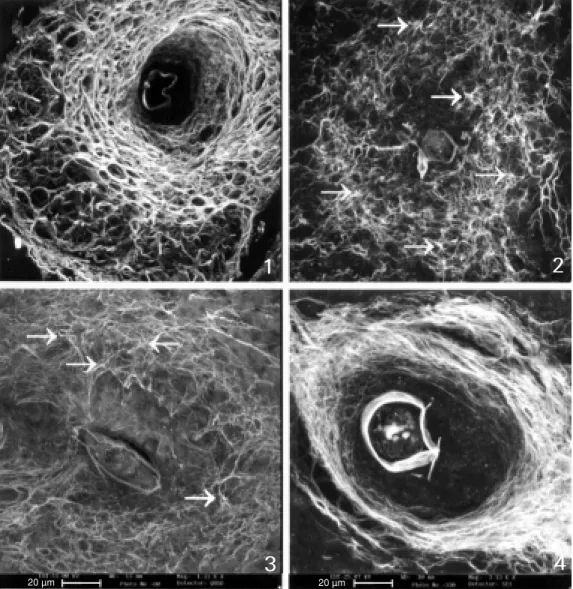
Figure 1 - (All the figures, except 7 and 8, represent black and w hite images of liver granulo-mas originated from 3-D recon-structions made by CSLM ). Exu-dat ive-product ive st age w it h three (internal, medial or para-central and external) defined lay-ers. The external layer is ex-panded and f orm s a loose stroma favoring perigranuloma-tous hem opoietic m etaplasia. The medial layer show s a honey-comb arrangement of collagen fibers, seen also in Figure 7. The central layer is devoid of collagen in the periovular subregion, and presents a smaller amount of fi-bers, w hich are thin.
Figures 2 and 3 - Initial exuda-tive-productive stage presenting a mesh-like pattern of collagen fibers, show ing several isolated fiber anchorage points (fiber ra-diation centers) (arrow s).
Figure 4 - Productive stage w ith a com pact concentric m edial layer.
Granuloma size in acute Schistosoma mansoni infection is determined primarily
by T cell reactivity, and is, therefore, sensi-tive to IL-10 (5,6), while the antibody has a minor effect (7). In contrast, in chronic in-fections, downregulation of granuloma size requires an antibody and downmodulated T cells (8). The regulation of fibrosis in the liver may differ from the regulation of granu-loma size (7,9-11).
The histoarchitecture of hepatic schisto-somal granulomas was examined by three-dimensional analysis of isolated or in situ
granulomas from Swiss Webster mice. The animals were infected by percutaneous ex-posure to 70 cercariae of the Belo Horizonte
1
2
3
4
(LSM 410, Zeiss, Jena, Germany). Deparaf-fined sections were desiccated in an incuba-tor at 37oC for 1 h, and analyzed by scanning
electron microscopy (SEM), low-vacuum mode (435-VP, Leo, Cambridge, England).
We observed, as previously described (15), that the granuloma stage in the liver is preceded by a lytic pre-granulomatous phase (exudative phase) which produces a spheri-cal space by destruction of the parenchyma for the final establishment and organization of the lesion. In the granulomatous stage, cellular adhesion and sorting play an impor-tant role. The effects of aggregation and sorting create different zones in the mature granuloma structure, where the combination of different types of cells consistently forms an internal layer (peri-ovular), enveloped by paracentral and external layers (Figure 1). Probably the cells with stronger mutual
ad-hesion aggregate in the center, whereas less cohesive cells remain on the surface. Cells appear to migrate from the less adhesive (external layer) to the more cohesive layer (internal layer rich in CD44+ cells) (data not shown) through an adhesive haptotactic gra-dient provoked by egg products. They fol-low a centripetal migration in a diffusion-generated radial concentration gradient (di-rect migration model) (16).
The three-dimensional tissue architecture of the granuloma, as observed by CLSM and SEM (variable vacuum), presents extracel-lular matrix (ECM) fibers which are initially arranged in a mesh pattern (Figures 2 and 3), evolving to a final and compact concentric arrangement (Figure 4). These two patterns of fiber arrangement correspond to two dif-ferent collagen populations which appear successively during granuloma evolution:
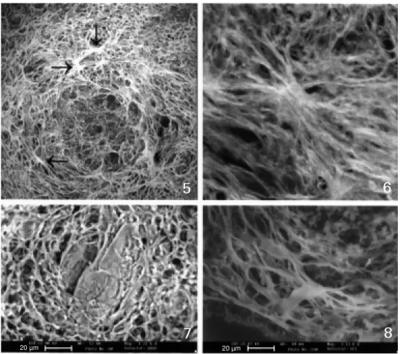
Figure 5 - Exudative-productive stage w ith intense fiber com-pactness, clearly show ing sev-eral fiber anchorage points (fiber radiation centers) (arrow s).
Figures 6 and 8 - Details of fiber anchorage points or fiber radia-tion centers seen by CSLM (Fig-ure 6) and by TEM (low vacuum) (Figure 8). From the anchorage points the fibers are emitted or projected under restricted an-gles.
Figure 7 - Honeycomb aspect of the m edial layer (TEM - low vacuum).
5
6
7
8
Re fe re nce s
1. Turk JL (1992). Granulomatous disease. In: M cGee JOD, Isaacson PG & Wright NA (Editors), Oxford Textbook of Patholo-gy. Oxford University Press, Oxford, 394-406.
2. Adams DO (1983). The biology of the granuloma. In: Ioachim HL (Editor), Pa-thology of Granulomas. Raven Press, New York, 1-20.
3. M ariano M (1995). The experim ental granuloma. A hypothesis to explain the persistence of the lesion. Revista do Instituto de M edicina Tropical de São Paulo, 37: 1-15.
4. Lenzi HL, Lenzi JA, Kerr IB, Antunes SLG, M ota EM & Oliveira DN (1991). Extracel-lular matrix in parasitic and infectious dis-eases. M emórias do Instituto Osw aldo
Cruz, 86 (Suppl III): 77-90.
5. Flores-Villanueva PO, Zheng XX, Strom TB & Stadecker M J (1996). Recombinant IL-10 and IL-10/ Fc treatment dow n-regu-lates egg antigen-specific delayed hyper-sensitivity reactions and egg granuloma formation in schistosomiasis. Journal of Immunology, 156: 3315-3320.
6. Wynn TA, M oraw etz R, Scharton-Kersten
collagen type III containing reticular fibers is later replaced by collagen type I contain-ing collagen fibers (17).
The arrangement and distribution of the collagen network, even in its initial mesh pattern, seems to be preferentially oriented toward the internal secretory cells (fibro-blasts and/or myofibro(fibro-blasts), aiming to hold in place the tissues surrounding the new organoid structure.
The curvature or angles between col-lagen fibers probably depends on the fibro-blast or myofibrofibro-blast organization. In fact, these cells form anchorage points, i.e. fiber radiation centers, from where collagen fi-bers are shed and self-assembled (Figures 5, 6 and 8) sometimes forming a honeycomb arrangement (Figures 1 and 7).
The large amount of ECM produced by granuloma cells appears to be independent of the surrounding forces, and thus empha-sizes the biological role, compared to the mechanical role, of ECM molecules in the formation of the granuloma. This is also supported by the observation that independ-ently of the organ (e.g. liver, lung, intestine, pancreas, milky spot) or site (superficial (sub-capsular or subpleural) or deep in the center of the parenchyma) where the granulomas are located, they always tend to be spherical. Moreover, when granulomas were mechani-cally cut, there was no extrusion of their content from the core to the periphery (data not shown), which indicates that their struc-ture does not present internal pressure coun-terbalanced by tension in the fibrous enve-lope (external layer).
The structure, which is formed by rear-rangement into concentric layers, seems to be based upon tension and/or adhesion-de-pendent forces. If so, due to a process of minimization of energy, the granuloma would develop in a spherical form, as this is the form in which opposing forces pull equally from all directions and are in balance (15).
The loose unstable lattice of the exuda-tive-productive stage metamorphoses into a highly packed, stable architecture as a result of progressive compactness. Probably, both matrix-specific and shape (or stress)-specif-ic effects also activate different genes volved in the granuloma maturation and in-volution.
In conclusion, a schistosomal granuloma is not a tangled web of individual cells and fibers, but an organized structure, where the three-dimensional arrangement of its fibers originating from anchorage points (or fiber radiation centers) serves to provide increased tissue integrity, efficient distribution of soluble compounds (growth factors and cy-tokines) and a haptotactic background to the cells. Thus, in granuloma development there is a synergistic process in that the function of the fully organized structure (little macro-phagic metazoa) is literally greater than the sum of the actions of its individual cells and ECM fibers or components.
Ackno wle dgm e nts
T, Hieny S, M orse III HC, Kuhn R, M uller W, Cheever AW & Sher A (1997). Analy-sis of granuloma formation in double cy-tokine deficient mice reveals a central role for IL-10 in polarizing both th1 and th2-type cytokine responses in vivo. Journal of Immunology, 159: 5014-5023. 7. Olds GR, M eneza S el, M ahmoud AAF &
Kresina TF (1989). Differential immuno-regulation of granulomatous inflamma-tion, portal hypertension and hepatic fi-brosis in murine schistosomiasis man-soni. Journal of Immunology, 142: 3605-3611.
8. Jankovic D, Cheever AW, Kullberg M C, Wynn TA, Yap G, Caspar P, Lew is FA, Clynes R, Ravetch JV & Sher A (1998). CD4+ cell-mediated granulomatous pa-thology in schistosomiasis is dow nregula-ted by a B cell-dependent mechanism re-quiring Fc receptor signaling. Journal of Experimental M edicine, 187: 619-629. 9. Phillips SM , Ramadan M A, Zekavat AM ,
Hilliard B, Sugaya H el & Rafaei M (1996). Regulation of the schistosome granuloma
and fibrosis by TGFß-1 and antifibrotic chemoterapy-safronil. American Journal of Tropical M edicine and Hygiene, 55: 191 (Abstract).
10. Cheever AW (1997). Differential regula-tion of granuloma size and hepatic fibro-sis in schistosome infections. M emórias do Instituto Osw aldo Cruz, 92: 689-692. 11. Cheever AW , Jankovic D, Yap GS,
Kullberg M C, Sher A & Wynn TA (1998). Role of cytokines in the formation and dow nregulation of hepatic circum oval granulomas and hepatic fibrosis in Schis-tosoma mansoni-infected mice. M emó-rias do Instituto Osw aldo Cruz, 93: 25-32. 12. Cotta-Pereira G, Lenzi HL, Oliveira DN, Wajsenzoni IJR & Lenzi JA (1991). On the presence of elastic microfibrils in liver granulom a of m urine schistosom iasis mansoni. M emórias do Instituto Osw aldo Cruz, 86 (Suppl III): 129-130.
13. Carson FL, M artin JH & Lynn JA (1973). Formalin fixation for electron microscopy: A re-evaluation. American Journal of Clini-cal Pathology, 59: 365-373.
14. Dolber PC & Spach M S (1993). Conven-tional and confocal fluorescence micros-copy of collagen fibers in the heart. Jour-nal of Histochemistry and Cytochemistry, 41: 465-469.
15. Lenzi HL, Kim m el E, Schechtm an H, Pelajo-M achado M , Rom anha W S, Pacheco RG, M ariano M & Lenzi JA (1998). Histoarchitecture of schistosomal granuloma development and involution: M orphogenetic and biomechanical ap-proaches. M emórias do Instituto Osw aldo Cruz, 93 (Suppl I): 141-151.
16. Steinberg M S (1996). Adhesion in devel-opment: Historical overview . Develop-mental Biology, 180: 377-388.