Variations of hepatic artery: anatomical study on cadavers
Variations of hepatic artery: anatomical study on cadavers
Variations of hepatic artery: anatomical study on cadavers
Variations of hepatic artery: anatomical study on cadavers
Variations of hepatic artery: anatomical study on cadavers
Variações da artéria hepática: estudo anatômico em cadáveres
Variações da artéria hepática: estudo anatômico em cadáveres
Variações da artéria hepática: estudo anatômico em cadáveres
Variações da artéria hepática: estudo anatômico em cadáveres
Variações da artéria hepática: estudo anatômico em cadáveres
GERALDO ALBERTO SEBBEN, TCBC-PR1; SÉRGIO LUIZ ROCHA, TCBC-PR2; MARCO AURÉLIO SEBBEN3; PLÁCIDO ROBERTO PARUSSOLO FILHO3;
BRUNO HENRIQUE HABU GONÇALVES3
A B S T R A C T A B S T R A C T A B S T R A C T A B S T R A C T A B S T R A C T
Objective Objective Objective Objective
Objective: to demonstrate the minutiae of hepatic arterial system, the incidence of anatomical variations and to compare data obtained from the literature, thus contributing with students and professionals working in this area. MethodsMethodsMethodsMethodsMethods: We prepared 45 corpses at the Department of Anatomy of the Pontifical Catholic University of Paraná, between July 2010 and April 2011, of which group 30 displayed integrity of structures. We analyzed the anatomical variations of the hepatic arteries, their main characteristics, such as origin, course, length and diameter. The overall result was expressed as frequency and percentage of cadavers with anatomic variations of the hepatic arterial system. The estimative of this percentage was done by constructing a confidence interval of 95%. ResultsResultsResultsResultsResults: There was some kind of anatomical variation in 40% (n = 12) of cadavers. We found 02 variations in the common hepatic artery, 03 in the gastroduodenal artery, 03 in the right hepatic artery, 01 in the left hepatic artery, 01 in the right gastric artery, and 02 in cystic artery. As for the celiac artery, there were variations in length, diameter and height in its origin, which was common on the aorta. The variation of right hepatic artery originating from the superior mesenteric artery was found in 10% (n = 3) of the specimens studied and it was considered the most prevalent type of variation in this study. Conclusion
Conclusion Conclusion Conclusion
Conclusion: Changes in hepatic arteries are frequently found and in this study their incidence was 40%, similar to the literature. The most significant change, observed in 10% (3 cases), was the right hepatic artery with its origin in the superior mesenteric artery.
Key words: Key words: Key words: Key words:
Key words: Hepatic artery. Liver. General surgery. Anatomy. Cadaver.
Work performed in the Anatomy Department at the Pontifical Catholic University of Paraná, Curitiba, Paraná State – PR, Brazil.
1. Assistant Professor, Anatomy Medical, Pontifical Catholic University of Paraná – PUCPR; Head, Department of General Surgery and Residency, Curitiba São Vicente Hospital; 2. Assistant Professor, Medical Anatomy, PUCPR and Federal University of Paraná – UFPR; 3. Medical School Graduate, PUCPR.
INTRODUCTION
INTRODUCTION
INTRODUCTION
INTRODUCTION
INTRODUCTION
T
he anatomical knowledge of the human body dates back to five hundred years before Christ in southern Italy with Alcmeón of Crotona, who performed the first dissections, even in animals. In the third century BC, the study of anatomy had advanced considerably in Alexandria and many discoveries made there can be attributed to Herophilus and Erasistratus, the first to perform a systematic human dissections.Galen was the first anatomist to examine in detail the arterial system from the celiac trunk, describing that the arteries leading to the stomach, liver and spleen did not come from a common trunk in the aorta as the artery destined to the gut, but rather in two distinct branches. Andreas Vesalius gave anatomical descriptions superior to Galen’s in the sixteenth century, commenting on a division into two branches of the celiac trunk: right, corresponding to the hepatic artery, and left corresponding to the splenic artery that provides a gastric branch, the left gastric artery. However, Jacques Benigne Winslow
and Albert Haller, considered the fathers of modern angiology, correctly defined the anatomy of the celiac trunk. Winslow completely described the trunk and its branches and Haller addressed the anatomical details of the anomalous hepatic artery 1.
As described by renowned anatomists as Testut, Moore, Sobotta and Netter, the anatomy of the hepatic artery occurs when the celiac trunk with origin of the aorta branches into the left gastric artery, splenic artery and common hepatic artery. The latter, after the emergence of the gastroduodenal artery, continues as the proper hepatic artery and branches into right and left hepatic artery in the hepatic hilum 2-5. This
configuration is adopted by most scholars of the subject, like Michels et al. and Soin et al. 6,7. According to the
Given the high incidence of variations in the hepatic arterial system and its influence on procedures involving the region, it is critical that it be thoroughly studied, detailed and known by students and health professionals. This knowledge is relevant mainly for liver transplantation surgery. A significant number of complications can be avoided when the recognition of possible anatomical variations, both in organ donation and in liver implant, either from a living-donor or from a cadaver.
In this context, the study and learning of the multiplicity of anatomical variations, particularly the liver’s, is fundamental. To recognize them and to handle them properly is very important for successful diagnosis and surgical therapy.
The objective of this study was to dissect human cadavers and to study the minutiae of hepatic arterial system, the incidence of anatomical variations and to compare the results with the literature, thus contributing with students and professionals working in this area.
METHODS
METHODS
METHODS
METHODS
METHODS
We dissected and analyzed 45 corpses in the laboratory of the Department of Anatomy, Faculty of Medicine, Pontifical Catholic University of Paraná – PUCPR. We discarded the damaged specimens.
The cadavers used in the study were of both sexes and various ethnicities, fixed and maintained for more than a year in 10% formaldehyde solution. They were used in accordance with Brazilian Federal Law 8501 of November 30th, 1992, which provides for the use of unclaimed corpses for study or scientific research.
This study was submitted to the Ethics in Research Committee of PUCPR and approved under protocol number 0005071/11.
In this study we used a sample of 30 mestizo cadavers, approximate age ranging between 20 and 70 years. Some anatomical specimens were pre-dissected, 83.33% (n = 25) were male and 16.66% (n = 5) were female. We analyzed the origin, position and course of the arteries, measured their length and diameter, as well as recorded the relations of all arteries from the celiac trunk, emphasizing, however, the hepatic arterial system.
In order to facilitate the study and comparison of data, we created a table that was used for data collection. We marked the arteries studied with colored thread, in order to differentiate arteries, veins and the ducts of the biliary tree (Figure 1). The findings were documented photographically and data were tabulated and compared with those reported in the literature.
Concurrently with data collection in the field research, we conducted an extensive literature review, and all data were compared with the ones from various authors. The review was conducted in the databases available at PUCPR in ITS Computer Labs and Central Library.
Regarding statistical analysis, the overall result was expressed as frequency and percentage of cadavers with anatomic variations of the hepatic arterial system. The estimation of this percentage was done by constructing a confidence interval of 95%.
This study was submitted to the Ethics Committee in Research of PUCPR and approved under Opinion No 0005071/11.
In the field research, data and the characteristics related to hepatic arterial system were obtained. We recorded length, diameter and origin of all arteries from the celiac trunk and some important anatomical variations. The data obtained were analyzed and are reproduced below.
RESULTS
RESULTS
RESULTS
RESULTS
RESULTS
We observed some kind of anatomical variation, considering the celiac trunk, common, proper, right and left hepatic arteries, gastroduodenal, right gastric, and cystic arteries in 40% (n = 12) of the 30 cadavers dissected, as shown in Figure 1. Statistically analyzing the overall result by the frequency and percentage of cadavers with anatomic variations of the hepatic arteries, and constructing a confidence interval of 95%, there is therefore a chance that the range of 22.5% to 57.5 % contains the true percentage of cadavers with variations.
Variations occurred in three of the female corpses and nine of the male. Anatomical variations were more prevalent in Caucasians, with 42.8%; there were 40% variations in browns; and in 17.2% of blacks.
In our study, all celiac trunks emerged from the abdominal aorta. Variations were found in their branches (Table 1). The average length of the celiac trunk, measured from its base to its trifurcation, was 0.68 cm. One of the corpses differed markedly, with celiac trunk of 2.9 cm in length. With respect to the diameter, the average was 1.33 cm, ranging from 0.4 to 2.
Two common hepatic arteries showed alterations. They originated directly from the superior mesenteric artery. The average length found in the common hepatic artery was 2.85 cm, ranging from 0.6 to 4.7. The mean diameter was 0.68 cm, ranging from 0.5 to 1.2.
Abnormalities were found in three gastroduodenal arteries and in one cadaver the gastroduodenal artery was not identified. Among the variations found: two emerged from the right hepatic artery and the other from the superior mesenteric artery. All others came from the common hepatic artery. The length was not identified by technical difficulties. The average diameter was found to be 0.464, from 0.3 to 0.7cm.
average length found in the common hepatic artery was 2.42 cm, ranging from 0.5 to 4,5, and its average diameter was 0.62 cm, ranging from 0.4 to 1.3.
In three cases we found the right hepatic artery originating from the superior mesenteric artery, corresponding to 10% of the cadavers and specimens with anatomical variations. Of these, all were male, two were black and one brown. The average length was 3.29 cm, ranging from 0.5 to 5.5, and its average diameter was 0.5 cm, ranging from 0.2 to 0.8.
Regarding the left hepatic artery, in one case it originated directly from the superior mesenteric artery. Its average length was 2.46 cm, ranging from 0.6 to 4.7 and its average diameter was 0.42 cm, ranging from 0.3 to 0.7.
We found a variant cystic artery in two corpses and in one of them it could not be identified. In one case the cystic artery had its origin in the bifurcation of the right and left hepatic arteries and in the other in the left hepatic artery. The average length was 2.45 cm, ranging from 0.1 to 4 and the average diameter was 0.17 cm, ranging from 0.1 to 0.3.
In 23 cases the right gastric artery originated from the proper hepatic artery; in three other cases, from the common hepatic artery. There was a special case where the right gastric artery emerged from the left hepatic artery. It was not identified in three cadavers.
DISCUSSION
DISCUSSION
DISCUSSION
DISCUSSION
DISCUSSION
The classic study of 200 dissections of Michels, published in 1966, defined the basic anatomical variations in hepatic artery supply and has served as reference to a vast majority of subsequent contributions in this area. This classification involves the common hepatic artery, proper hepatic artery and right and left hepatic arteries, where Class 1 is the normal arrangement of the hepatic arterial system; in Class 2, the left hepatic artery originates from the left gastric artery; Class 3 has the right hepatic artery originating from the superior mesenteric artery; in Class 4, there are multiple aberrant branches; and in Class 5
the common hepatic artery originates from the superior mesenteric artery 10. Our work has followed this
classification.
The analysis of papers that involve the hepatic arterial system shows that variations can be found between 20% and 50% of the population. As examples, we quote works such as from Hiatt et al. , who underwent revision of anatomy in 1,000 patients undergoing liver transplantation and found 24.3% of hepatic arterial changes 11. Soin et al.
found abnormalities in 30.6% of 527 liver donors 7. Chaib, 39%, studying 80 donors 12. Michels, in his series of 200
dissections of cadavers, showed 45% of anatomical anomalies 10. The study by Kemeny et al was the work that
demonstrated the highest level of variations, 50%. Their study was conducted with arteriography of the celiac trunk and superior mesenteric artery in 100 patients 13. In our
study, the rate of variations found in the analysis of 30
Figure 1 Figure 1 Figure 1 Figure 1
-Figure 1 - Right hepatic artery originating from the Superior Mesenteric Artery.
Caption: MS: Superior mesenteric artery; TC: Celiac Trunk; GE: Left Gastric Artery; E: Splenic artery; HC: Common hepatic artery; GD: Right gastric artery; HE: Left hepatic artery; GDL: Gastroduodenal artery; HD: Right hepatic artery; VB: Gallbladder, DC: Cystic duct; DHC: Common Hepatic Duct, CL: Choledochus.
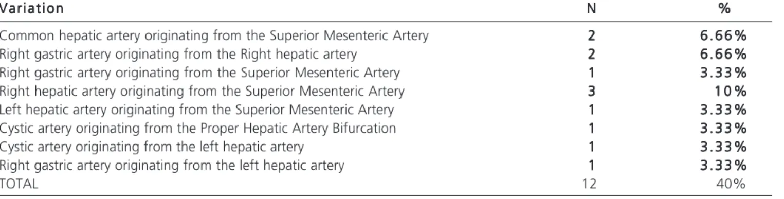
Table 1 Table 1 Table 1 Table 1
-Table 1 - Anatomical variations found in the study...
V a r i a t i o n V a r i a t i o n V a r i a t i o n V a r i a t i o n
V a r i a t i o n NNNNN %%%%%
Common hepatic artery originating from the Superior Mesenteric Artery 22222 6 . 6 6 %6 . 6 6 %6 . 6 6 %6 . 6 6 %6 . 6 6 % Right gastric artery originating from the Right hepatic artery 22222 6 . 6 6 %6 . 6 6 %6 . 6 6 %6 . 6 6 %6 . 6 6 % Right gastric artery originating from the Superior Mesenteric Artery 11111 3 . 3 3 %3 . 3 3 %3 . 3 3 %3 . 3 3 %3 . 3 3 % Right hepatic artery originating from the Superior Mesenteric Artery 33333 1 0 %1 0 %1 0 %1 0 %1 0 % Left hepatic artery originating from the Superior Mesenteric Artery 11111 3 . 3 3 %3 . 3 3 %3 . 3 3 %3 . 3 3 %3 . 3 3 % Cystic artery originating from the Proper Hepatic Artery Bifurcation 11111 3 . 3 3 %3 . 3 3 %3 . 3 3 %3 . 3 3 %3 . 3 3 % Cystic artery originating from the left hepatic artery 11111 3 . 3 3 %3 . 3 3 %3 . 3 3 %3 . 3 3 %3 . 3 3 % Right gastric artery originating from the left hepatic artery 11111 3 . 3 3 %3 . 3 3 %3 . 3 3 %3 . 3 3 %3 . 3 3 %
corpses and specimens was 40% (n = 12), a value similar to that found in most of the works reviewed.
The correct division of the proper hepatic artery in right and left hepatic arteries (Class 1) reached a value of 73.33%. Literature values ranged from 55% to 80%. we highlight Hiatt et al, with 75.7% 11.
Class 2, characterized by the left hepatic artery originating from the left gastric artery, was not found in our study and the literature shows a low prevalence of it. Bertevello and Chaib, who conducted the study in 60 livers from cadavers, showed only 2 cases (3.3%) of this variation 8,12. Hiatt et al obtained 9.7% 11.
Regarding the right hepatic artery originating from the superior mesenteric artery (Class 3), we found 3 cases (10%) and it was the most significant alteration recorded. In the literature, the values are between 8% and 18%. We believe that this type of variation presents with more relevance both for its greater prevalence and the potential to affect surgical procedures. As the vessel passes between the inferior vena cava and the portal vein at the level of the hepatobiliary triangle, one should be very careful in surgeries such as cholecystectomy and also pancreatectomy.
Class 4 Michels is intended for cases involving association of variants, for example, the right hepatic artery emerging from the superior mesenteric artery and the left hepatic artery originating from the left gastric artery. It is a most unusual situations and we found no such case in our research. Hiatt found some association in only 2.3% 11.
Class 5, which refers to the common hepatic artery originating from the superior mesenteric artery, was found in one cadaver (3.33%). Daseler et al performed a study that observed five hundred specimens of hepatic arterial system and found 4.4% of the common hepatic artery emerging from the superior mesenteric artery 14. Bertevello and Chaib, on their turn, found
this variation in only one case (1.6%) 8,12. Hiatt et al
found that variation in only 1.5% 11. In another study
with 150 liver transplants from the Liver Transplant Service of the Clinics Hospital of the Federal University of Paraná, Freitas et al demonstrated that the common hepatic artery originated from the superior mesenteric artery in 4 patients, totaling 1.62% 15.
Among the variations described, some deserve even greater attention for a higher risk of complications in surgical procedures. Chaib described an hepatic artery whose origin was in a stem connecting the celiac trunk and the superior mesenteric artery, and another case in which a left hepatic artery originated from the left gastric artery and also involved the esophagus 12. In our study
one case (3.33%) deserves attention, in which the common hepatic artery originated from the superior mesenteric artery, classified as Michels Class 5. When the left hepatic artery arises from the left gastric artery (Class 2), there will be a high possibility of ischemic
commitment of left lobe of the liver if this variation is not recognized during a radical gastrectomy. We believe that the variation of type 3, right hepatic artery originating from the superior mesenteric artery, has higher relevance due to its higher prevalence.
The use of spiral CT and three-dimensional CT arteriography assists greatly the surgeon in planning liver surgery, especially transplantation, previously identifying the diameter of the vascular lumen, abnormalities of the arterial supply of the liver, portal vein thrombosis and splenic artery aneurysms, since the splenic artery may the site for anastomosis in cases of receptors with hepatic arteries of narrow diameter. Such practices are not recommended for minor procedures, but the surgeon must be able to identify these changes at the time of operation.
Liver transplantation was introduced by Starzl et al. , and since then, many difficulties have been overcome, but it main limitation has been the lack of organ donors. To minimize the loss of organs, extrahepatic arteries must be accurately identified at the time of organ collection, preventing injuries that may hinder complete arterialization of the graft 16.
A normal liver can survive ligation of the hepatic artery due to collateral portal circulation. Therapeutic ligations are held for hepatic neoplasms, trauma and liver arterial lesions17. Such procedures are proscribed in liver
transplant patients, as all routes of collateral circulation are interrupted during the graft collection, thus not having the mechanism to compensate for an injury to the main arterial trunk, which would cause tissue necrosis with serious consequences 15.
The detailed study of the anatomy of liver structures, both intra-and extra-hepatic, allowed, in recent years, the development of alternate successful techniques of liver transplantation, such as reduced liver transplant, so called “split-liver” or splitting liver and, recently, the living-donor transplantation 9. In the particular case of splitting
the liver, an organ can originate two grafts (right lobe and left lobe), thus benefiting two receivers, a technique developed by Pilchlmayr et al. 18. According to Couinaud
and Houssin, the number of cases who display some type of anatomical variation that render the splitting of the liver, and its use in liver transplantation, impossible, is small (5%)
19. Therefore, adequate knowledge of all hepatic arterial
system is of utmost importance.
originating from the superior mesenteric artery. According to Ottone et al. in these cases it traverses a retro duodenal path until reaching the hepatic hilum 20.
We consider the study and research in the discipline of human anatomy a real need. It contributes unequivocally to the practice of medicine in general and in particular to surgery, with which it has a fundamental
relationship. We emphasize that further studies would bring valuable contributions.
Variations in the hepatic arteries, including its branches, and are frequently found, being 40% in this study, similar to the literature. The most significant variation, observed in 10% (3 cases), was the right hepatic artery with its origin in the superior mesenteric artery.
R E S U M O R E S U M O R E S U M O R E S U M O R E S U M O
Objetivo: Objetivo: Objetivo: Objetivo:
Objetivo: Demonstrar as minúcias do sistema arterial hepático, a incidência das variações anatômicas e comparar os dados obtidos com os da literatura. Métodos:Métodos:Métodos:Métodos:Métodos: Foram preparados 45 cadáveres do Departamento de Anatomia da Pontifícia Universi-dade Católica do Paraná, entre julho de 2010 e abril de 2011, sendo aproveitados 30 que possuíam integriUniversi-dade das estruturas. Analisaram-se as variações anatômicas das artérias hepáticas, suas principais características, como origem, trajeto, comprimento e diâmetro. O resultado global foi expresso por frequência e percentual de cadáveres com variações anatômicas do sistema arterial hepático. A estimativa deste percentual foi feita construindo-se um intervalo de confiança de 95%. Resultados:Resultados:Resultados:Resultados:Resultados: Observou-se algum tipo de variação anatômica em 40% (n=12) dos cadáveres estudados. Encontraram-se variações em duas artérias hepáticas comuns, três artérias gastroduodenais, três artérias hepáticas direita, uma artéria hepática esquerda, uma artéria gástrica direita e duas artérias císticas. Quanto ao tronco celíaco, verificaram-se variações em seu comprimento, diâmetro e altura de sua origem que foi comum na aorta. A variação da artéria hepática direita originando-se da artéria mesentérica superior foi encontrada em 10% (n=3) dos espécimes estudados e foi considerado o tipo de variação mais prevalente neste estudo. Conclusão: Conclusão: Conclusão: Conclusão: Conclusão: As variações nas artérias hepáticas são encontradas com frequência, e neste estudo foi 40%, valor semelhante ao da literatura. A variação mais significativa, observada em 10% (3 casos), foi a da artéria hepática direita com sua origem na artéria mesentérica superior.
Descritores: Descritores: Descritores: Descritores:
Descritores: Artéria hepática. Fígado. Cirurgia geral. Anatomia. Cadáver.
REFERENCES
REFERENCES
REFERENCES
REFERENCES
REFERENCES
1. Haller A. Methodus studi medici. In: Rio-Branco P. Essaisur l’anatomie ET La médecine óperatoire Du tronc coeliaque et de sés branches de l’ártère hépatique em particulier. Paris: G. Steinheil; 1912. 2. Testut L, Latarget A. Tratado de anatomia humana. 9a ed.
Barcelona: Salvat; 1968.
3. Moore KL, Arthur FD, Anne MRA. Anatomia Orientada para a Clínica. 6ª ed. Rio de Janeiro: Elsevier; 2010.
4. Sobotta J. Atlas de Anatomia Humana. 22a ed. Rio de Janeiro:
Guanabara Koogan; 2008.
5. Netter FH. Atlas de Anatomia Humana. 5a ed. Rio de Janeiro:
Elsevier; 2011.
6. Michels NA. Newer anatomy of the liver and its variant blood supply and collateral circulation. Am J Surg. 1966;112(4):337-47. 7. Soin AS, Friend PJ, Rasmussen A, Saxena R, Tokat Y, Alexander GJ, et al. Donor arterial variations in liver transplantation: management and outcome of 527 consecutive grafts. Br J Surg. 1996;83(4):637-41.
8. Bertevello PL, Chaib E. Variações do sistema arterial hepático e sua aplicabilidade na bipartição do fígado. Arq gastroenterol. 2002;39(2):81-5.
9. Soares RV, Coelho JCU, Matias JEF, Zeni Neto C, Freitas ACT, Godoy JL. Anatomia da artéria hepatica em doadores e receptors de transplante hepático intervivos. Rev Col Bras Cir. 2006;33(2):63-7.
10. Michels NA. Variational anatomy of the hepatic, cystic, and retroduodenal arteries; a statistical analysis of their origin, distribution, and relations to the biliary ducts in two hundred bodies. AMA Arch Surg. 1953;66(1):20-34.
11. Hiatt JR, Gabbay J, Busuttil RW. Surgical anatomy of the hepatic arteries in 1000 cases. Ann Surg. 1994;220(1):50-2. 12. Chaib E. Transplante de fígado: alterações da artéria hepática
e do fígado em 80 doadores. Arq gastroenterol. 1993;30(4):82-7.
13. Kemeny MM, Hogan JM, Goldberg DA, Lieu C, Beatty JD, Kokal W, et al. Continuous hepatic artery infusion with an implantable pump; problems with hepatic artery anomalies. Surgery. 1986;99(4):501-4.
14. Daseler EH, Anson BJ, Hambley WC, Reimann AF. The cystic artery and constituents of the hepatic pedicle; a study of 500 specimens. Surg Gynecol Obstet. 1947;85(1):47-63.
15. Freitas ACT, Coelho JCU, Matias JEF, Zeni Neto C, Martins EL, Druszcz CC. Anatomia arterial hepática: estudo em 150 transplantes hepáticos. Rev Col Bras Cir. 2001;28(1):13-6. 16. Starzl TE, Marchioro TL, Vonkaulla KN, Hermann G, Brittain RS,
Waddell WR. Homotransplantation of the liver in humans. Surg Gynecol Obstet. 1963;117:659-76.
17. Suzuki T, Nakayasu A, Kawabe K, Takeda H, Honjo I. Surgical significance of anatomic variations of the hepatic artery. Am J Surg. 1971;122(4):505-12.
18. Pichlmayr R, Ringe B, Gubernatis G, Hauss J, Bunzendahl H. Transplantation einer spenderleber auf zweiempfanger (splitting-transplantation), eine neue methodein der weiterentwicklung der leber segment transplantation. Langenbecks Arch Chir. 1988;373(2):127-30.
20. Ottone NE, Arrotea Molina A, Domínguez ML, Lo Tartaro M, García de Quiros N, Medan C, et al. Arterias hepáticas aberrantes: estudio en 64 cadáveres disecados. Int j morphol. 2006;24(4):581-5.
Received on 15/07/2012
Accepted for publication 30/08/2012 Conflict of interest: none
Source of funding: none
How to cite this article: How to cite this article:How to cite this article: How to cite this article:How to cite this article:
Sebben GA, Rocha SL, Sebben MA, Parussolo Filho P, Gonçalves BHH. Variations of hepatic artery: anatomical study on cadavers. Rev Col Bras Cir. [periódico na Internet] 2013;40(3). Disponível em URL: http:/ /www.scielo.br/rcbc
Address correspondence to: Address correspondence to:Address correspondence to: Address correspondence to:Address correspondence to: Geraldo Alberto Sebben