Ultraso no graphic de te rminatio n o f
go ite r pre vale nce in so uthe rn
Brazilian scho o lchildre n
1Departamento de Medicina, Faculdade de Medicina,
Universidade de Passo Fundo, Passo Fundo, RS, Brasil
2Divisão de Endocrinologia, Hospital de Clínicas de Porto Alegre,
Universidade Federal do Rio Grande do Sul, Porto Alegre, RS, Brasil H.R.K. Lisbôa1
and J.L. Gross2
Abstract
The aim of the present study was to estimate the prevalence of goiter in schoolchildren in a formerly iodine-deficient region in southern Brazil by assessing the relationship between body surface area (m2)
and thyroid volume (ml) measured by ultrasonography. A population-based sample of 1,094 randomly selected schoolchildren (6 to 14 years; 556 boys and 538 girls) underwent clinical evaluation. A total of 119 (10.9%) children were diagnosed with goiter upon clinical exami-nation according to WHO criteria (grade Ia: 65, grade Ib: 24, grade II: 29, grade III: 1). Of these, 85 underwent ultrasonography. In order to ascertain the absence of goiter in the 975 schoolchildren with a negative result upon clinical examination, one of ten children was randomly selected for ultrasonography. Sixty-two children agreed to be submitted to the exam. Thus, 147 schoolchildren were evaluated by ultrasonography (7.5-MHz transducer). Goiter was considered to be present when the thyroid volume:body surface area index was >6.2 ml/ m2. The estimated prevalence of goiter if all schoolchildren had been
submitted to thyroid volume measurement by ultrasound was 7.2%; it was higher in the lower socioeconomic class (8.2%) than in the upper (7.8%) and middle classes (6.5%). In conclusion, the prevalence of goiter in schoolchildren of this region was higher than in other iodine-sufficient areas, especially in lower socioeconomic classes. Goiter in this region may be associated with naturally occurring goitrogens that operate more intensively among less privileged individuals.
Co rre spo nde nce
J.L. Gross
Serviço de Endocrinologia Hospital de Clínicas de Porto Alegre Rua Ramiro Barcelos, 2350 Prédio 12, 4º andar 900035-003 Porto Alegre, RS Brasil
Fax: + 55-51-330-9100/332-5188 E-mail: gross@ hotnet.net
Research partially supported by CNPq and Hospital de Clínicas de Porto Alegre. H.R.K. Lisbôa was the recipient of a scholarship from CAPES.
Received May 16, 2001 Accepted July 10, 2002
Ke y words
·Goiter
·Prevalence
·Thyroid gland
·Ultrasonography
Intro ductio n
The diagnosis of goiter has been tradi-tionally based on inspection and palpation of the thyroid. According to the World Health Organization (WHO) (1), the presence of goiter is established when the thyroid lobes are larger than the terminal phalanges of the thumb of the person being examined. How-ever, the clinical examination of thyroid vol-ume often overestimates the presence of
in-dex) is a reliable measure for establishing a normal reference range for thyroid volume (4). Others have also observed an associa-tion between thyroid volume and body sur-face area in both children (5) and adults (6), and the variation of body surface area has been considered to be the main determinant of thyroid volume (7).
In addition to body surface area, several other factors are associated with thyroid volume, the best known being iodine defi-ciency. The recommended intake of iodine is 150 µg/day for adults. This corresponds to a urinary concentration of approximately 10 µg/dl (8). Before the salt iodination program was fully implemented in Brazil in 1983 (9), the highland region of the State of Rio Gran-de do Sul was consiGran-dered to be iodine Gran- defi-cient. A study performed in 1955 (10) showed that 31.4% of young adults from the region of Passo Fundo were not accepted for mili-tary compulsory service due to the presence of goiter.
Therefore, the objective of the present study was to estimate the prevalence of goi-ter by ultrasonography in this city located on the highlands of southern Brazil. Thyroid volume was determined by the Echobody index as previously reported (4).
Patie nts and Me thods
The study was carried out in 1994 in the city of Passo Fundo, located in the highland region (800 m altitude) of southern Brazil. In 1998, the average urinary iodine excretion (Sandell-Kolthoff method) detected in ran-dom urinary samples of 100 schoolchildren was 22 µg/dl (11).
A total of 1,127 schoolchildren aged 6 to 14 years were selected randomly among the entire population of 21,544 schoolchildren. The sample size calculation for a confidence level of 95% and assuming a proportion of 5 and 15% of schoolchildren with goiter indi-cates that 377 students should be representa-tive of the population. The sample was
se-lected on the basis of data from the City Department of Education, which provided a list of the 66 public and private schools and of the schoolchildren enrolled in them. Ac-cording to the Brazilian Geography and Sta-tistics Institute, the population living in the city was 165,000 inhabitants. The distribu-tion of the schools according to socioeco-nomic status was: 39 lower-class schools, 16 middle-class schools, and 11 upper-class schools. Seven of these schools were studied (three lower-class, two middle-class, and two upper-class ones).
Schools, shifts, and students in each class-room were all chosen randomly by drawing lots. In the classroom, each student was as-signed a number (starting with 1). Then, the teacher was asked to select a number from 1 to 10. The students who had been assigned numbers that were multiples of that selected by the teacher took part in the study. Twenty-one children with a history of thyroid disease and 12 who had used drugs containing io-dine (potassium ioio-dine syrup or topical solu-tions) during the three months before the study were excluded. Therefore, 1,094 schoolchildren (556 boys and 538 girls) un-derwent clinical examination to detect the presence of goiter.
For each subject, an identification form was completed and the following data were obtained: name of the school, grade, name of the student, place of birth, age, and sex. Weight, height, and triceps skinfold thick-ness (measured with a Harpenden caliper at the midpoint between olecranon and acro-mion) were determined. Body surface area was obtained using a normogram based on weight and height. A total of 147 individuals agreed to undergo ultrasonography. Informed consent was obtained from the children’s parents or legal representatives. The study was approved by the Ethics Committee of Hospital de Clínicas de Porto Alegre.
tissue; grade Ia, palpable thyroid with lateral lobes larger than the distal phalanx of the thumb of the child who was being examined; grade Ib, thyroid visible when the subject swallows with head extended; grade II, thy-roid visible with head in normal position; grade III, thyroid bulky and visible at a dis-tance. The child was asked to swallow with the head in normal position, with the doctor standing in front of him/her. Then, with the head extended, the gland was inspected and the thyroid was palpated with the thumbs while the child was asked to swallow again. All children were examined by one of the authors (H.R.K. Lisbôa).
Ultrasonography was performed using an Aloka 630 instrument with a 7.5-MHz transducer, always by the same physician, who was not aware of the results of clinical examination. The volume of each lobe was calculated according to the formula antero-posterior x transversal x longitudinal diam-eters x 0.52. The results for each lobe were summed to obtain the total volume. The mean coefficient of variation of two meas-urements separated by a 15-day interval in eight young adults was 3.9%.
The ratio of thyroid volume to body sur-face area (Echobody index) was considered as the standard criterion to define thyroid size in this sample of schoolchildren. Thus, all chil-dren with an Echobody index equal to or higher than 6.2 ml/m2 (upper limit of
normal-ity) were considered to have an increased thyroid volume. This value was calculated on the basis of the 95th percentile of the distribu-tion curve for 62 children without goiter upon clinical examination that underwent ultra-sonography in the present study.
The prevalence of goiter in the popula-tion was estimated by calculating the num-ber of schoolchildren that would probably have goiter if all the population had under-gone ultrasound examination of the thyroid. The distribution of schoolchildren with goi-ter in each socioeconomic class was calcu-lated with a formula (12) that employs
posi-tive and negaposi-tive predicposi-tive Echobody index values: actual prevalence = {(positive test x positive predictive value) + [negative test x (1-negative predictive test value)]}. We have previously demonstrated that clinical exami-nation has a sensitivity of 41% for diagnos-ing goiter, a specificity of 91%, a positive predictive value of 27% and a negative pre-dictive value of 95% (4).
Statistical analysis
Data are reported as means ± SD. The adopted level of significance was 5%. The unpaired Student t-test was used for
compar-ison between means and the chi-square test was used to compare proportions.
Re sults
One hundred and nineteen (10.9%) of the 1,094 schoolchildren examined were con-sidered to have goiter upon clinical exami-nation. The distribution of goiter size ac-cording to WHO criteria is shown in Table 1. Most goiters were classified as grade Ia. Eighty-five children in this group agreed to undergo ultrasonography. Sixty-two children with a negative clinical examination agreed to be submitted to the exam. Therefore, 147 schoolchildren (85 with goiter and 62 with-out goiter upon clinical examination) were evaluated by ultrasonography.
According to the Echobody index, three schoolchildren (4.8%) with goiter were
iden-Table 1. Distribution of goiter size according to WHO criteria in 119 schoolchildren.
Goiter size Number and percentage
of schoolchildren
Ia 65 (55% )
Ib 24 (20% )
II 29 (24% )
III 1 (1% )
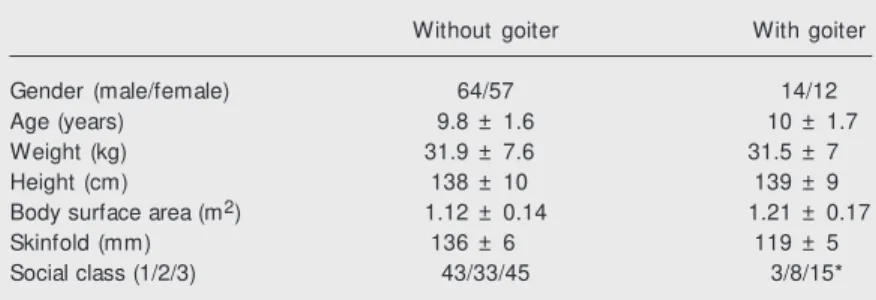
tified among the 62 thought not to have goiter upon clinical examination, and 23 (27.05%) in the group of 85 individuals with goiter upon clinical examination. The group with goiter according to the Echobody index (N = 26) did not differ from the group with-out goiter (N = 121) regarding age, distribu-tion of sex, body surface area, or skinfold thickness. The number of lower-class chil-dren with goiter was significantly higher (chi-square = 6.24, P = 0.04) (Table 2).
Based on these data, if all the 1,094 schoolchildren had been submitted to ultra-sonography, it would be possible to estimate that 47 children (4.8% of 975) would have been diagnosed as having goiter according to the Echobody index in the group thought not to have goiter on the basis of clinical examination, and 32 (27.05% of 119) in the group found to have goiter upon clinical examination. According to this inference, 79 individuals would have goiter on ultrasound assessment. Extrapolating these findings to all schoolchildren (1,094/79), we estimate that 7.2% probably had an enlarged thyroid. When the students were classified ac-cording to socioeconomic status, the esti-mated prevalence of goiter was 8.2, 7.8, and 6.5% in the lower, middle and upper classes, respectively. This estimate was obtained us-ing the prevalence found in the sample ex-trapolated to the 1,094 school children.
D iscussio n
The estimated goiter prevalence of 7.2% in the schoolchildren of Passo Fundo, Brazil, is higher than the 5% value detected in re-gions where urinary iodine is above 10 µg/dl (8). Epidemiological studies measuring thy-roid volume by ultrasonography and taking into account height, body weight and age have reported a goiter prevalence of 3.9% in iodine-sufficient areas (13). In iodine-defi-cient areas in Italy (13) and around Chernobyl in the former Soviet Union (14), the reported prevalence of goiter was 25.3 and 35.9%, respectively.
Current iodine deficiency alone does not explain the increased prevalence of goiter detected in this study. Since the salt iodina-tion program in Brazil was implemented in 1983 (9), the average urinary iodine excre-tion in this region has reached 22 µg/dl, above the value of 10 µg/dl established as the minimum for iodine sufficiency (8). In addition, the normal reference range for thy-roid volume in the region (6.2 ml/m2) is very
similar to that reported for iodine-sufficient regions using ultrasonography and taking into account body surface area (5). Even considering that in order to reduce thyroid weight in endemic areas it is necessary to have an adequate supply of iodine for at least 25 years (15), other factors may be associ-ated with the increased occurrence of goiter in Passo Fundo (16).
Several goiter endemias have been asso-ciated with exposure to naturally occurring goitrogenic and antithyroid agents in food-stuffs and drinking water (17). These goitro-genic agents act directly on the thyroid gland, interfering with the main steps of thyroid hormone production or altering the excre-tion of thyroid hormones. Several foodstuffs have been implicated in goiter endemias (such as cassava, millet, babassu, seaweeds, piñon nut) due to the presence of compounds with antithyroid action, e.g., thiocyanate, thio-oxazolidone, and flavonoids. Soybean has
Table 2. Clinical characteristics of schoolchildren w ith and w ithout goiter according to the Echobody index.
Without goiter With goiter
Gender (male/female) 64/57 14/12
Age (years) 9.8 ± 1.6 10 ± 1.7
Weight (kg) 31.9 ± 7.6 31.5 ± 7
Height (cm) 138 ± 10 139 ± 9
Body surface area (m2) 1.12 ± 0.14 1.21 ± 0.17
Skinfold (mm) 136 ± 6 119 ± 5
Social class (1/2/3) 43/33/45 3/8/15*
Data are reported as means ± SD or number of individuals w ith the characteristic. Numbers 1, 2 and 3 represent the upper, middle, and low er socioeconomic classes, respectively.
also been implicated in the development of goiter, probably due to the inhibition of thy-roid peroxidase by the isoflavones genistein and daidzein (18). It is interesting to note that Passo Fundo is located within a soybean growing area and, although the intake of soybean products was not evaluated, it could be higher there than in other areas.
Increased prevalence of goiter has also been related to undernutrition and bacterial pollution of drinking water (19). The pres-ence of Escherichia coli may decrease
io-dine uptake; furthermore, there is evidence of mimicry of antigens from this bacterium and human thyroid plasma membrane which can trigger an autoimmune reaction (16). Recently it was reported that the molecular mimicry of thyroid antigens by bacteria was a local sequence homology between sodium/ iodide symporters and a number of both bacterial and viral proteins. The authors sug-gested that cross-reactivity with sodium/io-dide symporters by antibodies originally against microorganisms may compromise iodide uptake by the thyroid, thus increasing the prevalence of goiter in a polluted envi-ronment (20). Probably, water pollution was also a relevant factor in our study, especially for the lower social class, in which the preva-lence of goiter was higher than in the middle and upper classes.
Goiter in these schoolchildren also could have been due to chronic autoimmune thy-roiditis. However, although antithyroid anti-bodies were not measured, this is an unlikely explanation for the presence of goiter in this study, since in only one patient did we observe a thyroid with an irregular surface and firm consistency, characteristic of Hashimoto’s thyroiditis. Furthermore, the prevalence of autoimmune thyroiditis in adolescents is only 0.35% (21).
This study has two potential limitations. The interval between thyroid examination and the measurement of urinary iodine in the population was of approximately 4 years. However, this probably did not constitute a
major caveat because a sufficient iodine sup-ply for the Brazilian population has been assured since 1983 (22).
Secondly, the definition of goiter used was based on the 95th percentile of the dis-tribution curve for 62 children without goiter upon clinical examination. However, accord-ing to recommendations of the WHO (8), the cutoff value should be 97% for a population of children with normal iodine intake. When the 97% cutoff of the distribution of children without goiter is used we obtain 7.2 ml/m2 as
the upper limit of the reference range for our study group. Using this value rather than the 95th percentile cutoff value (6.2 ml/m2)which
was used in the present study, we obtain 4% of children with goiter for the entire popula-tion (extrapolated to 1,094 children) and 4.2, 3.5, and 4% for children of lower, middle and upper socioeconomic classes, respec-tively (extrapolated to 404, 506 and 186 children of the above socioeconomic classes) based on the Echobody index. As expected, using the higher cutoff value decreased the percentage of schoolchildren having goiter. However, the WHO recommendation was based on a European study which was recog-nized to have a systematic measurement bias resulting in an overestimation of the thyroid volume (23). Furthermore, the 95% cutoff value adopted in our study (6.2 ml/m2) agrees
better with other accepted standards (6.5 ml/ m2) than the 97% cutoff value of 7.2 ml/m2
(5,23).
The increased prevalence of goiter diag-nosed by ultrasonography in the schoolchil-dren of this currently iodine-sufficient re-gion may be related to the presence of natu-rally occurring goitrogens operating more intensively among less privileged individu-als.
Ackno wle dgm e nts
Re fe re nce s
1. Delange F, Bastiani S, Benmiloud M , DeM ayer E, Isayama M G, Koutras D, M uzzo S, Niepomniszcze H, Pandav CS & Riccabona G (1986). Definitions of en-demic goiter and cretinism, classification of goiter size and the severity of ende-mias, and survey techniques. In: Dunn JT, Pretell EA, Daza CH & Viteri SE (Editors), Tow ards the Eradication of Endemic Goi-ter, Cretinism, and Iodine Deficiency. Pan American Health Organization, Scientific Publication No. 502, Washington, DC, USA, 373.
2. Rasmussen SN & Hjorth L (1974). Deter-mination of thyroid volume by ultrasonic scanning. Journal of Clinical Ultrasound, 2: 143-146.
3. Berghout A, Wiersinga WM , Smits NJ & Touber JL (1987). Determinants of thy-roid volume as measured by ultrasonogra-phy in healthy adults in a non-iodine defi-cient area. Clinical Endocrinology, 26: 273-280.
4. Lisbôa HRK, Gross JL, Orsolin A & Fuchs S (1996). Clinical examination is not an accurate method of defining the presence of goitre in schoolchildren. Clinical Endo-crinology, 45: 471-475.
5. Delange F, Benker G, Caron PH, Eber O, Ott W, Peter F, Podoba J, Simescu M , Szybinsky Z, Vert ongen F, Vit t i P, Wiersinga W & Zamrazil V (1997). Thyroid volume and urinary iodine in European schoolchildren: standardization of values for assessment of iodine deficiency. Eu-ropean Journal of Endocrinology, 136: 180-187.
6. Barrère X, Valeix P, Preziosi P, Bensimon M , Pelletier B, Galan P & Hercberg S (2000). Determinants of thyroid volume in healthy French adults participating in the SU.VI.M AX cohort. Clinical Endocrinology, 52: 273-278.
7. Gómez JM , M aravall FJ, Gómez N, Gumà A & Soler J (2000). Determinants of thy-roid volume as measured by
ultrasonogra-phy in healthy adults randomly selected. Clinical Endocrinology, 53: 629-634. 8. World Health Organization, United
Na-tions Children’s Fund and International Council for Control of Iodine Deficiency Disorders (1994). Indicators for Assess-ing Iodine Deficiency Disorders and their Control through Salt Iodination. World Health Organization, Geneva, Sw itzerland. 9. M edeiros Neto GA (1988). Tow ards the eradication of iodine deficiency disorders in Brazil t hrough salt iodinat ion pro-gramme. Bulletin of the World Health Or-ganization, 66: 637-642.
10. Oliveira PP, M aldonado G, M ontedonio JM & Lacerda EBF (1955). O bócio endê-mico no sul do Brasil. Hospital, 47: 91-125.
11. Lisbôa HRK (1999). Thyroid volum e change during puberty. Doctoral thesis, Universidade Federal do Rio Grande do Sul, Porto Alegre, RS, Brazil.
12. Ahlbon A & Norell S (1990). Introduction to M odern Epidemiology. Epidemiology Resources Inc., Chestnut Hill, M A, USA, 24.
13. Vitti P, M artino N, Lombardi FA, Rago T, Antonangeli L, M accherini D, Nanni P, Loviseli A, Balestrieri A, Araneo G & Pinchera A (1994). Thyroid volume meas-urement by ultrasound in children as a tool for the assessment of mild iodine deficiency. Journal of Clinical Endocrinol-ogy and M etabolism, 79: 600-603. 14. Ashizaw a K, Shibata Y, Yamashita S,
Namba H, Hoshi M , Yokoyama N, Izumi M & Nagataki S (1997). Prevalence of goi-ter and urinary iodine excretion levels in children around Chernobyl. Journal of Clinical Endocrinology and M etabolism, 82: 3430-3433.
15. Lindberg O, Andersson LC & Lamberg BA (1989). The impact of 25 years of iodine prophylaxis on the adult thyroid w eight in Finland. Journal of Endocrinological Inves-tigation, 12: 789-793.
16. Gaitan E (1990). Goitrogens in food and w ater. Annual Review of Nutrition, 10: 21-39.
17. M edeiros-Neto G (2001). Iodine defi-ciency disorders. In: DeGrot t LL & Jameson JL (Editors), Endocrinology. Vol. 2. W.B. Saunders Co., Philadelphia, PA, USA, 1529.
18. Divi RL, Chang HC & Doerge DR (1997). Anti-thyroid isoflavones from soybean: isolation, characterization and mechan-isms of action. Biochemical Pharmacolo-gy, 54: 1087-1096.
19. Gaitan E, Cooksey RC & Lindsay RH (1986). Factors other than iodine defi-ciency in endemic goiter: Goitrogens and protein-calorie malnutrition. In: Dunn JT, Pretell EA, Daza CH & Viteri SE (Editors), Tow ards the Eradication of Endemic Goi-ter, Cretinism, and Iodine Deficiency. Pan American Health Organization, Scientific Publication No. 502, Washington, DC, USA, 28.
20. Benvenga S, Alesci S, Trimarchi F & Facchiano A (1999). Homologies of the thyroid sodium-iodide symporter w ith bac-terial and viral proteins. Journal of Endo-crinological Investigation, 22: 535-540. 21. Jaksic J, Dumic M , Filipovic B, Ille J,
Cvijetic M & Gjuric G (1977). Thyroid dis-eases in a school population w ith thyro-megaly. Archives of Disease in Childhood, 70: 103-106.
22. Pretell EA & Noguera A (1994). Current status of iodine deficiency disorders in Latin America. Iodine Deficiency Disor-ders New sletter, 10: 15-22.