Influe nce o f e xe rcise o n the activity
and the distributio n be twe e n fre e and
bo und fo rm s o f glyco lytic and associated
e nzym e s in tissue s o f ho rse m acke re l
1Department of Natural Sciences, Precarpathian University, Ivano-Frankivsk, Ukraine 2Institute of Biochemistry and Department of Biology, Carleton University, O ttawa, O ntario, Canada
V.I. Lushchak1,*, T.V. Bagnyukova1,*, J.M. Storey2 and K.B. Storey2
Abstract
The effects of short-term burst (5 min at 1.8 m/s) swimming and long-term cruiser (60 min at 1.2 m/s) swimming on maximal enzyme activities and enzyme distribution between free and bound states were assessed for nine glycolytic and associated enzymes in tissues of horse mackerel, Trachurus mediterraneus ponticus. The effects of exercise were greatest in white muscle. The activities of phosphofructokinase (PFK), pyruvate kinase (PK), fructose-1,6-bisphosphatase (FBPase), and phosphoglucomutase (PGM) all decreased to 47, 37, 37 and 67%, respectively, during 60-min exercise and all enzymes except phospho-glucoisomerase (PGI) and PGM showed a change in the extent of binding to subcellular particulate fractions during exercise. In red muscle, exercise affected the activities of PGI, FBPase, PFK, and lactate dehydrogenase (LDH) and altered percent binding of only PK and LDH. In liver, exercise increased the PK activity 2.3-fold and reduced PGI 1.7-fold only after 5 min of exercise but altered the percent binding of seven enzymes. Fewer effects were seen in brain, with changes in the activities of aldolase and PGM and in percent binding of hexokinase, PFK and PK. Changes in enzyme activities and in binding interactions with subcellular particulate matter appear to support the altered demands of tissue energy metabolism during exercise.
Co rre spo nde nce
V.I. Lushchak
Department of Natural Sciences Precarpathian University 57 Shevchenko str. Ivano-Frankivsk, 76000 Ukraine
E-mail: lushchak@ pu.if.ua *O n leave from Karadag Natural Reserve, National Academy of Sciences of Ukraine, Kurortne, Feodosia, Crimea, Ukraine
Received April 17, 2000 Accepted May 15, 2001
Ke y wo rds
·Trachurus m ed iterraneus ·Swimming exercise
·Glycolysis
·Subcellular enzyme binding
Intro ductio n
Horse mackerel (Trachurus
mediterra-neus ponticus) are highly active, pelagic fish
that can swim at intermediate speed for long periods of time as well as in brief high-speed bursts for shorter periods (1). Intensification of swimming is powered by creatine phos-phate hydrolysis and by ATP synthesis via
AMP and IMP (1-9). In addition, exercise increased both total glycogen phosphorylase activity and the percentage of enzyme in the
active, a form for an overall 6-fold increase
in the amount of active phosphorylase in trout white muscle (8). The activation of energy metabolism in fish involves several regulatory mechanisms acting in concert; allosteric regulation, protein phosphoryla-tion and enzyme binding to subcellular mac-romolecules all contribute to the activation of different enzymes (10). The most contro-versial of these mechanisms is reversible enzyme binding to subcellular macromol-ecules.
Enzyme binding to cellular macromol-ecules has been reported extensively. For example, enzymes of glycogen metabolism bind to glycogen particles, AMP-deaminase binds to myosin, and various glycolytic en-zymes bind to filamentous actin (11-14). The distribution of enzymes between free and bound forms may, in turn, be modulated by changes in metabolite and ion levels (15-17) or by reversible protein phosphorylation (18). The physiological relevance of some interactions is obvious (e.g., glycogen phos-phorylase or synthetase binding to glyco-gen), but the functional significance of gly-colytic enzyme association with F-actin has been the subject of debate (13,17,19). These interactions can change enzyme functional properties. For example, bound lactate dehy-drogenase (LDH) is not inhibited by high concentrations of pyruvate (20,21), and is more stable to trypsin hydrolysis than the free enzyme (22). Catalytic properties can also be modified by enzyme interaction with structural elements (21-25).
A redistribution of glycolytic enzymes between bound and soluble fractions has been noted in several systems as a response to stimuli that change glycolytic flux (e.g., exercise, anoxia, hypoxia) (26-28), includ-ing trout white muscle (12,29). In general, an increase in enzyme binding correlates with an increase in glycolytic flux and vice versa.
But in our recent studies with a teleost fish, the sea scorpion, subjected to hypoxia we found that binding of some enzymes in-creased whereas binding of other enzymes decreased under the same stress conditions (30). Therefore, to determine whether this was an isolated instance or a common re-sponse by fish muscle, we undertook a com-parable study with another teleost species.
The present study analyzes the effects of swimming exercise under two regimens, short-term burst exercise and long-term cruiser exercise, on the maximal activities and free versus bound distribution of en-zymes in four tissues (brain, liver, and red and white muscle) of the Black Sea teleost fish, the horse mackerel. The results show that activities and distribution of enzymes are modified during exercise and that the effects are dependent on the intensity of swimming.
Mate rial and Me tho ds
Expe rim e ntal anim als
Horse mackerel (Trachurus
mediterra-neus ponticus Aleev) weighing 25-40 g were
caught in October 1995 at Karadag Bay, Black Sea (Crimea, Ukraine) and were held in tanks provided with flowing sea water on a native dark/light cycle and temperature of
16 ± 1oC. Fish were not fed and were used
the next day.
Bio che mical re age nts and co upling e nzyme s
All chemicals were obtained from Boeh-ringer Mannheim (Montreal, PQ, Canada), Sigma Chemical Co. (St. Louis, MO, USA) or Reachim (Russia, USSR).
Fish e xe rcise
flow of water and equipped with an electric engine as described earlier (1). Eight fish were used in one experiment which was started about 6-7 am. Initially, water flow was held at low speed (0.1-0.3 m/s) for 1 h. After that, four fish were removed as con-trols and the rest were exercised for 60 min at a speed of 1.2 m/s (cruiser exercise) be-fore sampling. In previous work from our laboratory it was shown that under this regi-men fish may swim for several hours (1). Other fish were exposed to the same 1-h low speed swimming but this was then followed by 5 min at a speed of 1.8 m/s (burst exer-cise); fish in this group were exercised and sampled individually. All fish were killed quickly by trans-spinal dissection and tissue samples were immediately removed and used for the preparation of homogenates; this pro-cess took about 2 min. Whole brain and liver, and portions of red and white muscle were used for analysis. White muscle samples were always removed from the same posi-tion in the body, just below the first spinal fin.
Pre paratio n o f tissue e xtracts
Exercised tissues were quickly washed in ice-cold 0.9% NaCl, blotted on filter paper, weighed and homogenized in a Potter ho-mogenizer with a glass pestle in 10 volumes of homogenization solution containing 0.25 M sucrose, 10 mM dithiothreitol (DTT), 20 mM NaF, 1 mM EDTA, and 20 mM
imidaz-ole-HCl, pH 7.0, at 20o
C; 0.1 mM phenyl-methylsulfonyl fluoride (PMSF, a protease inhibitor) was added immediately before homogenization (30). The homogenate was
centrifuged for 20 min at 45,000 g at 4oC and
the supernatant was removed and stored on ice. Enzyme activity in the supernatant rep-resents the free enzyme fraction. The pellet was rehomogenized in 5 volumes (relative to initial tissue weight) of the second homog-enization solution with a high salt content to release bound enzymes (0.25 M sucrose, 10
mM DTT, 20 mM NaF, 1 mM EDTA, 20 mM imidazole-HCl and 500 mM KCl). After centrifugation, the supernatant was removed and the pellet was extracted a second time and centrifuged and the second supernatant was combined with the first. Enzyme activ-ity in this fraction represents the bound en-zyme fraction. Enen-zyme activities were quan-tified in both free and bound fractions and the percent binding was calculated. Recov-ery of total enzyme activity was checked in all cases by comparing the sum of enzyme activities in free and bound fractions with total enzyme activities measured in tissue homogenates. Recovery was close to 100% in most cases, ranging from 80 to 120%. The maximal enzyme activities (units/g wet weight) shown in the tables are those meas-ured in whole homogenates.
Me asure m e nt o f e nzym e activitie s
Maximal activities were assayed at 25o
C using a KFK-324 spectrophotometer (Lomo, Leningrad, Russia) by monitoring the change in absorbance of NADH or NADPH at 340 nm. All reaction mixtures contained 50 mM imidazole-HCl, pH 7.0, in a final volume of 1.0 ml including the added extract. Blanks did not contain the most specific substrate (indicated by an asterisk in the assays be-low). Reactions were started by the addition of the specific substrate but, if no spontane-ous change in absorbance was observed, subsequent assays were started by the addi-tion of homogenate. Optimal assay condi-tions were based on those described earlier (30) and adapted to our experiments. Hex-okinase (HK, EC 2.7.1.1): 10 mM glucose (*), 0.2 mM NADP, 2 mM ATP, 5 mM
MgCl2, 0.5 unit (U) glucose-6-phosphate
de-hydrogenase (G6PDH), and 50 µl superna-tant. Phosphoglucoisomerase (PGI, EC 5.3.1.9): 2.5 mM fructose-6-phosphate (*),
0.2 mM NADP, 5 mM MgCl2, 0.5 U G6PDH,
fructose-6-supernatant. LDH (EC 1.1.1.27): 1 mM pyru-vate (*), 0.15 mM NADH, 1 mM EDTA, and 1-50 µl supernatant. G6PDH (EC 1.1.1.49): 2.0 mM glucose-6-phosphate (*), 5 mM
MgCl2, 0.2 mM NADP, and 20-50 µl
super-natant. Phosphoglucomutase (PGM, EC 2.7.5.1): 15 mM glucose-1-phosphate (*),
0.20 mM NADP, 5.0 mM MgCl2, 0.5 U
G6PDH, and 20-50 µl supernatant. Fruc-tose-1,6-bisphosphatase (FBPase, EC 3.1.3.11): 0.1 mM fructose-1,6-bisphosphate
(*), 0.2 mM NADP, 5 mM MgCl2, 0.5 U
G6PDH, 0.5 U PGI, and 30-50 µl superna-tant. One unit of enzyme activity is defined as the amount of enzyme that utilizes 1 µmol
of substrate per 1 min at 25oC. All values are
expressed as means ± SEM. Differences be-tween means were determined by analysis of variance followed by the two-tailed Dunnett test. For technical reasons, the number of animals was limited (3-4 per set) but, even so, in many cases we obtained statistically significant differences between experimen-tal groups.
Re sults
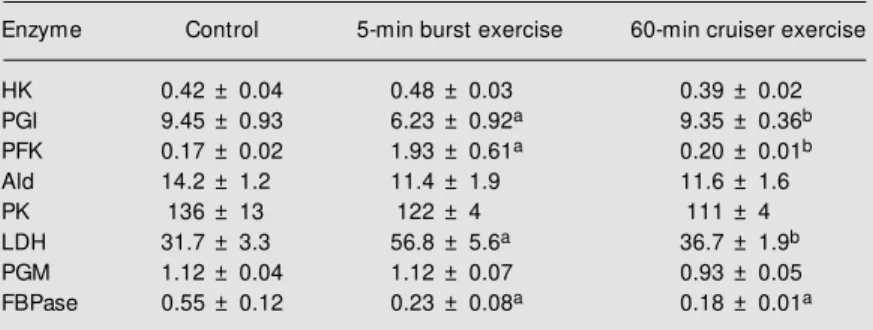
The effect of exercise under both regi-mens on the activities of glycolytic and asso-ciated enzymes is shown in Tables 1-4. In white muscle, exercise resulted in a signifi-cant decrease in the activities of several enzymes (Table 1). Activities of PFK, PK and FBPase decreased under both exercise regimens, whereas PGM was reduced by 33% only after long-term cruiser swimming. After 5-min burst exercise, PFK, PK and FBPase activities were reduced to 72, 46, and 75% of their corresponding control val-ues, but 60 min of sustained exercise had an even greater effect, lowering activities to 47, 36 and 37% of the controls, respectively. The activities of four enzymes were affected by exercise in red muscle (Table 2). The activities of PGI and FBPase decreased sig-nificantly after 5-min burst exercise falling to 66 and 42%, respectively, of their control Table 2. Effect of exercise on the activities (units/g w et w eight) of glycolytic and
associated enzymes in red muscle of horse mackerel.
Enzyme Control 5-min burst exercise 60-min cruiser exercise
HK 0.42 ± 0.04 0.48 ± 0.03 0.39 ± 0.02
PGI 9.45 ± 0.93 6.23 ± 0.92a 9.35 ± 0.36b
PFK 0.17 ± 0.02 1.93 ± 0.61a 0.20 ± 0.01b
Ald 14.2 ± 1.2 11.4 ± 1.9 11.6 ± 1.6
PK 136 ± 13 122 ± 4 111 ± 4
LDH 31.7 ± 3.3 56.8 ± 5.6a 36.7 ± 1.9b
PGM 1.12 ± 0.04 1.12 ± 0.07 0.93 ± 0.05
FBPase 0.55 ± 0.12 0.23 ± 0.08a 0.18 ± 0.01a
Data are reported as means ± SEM of 3-4 determinations in tissue from separate individuals. aP<0.05 compared to the corresponding control value and bP<0.05 com-pared to the 5-min burst exercise group (tw o-tailed Dunnett test).
For abbreviations, see legend to Table 1.
phosphate (*), 5 mM ATP, 0.15 mM NADH,
10 mM MgCl2, 50 mM KCl, 0.5 U aldolase
(Ald), 0.5 U triosephosphate isomerase, 2 U glycerol-3-phosphate dehydrogenase, and 30-50 µl supernatant. Ald (EC 4.1.2.13): 0.5 mM fructose-1,6-bisphosphate (*), 0.15 mM NADH, 0.5 U triosephosphate isomerase, 2 U glycerol-3-phosphate dehydrogenase, and 2-20 µl supernatant. Pyruvate kinase (PK, EC 2.7.1.40): 10 mM phosphoenolpyruvate
(*), 2.5 mM ADP, 50 mM KCl, 5 mM MgCl2,
0.15 mM NADH, 0.5 U LDH, and 1-10 µl
Table 1. Effect of exercise on the activities (units/g w et w eight) of glycolytic and associated enzymes in w hite muscle of horse mackerel.
Enzyme Control 5-min burst exercise 60-min cruiser exercise
HK 0.28 ± 0.09 0.25 ± 0.06 0.17 ± 0.02
PGI 14.5 ± 0.4 15.6 ± 0.7 13.9 ± 0.2
PFK 0.76 ± 0.03 0.55 ± 0.08a 0.36 ± 0.04a,b
Ald 52.4 ± 8.7 54.3 ± 4.5 70.4 ± 7.1
PK 241 ± 24 112 ± 13a 88.0 ± 7.7a
LDH 140 ± 4 131 ± 17 121 ± 9
PGM 3.01 ± 0.16 2.41 ± 0.15 2.01 ± 0.32a
FBPase 0.57 ± 0.04 0.43 ± 0.05a 0.21 ± 0.01a,b
Data are reported as means ± SEM of 3-4 determinations in tissue from separate individuals. aP<0.05 compared to the corresponding control value and bP<0.05 com-pared to the 5-min burst exercise group (tw o-tailed Dunnett test).
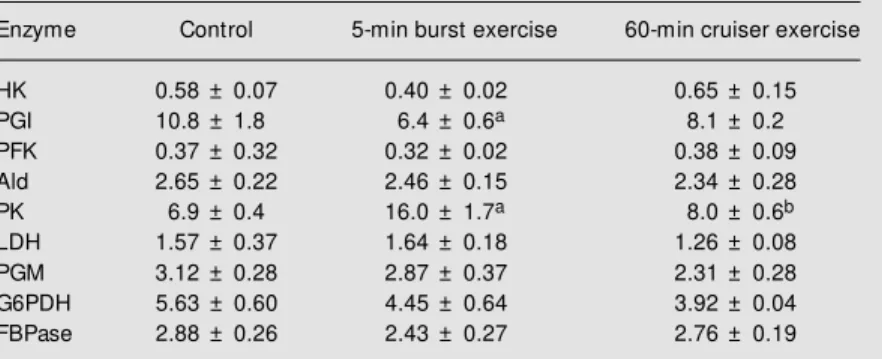
values. FBPase activity was also reduced after 60-min cruiser swimming to 33% of the control value. By contrast, PFK activity rose by 11-fold and LDH by 1.8-fold after 5-min burst exercise. However, neither enzyme was affected by sustained 60-min swimming. In liver, stimulation of swimming affected only PGI and PK during 5-min burst swimming (Table 3). PGI activity fell to 59% of the control value whereas PK activity rose by 2.3-fold. No enzymes were affected during sustained cruiser swimming. In brain, Ald activity increased by 62% during both forms of exercise, whereas PGM activity was sharply reduced to 32% of control after 60 min sustained exercise and was undetectable in the tissue from fish submitted to 5-min burst exercise (Table 4). FBPase was also undetectable in brain of exercised fish.
Figures 1-4 show the distribution of en-zymes between free and bound forms in horse mackerel tissues. In white muscle, swimming exercise led to the redistribution of six of eight enzymes, but did not affect PGI or PGM (Figure 1). Short-term burst swimming significantly reduced the percent-age of bound PFK, Ald and FBPase and released all HK into the free fraction. In contrast, bound PK rose from about 15 to 54% of the total and the percentage of bound LDH also doubled. Sustained cruiser swim-ming had different effects on the distribution of enzymes in white muscle. Although PFK and PK responded similarly to both forms of exercise, the effect on FBPase was much more pronounced with no bound FBPase found in white muscle during this type of swimming. HK, Ald and LDH were unaf-fected during cruiser swimming.
Red muscle showed fewer changes in the proportions of bound enzymes during swim-ming exercise. The only effect of burst exer-cise on this tissue was that bound LDH dropped by one-half. By contrast, sustained cruiser exercise affected the bound propor-tion of four enzymes - PFK, PK, LDH and PGM. Bound PGM activity was not found at
Table 3. Effect of exercise on the activity (units/g w et w eight) of glycolytic and associated enzymes in liver of horse mackerel.
Enzyme Control 5-min burst exercise 60-min cruiser exercise
HK 0.58 ± 0.07 0.40 ± 0.02 0.65 ± 0.15
PGI 10.8 ± 1.8 6.4 ± 0.6a 8.1 ± 0.2
PFK 0.37 ± 0.32 0.32 ± 0.02 0.38 ± 0.09
Ald 2.65 ± 0.22 2.46 ± 0.15 2.34 ± 0.28
PK 6.9 ± 0.4 16.0 ± 1.7a 8.0 ± 0.6b
LDH 1.57 ± 0.37 1.64 ± 0.18 1.26 ± 0.08
PGM 3.12 ± 0.28 2.87 ± 0.37 2.31 ± 0.28
G6PDH 5.63 ± 0.60 4.45 ± 0.64 3.92 ± 0.04
FBPase 2.88 ± 0.26 2.43 ± 0.27 2.76 ± 0.19
Data are reported as means ± SEM of 3-4 determinations in tissue from separate individuals. aP<0.05 compared to the corresponding control value and bP<0.05 com-pared to the 5-min burst exercise group (tw o-tailed Dunnett test).
G6PDH, glucose-6-phosphate dehydrogenase. For other abbreviations, see legend to Table 1.
Figure 1. Effect of exercise on the distribution of glycolytic and associated enzymes betw een free and bound fractions in w hite muscle of horse mackerel. Con-trol (w hite bars), 5-min burst sw imming (grey bars) and 60-m in cruiser sw i60-m 60-m ing (black bars). Dat a are report ed as means ± SEM (N = 3-4 individual fishes). aP<0.05 compared to the corresponding control value and bP<0.05 compared to the 5-min
burst exercise group (tw o-tailed
Dunnett test). HK, hexokinase; PGI, phosphoglucoisomerase; PFK, phosphofructokinase; Ald, aldolase; PK, pyruvate kinase; LDH, lactate dehydrogenase; PGM , phosphoglucomu-tase; FBP, fructose-1,6-bisphosphatase.
Table 4. Effect of exercise on the activities (units/g w et w eight) of glycolytic and associated enzymes in brain of horse mackerel.
Enzyme Control 5-min burst exercise 60-min cruiser exercise
HK 0.21 ± 0.04 0.21 ± 0.01 0.18 ± 0.01
PGI 3.72 ± 0.11 3.97 ± 0.11 3.72 ± 0.11
PFK 0.18 ± 0.03 0.24 ± 0.03 0.25 ± 0.01
Ald 1.66 ± 0.11 2.69 ± 0.18a 2.69 ± 0.12a
PK 36.5 ± 1.8 34.5 ± 1.7 33.3 ± 1.2
LDH 9.20 ± 0.43 7.49 ± 0.36 8.24 ± 0.83
PGM 0.22 ± 0.06 0 0.07 ± 0.03a
G6PDH 0.07 ± 0.01 0 0.08 ± 0.02
FBPase 0.05 ± 0.02 0 0
Data are reported as means ± SEM of 3-4 determinations in tissue from separate individuals. aP<0.05 compared to the corresponding control value (tw o-tailed Dunnett test).
G6PDH, glucose-6-phosphate dehydrogenase. For other abbreviations, see legend to Table 1.
B
o
u
n
d
e
n
zy
m
e
(
%
)
60
50
40
30
20
10
0
HK PGI PFK Ald PK LDH PGM FBP
b
a
a a a
a
b a
a a
about 61% after burst exercise. Sustained exercise had the same effects as burst exer-cise on liver HK, PK and FBPase. However, the percentage of bound PGI and G6PDH did not change during sustained swimming. The proportion of bound LDH was reduced during cruiser swimming in liver compared to both the control and burst exercise situa-tion and, in addisitua-tion, no bound PGM was found in liver after sustained exercise.
In brain both exercise regimens increased the percentage of bound HK but PGI, Ald and LDH were unaffected by either form of exercise (Figure 4). PFK binding was not affected during burst exercise, but percent binding fell from about 53% in control to 11% after sustained 60-min swimming. In contrast, the percentage of bound PK in brain increased from about 8 to 16% during sus-tained swimming.
D iscussio n
The effect of exercise on intermediary metabolism and energetics of organisms has been well studied (31), including numerous studies of metabolic responses to exercise by fish species (4,5,8). Two areas of exercise metabolism in fish that are still not fully resolved, however, are the effects of exer-cise on enzyme activities and on the distribu-tion of enzymes between free and bound states. Therefore, in this study we focused on these two phenomena, monitoring the ef-fects of burst high-speed swimming (5 min at 1.8 m/s) and of sustained moderate-speed swimming (60 min at 1.2 m/s) on the maxi-mal activities of enzymes in four organs and on the subcellular distribution of enzymes between free and bound states. Short-term burst exercise is typically powered by white muscle in fish, whereas long-term cruiser exercise is primarily a function of red muscle. Previous work from our Karadag laboratory has analyzed the different modes of swim-ming and their effects on metabolite levels in horse mackerel tissues. These fish can swim
B o u n d e n zy m e ( % ) 100 90 70 50 40 30 0 60 a a a,b a,b 80 20 10
HK PGI PFK Ald PK LDH
B o u n d e n zy m e ( % ) 70 60 50 30 20 10 0 40
HK PGI PFK Ald PK LDH PGM FBP G6PDH
a a a b a a,b a a a b b a b a a B o u n d e n zy m e ( % ) 60 50 40 30 20 10 0
HK PGI PFK Ald PK LDH PGM FBP
Figure 2. Effect of exercise on the distribution of enzymes be-tw een free and bound fractions in red muscle of horse mackerel. Other information as in the leg-end to Figure 1.
Figure 3. Effect of exercise on the distribution of enzymes be-tw een free and bound fractions in liver of horse m ackerel. G6PDH, glucose-6-phosphat e dehydrogenase. Other informa-tion as in the legend to Figure 1.
Figure 4. Effect of exercise on the distribution of enzymes be-tw een free and bound fractions in brain of horse mackerel. Other information as in the legend to Figure 1.
all, bound PK activity fell compared to con-trol and bound LDH and PFK rose compared to burst exercise in red muscle (Figure 2).
In liver, both exercise regimens led to changes in percent binding of several en-zymes but did not affect PFK or Ald distribu-tion (Figure 3). After 5-min burst swimming the percentages of bound HK, PK, FBPase and G6PDH all increased significantly, whereas bound PGI decreased. The increase in bound G6PDH was particularly large, ris-ing from only about 1% bound in controls to
a
for 7-10 h at a speed of 1.2 m/s (1). After 1 h of swimming at this speed the levels of phos-phocreatine were reduced by about 50% in white muscle and ATP and AMP fell by about 10-20%, whereas ADP and inorganic phosphate levels were increased about 40%. The effects of exercise on the carbohydrate content of horse mackerel tissues were also studied. Glycogen concentration decreased and lactic acid concentration increased dur-ing the first 10-30 min of cruiser swimmdur-ing in white muscle. But even though glycogen had fallen further after 60 min of exercise, lactate concentration was reduced well be-low the initial level, suggesting a possible lactate export from the muscle during sus-tained swimming. Red muscle responded to exercise similarly, although the rates of change in metabolite levels were different.
The results of the present study show that exercise also modifies the activities of en-zymes of intermediary metabolism in horse mackerel tissues. The enzymes chosen for study were several glycolytic enzymes as well as the gluconeogenic enzyme FBPase, and G6PDH, the first enzyme of the hexose monophosphate shunt. With the exception of HK, all enzymes showed some exercise-related change in maximal activity in at least one of the four tissues tested. Both burst and cruiser exercise resulted in reduced activi-ties of PFK, PK and FBPase in white muscle but a relatively constant PFK:FBPase ratio was maintained, suggesting that exercise did not change the relative rates of glycolytic versus gluconeogenic flux in the muscle. The mechanism(s) regulating the coordinated decrease in the maximal activities of PFK, PK and FBPase remain(s) to be determined but notably these three enzymes are typically subject to multiple regulatory controls by a variety of factors. One of these is reversible phosphorylation via protein kinases and phos-phatases, which is not a mechanism known to control any of the other five enzymes. Hence, it would be interesting to determine whether exercise stimulated changes in the
phosphorylation state of these enzymes. Exercise-induced changes in the activi-ties of enzymes in red muscle were quite different except that, as in white muscle, the activity of FBPase decreased in exercised muscle. In this case, however, PFK activity did not decrease during exercise (indeed, it rose substantially during burst exercise) so that the net consequence would be a de-crease in the potential for gluconeogenic flux during exercise. Indeed, the PFK:FBPase ratio rose from a control value of 0.31 to values during burst and cruiser exercise of 8.39 and 1.11, respectively, showing that glycolysis is clearly favored during exercise. However, the very large increase in PFK activity during burst work is surprising, since it occurred within 5 min. Hence, this cannot be synthesis of new enzyme, but must repre-sent a change of the existing enzyme either due to effects of very powerful allosteric effectors (whose influence is still felt when the homogenate is diluted about 20-fold in the assay mixture) or to a stable change of enzyme properties such as those induced by reversible phosphorylation, polymerization or alteration of bound/free status, all known mechanisms of PFK control (32). However, the results illustrated in Figure 2 indicate that a change in the relative amounts of bound versus free PFK could not account for the change in enzyme activity.
which converts glucose-6-phosphate into fructose-6-phosphate for use by PFK.
Reversible enzyme binding to subcellu-lar structures and other macromolecules is well known but its importance in the control of enzymes and pathways still remains con-troversial. Previous studies from our labora-tories found that the distribution of enzymes of carbohydrate metabolism between free and bound fractions may be modified by exercise in trout (12,29), anoxia in goldfish (28), and hypoxia in sea scorpion (30). How-ever, whereas in the two first species an activation of glycolysis led to an increase or no change in enzyme binding, in sea scor-pion tissues the situation was more compli-cated. With increased glycolytic rate, the bound portion increased in some enzymes, decreased in some and did not change in others (30).
Exercise significantly changed the distri-bution of various enzymes between free and particle-bound states in horse mackerel tis-sues. In white muscle, for example, the dis-appearance of the bound form of HK during burst work and of FBPase after 60-min exer-cise could significantly change the function of these enzymes. The bound portion of PFK in white muscle also decreased by about one-half in both exercised groups. Detailed studies have suggested that PFK binding may be one of the few interactions between glycolytic enzymes and subcellular
struc-tures that have real significance in vivo. This
is because PFK binding holds up under physi-ological conditions of ions and metabolites, whereas interactions between various other glycolytic enzymes and F-actin (believed to be a chief binding site for glycolytic en-zymes) frequently disappear at physiologi-cal ionic strength (17). However, the present results (reduced PFK binding in exercised muscle) are contrary to results for trout where PFK binding increased during exercise (12, 29). The reason for this contradiction is not apparent from the current data but might be related to differences in the dependence of
white muscle from different species on phos-phagen versus anaerobic glycolysis for the support of muscle work. PK and LDH re-sponded in opposite ways to PFK during exercise, the percent binding of both en-zymes rising during burst exercise and bound PK also rising during cruiser exercise. This may serve to localize these two terminal enzymes of glycolysis at positions that could facilitate sustained anaerobic glycolysis in-cluding ATP production by PK and the re-oxidation of NADH by LDH.
Contrary to white muscle, red muscle showed very few changes in enzyme distri-bution during exercise. This type of muscle is used primarily in sustained cruiser exer-cise but enzyme binding changes during cruiser swimming consisted of only a small decrease in percent PK binding and a com-plete loss of PGM binding. Burst swimming only changed LDH binding in this muscle. This may not be surprising as long-term cruiser swimming powered by red muscle may be primarily fueled by the oxidation of lipids and hence only small changes in gly-colytic rate may occur during this type of exercise.
a substrate for aerobic oxidation, in a posi-tion that better facilitates pyruvate uptake by mitochondria.
There is no doubt that reversible binding of glycolytic and other enzymes to cellular structures can play a role in the precise regu-lation of metabolism. Unfortunately, to date, the mechanisms involved in this regulation are not well understood (14) and redistribu-tion of different enzymes in different direc-tions, as seen in the present study or in our previous work with sea scorpions (30), makes the picture even more complicated. Such redistribution indicates that this mechanism
may be involved in the regulation of glycoly-sis in its organs, providing space-time com-partmentation of the pathway to serve differ-ent metabolic needs. The specific enzymes affected in different tissues could result in tissue-specific changes in flux through dif-ferent pathways in a manner that serves the metabolic needs of each organ.
Ackno wle dgm e nts
The authors thank N. Glibina for techni-cal assistance.
Re fe re nce s
1. Shulman GE (Editor) (1978). Elements of Physiology and Biochemistry of General and Active M etabolism in Fishes. Naukova Dumka, Kyiv.
2. Rahim ZHA, Lutaya G & Griffiths JR (1979). Activation of AM P aminohydrolase during skeletal-muscle contraction. Bio-chemical Journal, 184: 173-176. 3. Wokoma A & Johnston IA (1983).
Anaero-bic metabolism during activity in rainbow trout (Salmo gairdneri). Experientia, 39: 1366-1367.
4. Parkhouse WS, Dobson GP, Belcastro AN & Hochachka PW (1987). The role of inter-mediary metabolism in the maintenance of proton and charge balance during exer-cise. M olecular and Cellular Biochemis-try, 77: 37-47.
5. Parkhouse WS, Dobson GP & Hochachka PW (1988). Control of glycogenolysis in rainbow trout muscle during exercise. Ca-nadian Journal of Zoology, 66: 345-351. 6. M ommsen TP & Hochachka PW (1988).
The purine nucleotide cycle as tw o tem-porally separated metabolic units: a study on trout muscle. M etabolism, 37: 552-558.
7. Van den Thillart G, Van Waarde A, M uller HJ, Erkelens C, Addink A & Lugtenburg J (1989). Fish muscle energy metabolism measured by in vivo31P-NM R during an-oxia and recovery. American Journal of Physiology, 256: R922-R929.
8. St orey KB (1991). M et abolic conse-quences of exercise in organs of rainbow trout. Journal of Experimental Zoology, 260: 157-164.
9. Schulte PM , M oyes CD & Hochachka PW
(1992). Integrating metabolic pathw ays in post-exercise recovery of w hite muscle.
Journal of Experimental Biology, 166: 181-195.
10. Lushchak VI (1995). Role of phosphoryla-tion and redistribuphosphoryla-tion of glycolytic en-zymes in adaptation of hydrobiontes to enviromental conditions. Hydrobiological Journal, 31: 18-29.
11. M asters CJ, Reid S & Don M (1987). Gly-colysis - new concepts in an old pathw ay.
M olecular and Cellular Biochemistry, 76: 3-14.
12. Brooks SPJ & Storey KB (1988). Subcellu-lar enzyme binding in glycolytic control: in vivo studies w ith fish muscle. American Journal of Physiology, 255: R289-R294. 13. Lushchak VI (1996). AM P-deam inase:
functions, molecular properties and local-ization in the cell. Biochemistry (M oscow ), 61: 195-211.
14. Lushchak VI (1996). Participation of su-pramolecular complex formation in the regulation of glycolytic enzymes. Ukrai-nian Biochemical Journal, 68: 20-28. 15. Hsu SC & M olday RS (1990).
Glyceralde-hyde-3-phosphate dehydrogenase is a major protein associated w ith the plasma membrane of retinal photoreceptor outer segments. Journal of Biological Chemis-try, 265: 13308-13313.
16. Lushchak VI (1991). Properties of mem-brane-bound lactate dehydrogenase from w hite skate muscle. Biokhimiia, 56: 2173-2180.
17. Brooks SPJ & Storey KB (1993). Control of glycolytic enzyme binding: effect of changing enzyme substrate
concentra-tions on in vivo enzyme distribution. M o-lecular and Cellular Biochemistry, 122: 1-7.
18. Luther M A & Lee JC (1986). The role of phosphorylation in the interaction of rab-bit muscle phosphofructokinase w ith F-actin. Journal of Biological Chemistry, 261: 1753-1759.
19. Kurganov BI (1986). The role of multien-zyme complexes in integration of cellular metabolism. Journal of Theoretical Biol-ogy, 119: 445-455.
20. Ehmann JD & Hultin HO (1973). Substrate inhibition of soluble and bound lactate de-hydrogenase (isoenzyme 5). Archives of Biochemistry and Biophysics, 154: 471-475.
21. Lushchak VI (1992). Lactate dehydrogen-ase interaction w ith the structural cell components: the possible physiological significance. Biokhimiia, 57: 1142-1154. 22. Lushchak VI (1992). Free and
membrane-bound lactate dehydrogenase from w hite driving muscles of skate. Biochemistry In-ternational, 26: 905-912.
23. Liou R-S & Anderson S (1980). Activation of rabbit muscle phosphofructokinase by F-actin and reconstituted thin filaments.
Biochemistry, 19: 2684-2688.
24. Kuo H-J, M alencik DA, Liou RS & Ander-son SR (1986). Factors affecting the acti-vation of rabbit muscle phosphofructoki-nase by actin. Biochemistry, 25: 1278-1286.
1103-1109.
26. Clarke FM , Shaw FD & M orton DJ (1980). Effect of electrical stimulation post mor-tem of bovine muscle on the binding of glycolytic enzymes. Functional and struc-tural implications. Biochemical Journal, 186: 105-109.
27. Brooks SPJ & Storey KB (1991). Studies on the regulation of enzyme binding dur-ing anoxia in isolated tissues of Busycon canaliculatum. Journal of Experimental Bi-ology, 156: 467-481.
28. Duncan JA & Storey KB (1991). Role of enzyme binding in muscle metabolism of the goldfish. Canadian Journal of Zoology, 69: 1571-1576.
29. Lushchak VI & Storey KB (1994). Effect of
exercise on the distribution of enzymes in trout w hite muscle and kinetic properties of AM P-deaminase from free and bound fractions. Fish Physiology and Biochemis-try, 13: 226-239.
30. Lushchak VI, Bahnjukova TV & Storey KB (1998). Effect of hypoxia on the activity and binding of glycolytic and associated enzymes in sea scorpion tissues. Brazil-ian Journal of M edical and Biological Re-search, 31: 1059-1067.
31. Booth FW & Thompson DB (1991). M o-lecular and cellular adaptation of muscle in response to exercise: perspectives of various models. Physiological Review s, 71: 541-585.
32. Storey KB & Brooks SPJ (1995). Is
glyco-lytic rate controlled by the reversible bind-ing of enzymes to subcellular structures? In: Hochachka PW & M ommsenTP (Edi-tors), Biochemistry and M olecular Biology of Fishes. Vol. 4. Elsevier, Amsterdam, 291-307.
33. Depinto V, Aljamal JA & Palmieri F (1993). Hexokinase-binding protein. Location of the dicyclohexylcarbodiimide-reactive glu-tamate residue in the bovine heart mito-chondrial porin. Journal of Biological Chemistry, 268: 12977-12982.