Myosin light chain kinase is necessary for post-shock
mesenteric lymph drainage enhancement
of vascular reactivity and calcium
sensitivity in hemorrhagic-shocked rats
Y.P. Zhang, C.Y. Niu, Z.G. Zhao, L.M. Zhang and Y.H. Si
Institute of Microcirculation, Hebei North University, Hebei, China
Abstract
Vascular hyporeactivity is an important factor in irreversible shock, and post-shock mesenteric lymph (PSML) blockade improves vascular reactivity after hemorrhagic shock. This study explored the possible involvement of myosin light chain kinase (MLCK) in PSML-mediated vascular hyporeactivity and calcium desensitization. Rats were divided into sham (n=12), shock (n=18), and shock++drainage (n=18) groups. A hemorrhagic shock model (40±2 mmHg, 3 h) was established in the shock and shock++drainage groups. PSML drainage was performed from 1 to 3 h from start of hypotension in shock++drainage rats. Levels of phospho-MLCK (p-MLCK) were determined in superior mesenteric artery (SMA) tissue, and the vascular reactivity to norepinephrine (NE) and sensitivity to Ca2++were observed in SMA rings in an isolated organ perfusion system. p-MLCK was significantly decreased in the shock group compared with the sham group, but increased in the shock++drainage group compared with the shock group. Substance P (1 nM), an agonist of MLCK, significantly elevated the decreased contractile response of SMA rings to both NE and Ca2++at various concentrations. Maximum contractility (Emax) in the shock group increased with NE (from 0.179±0.038 to 0.440±0.177 g/mg, P,0.05) and Ca2++ (from 0.515±0.043 to 0.646±0.096 g/mg, P,0.05). ML-7 (0.1 nM), an inhibitor of MLCK, reduced the increased vascular response to NE and Ca2++at various concentrations in the shock++drainage group (from 0.744±0.187 to 0.570±0.143 g/mg in Emaxfor NE and from 0.729±0.037 to 0.645±0.056 g/mg in Emaxfor Ca2++, P,0.05). We conclude that MLCK is an important contributor to PSML drainage, enhancing vascular reactivity and calcium sensitivity in rats with hemorrhagic shock.
Key words: Hemorrhagic shock; Mesenteric lymph; Drainage; Myosin light chain kinase (MLCK); Vascular reactivity; Calcium sensitivity
Introduction
Vascular hyporeactivity to vasoconstrictors occurs during sepsis and trauma. Hemorrhage is the major underlying mechanism of microcirculation failure, refrac-tory hypotension, no-reflow phenomenon and vital-organ hypoperfusion. It is also considered to be a major cause of a persistent severe shock condition (1). A number of studies showed that receptor desensitization (2,3), hyper-polarization of membrane potential (4-6) and decreased sensitivity of contractile elements to Ca2++ in vascular smooth muscle cells (VSMCs) (7-10) all contribute to the development of vascular hyporeactivity.
Studies in recent years have shown that post-shock mesenteric lymph (PSML) has a pivotal function in endothelial cell injury and multiple organ dysfunction induced by gut-derived infections (11-14). Findings from
our laboratory suggested that mesenteric duct ligation and PSML drainage both improved the reactivity and calcium sensitivity of vascular rings (i.e., cross-sections) isolated from severely shocked rats (15). Furthermore, in vitro
experiments demonstrated that the mesenteric lymph harvested from 1-3 h after shock decreased the contractile activity and calcium sensitivity of normal vascular rings (15). However, the mechanism by which the mesenteric lymph of severe shock conditions blunts vascular reactivity is not clear.
Myosin light chain kinase (MLCK) is a key enzyme that determines the phosphorylation levels of 20-kDa myosin light chain (MLC20) (16-18). Whether MLCK is involved in
PSML-mediated vascular hyporeactivity is worthy of investigation. This study explored the mechanism by
Correspondence: C.Y. Niu, Institute of Microcirculation, Hebei North University, Diamond South Road 11, Hebei, Zhangjiakou 075000, China. Fax: ++86-313-402-9168. E-mail: [email protected]
which PSML decreases vascular reactivity. The function of MLCK in the improved vascular reactivity and calcium sensitivity associated with PSML drainage was investi-gated using an MLCK agonist and an inhibitor.
Material and Methods
Animals and study groups
Forty-eight adult male Wistar rats weighing 260-280 g were purchased from the Animal Breeding Center of the Chinese Academy of Medical Sciences (Beijing, China). The rats were randomly divided into sham (n=12), shock (n=18), and shock++drainage (n=18) groups. All animal experiments performed in this study were reviewed and approved by the Institutional Animal Care and Use Committee of Hebei North University. All experiments conformed to the guidelines for the ethical use of animals, and every effort was made to minimize animal suffering and to reduce the number of animals used. Prior to experimentation, all rats were fasted for 12 h, but allowed free access to water.
Surgical procedures and preparation of a hemorrhagic shock model
Rats were anesthetized with pentobarbital sodium (1%, 50 mg/kg). After the right femoral vein and artery were isolated, heparin sodium (500 U/kg) was injected intrave-nously to prevent systematic blood clot formation. A polyethylene tube was inserted into the femoral artery for continuous mean arterial pressure (MAP) monitoring during the experimental process, using a biological signal acquisi-tion system (RM6240BD, Chengdu Instrument, China). The left femoral artery was also isolated, cannulated and attached in-line to an NE-1000 automatic withdrawal-infusion machine (New Era Pump Systems Inc., USA) for bleeding. Abdominal operations were performed on all rats to separate the mesenteric lymph duct from the surround-ing connective tissues. After laparotomy, all rats were allowed to stabilize for 30 min. Rats in the shock and shock++drainage groups were hemorrhaged slowly at a constant rate from the left femoral artery to produce an MAP of 40 mmHg within 10 min. The MAP was maintained at 40±2 mmHg for 3 h by withdrawing or reperfusing shed blood as required for the preparation of the hemorrhagic shock model. For lymph drainage in the shock++drainage group, the mesenteric lymph duct was cannulated from 1 to 3 h after shock was produced using a homemade flexible needle. The rats in the sham group received identical treatment as those for the shock group, except for the attachment to the automatic withdrawal-infusion machine, because no blood was withdrawn.
Preparation of vascular tissue and measurement of phospho-MLCK (p-MLCK) levels
After the in vivo experiments previously described, the superior mesenteric artery (SMA) was obtained from
6 rats in each group. Adhering tissues were removed, the SMA tissue was triturated in liquid nitrogen and then transferred to an EP tube with 0.2 mL lysis buffer [100mL
Triton X-100 (stock solution); 100mL (10mg/mL) PMSF;
10mL (10 mg/mL) aprotein; 10.1mL (1 mg/mL) leupeptin;
0.707mL (1 mg/mL) pepstatin]. Phosphate-buffered
saline (0.01 M) was added to a 10-mL total volume, and the tissue was homogenized using an SM-6500 ultra-sonic cell disruptor (Shunma Instrument Equipment Inc., China) for 15 min. Then, the homogenate was centri-fuged at 14,000 g for 5 min at 46C using a Labofuge 400R supercentrifuge (Thermo Fisher Scientific, USA), and the supernatant was collected. The p-MLCK level in the SMA homogenate was determined using a rat ELISA kit (R&D Systems, USA) after a standard curve was p l o t t e d ( y = 0 . 0 5 6 9 7 x ++ 0 . 0 0 5 1 x2+ 0 . 0 0 0 1 5 7 x3, r2=0.998). The protein content in the homogenate was quantified by the Coomassie brilliant blue colorimetric method.
Preparation of vascular rings and measurement of vascular reactivity and calcium sensitivity
SMA was harvested from the treated rats, and each was cut into two rings of 2 to 3 mm in length for the experiments. One ring was used to measure vascular reactivity, and the other was used to measure calcium sensitivity. An SMA ring was transferred to the chamber of a wire myograph system, and two stainless-steel wire hooks were cannulated through the SMA ring lumen. One hook was connected to a micrometer, and the other was linked to a force transducer (ADInstruments, Australia). Then, the SMA ring was immersed into Krebs-Hensley (K-H) solution: 118 mM NaCl, 4.7 mM KCl, 1.2 mM MgSO4, 25 mM NaHCO3, 1.2 mM KH2PO4, 2.5 mM
CaCl2, and 11 mM glucose at pH 7.3-7.4. This solution
was continuously bubbled with 95% O2-5% CO2, and its
temperature was maintained at 376C. A 0.5-g preload was exerted, and the K-H solution was replaced every 20 min. The tension of the SMA ring was determined using a Power Lab System (ADInstruments).
After 1.5 h of equilibration, the contractile responses of the SMA rings to norepinephrine (NE) (1610-9, 1610-8,
1610-7, 1610-6, 1610-5, and 1610-4M) in each group
(n=6) were measured as previously described (7,8,19). Tension/vascular ring wet weight (g/mg) was calculated, and cumulative concentration-response curves for the responses of artery rings to NE were plotted. The values of maximal contraction (Emax) and pD2(-log 50% effective
concentration) values for the agonists were obtained from the concentration-response curves and used to compare vascular reactivity.
SP and ML-7 dosages used in the present study were based on previous reports (17,20,21).
SMA rings were incubated and equilibrated in K-H solution for 1.5 h as previously described. Then, the solution was replaced with depolarizing solution contain-ing 2.7 mM NaCl, 120 mM KCl, 1.2 mM MgSO4, 25 mM
NaHCO3, 1.2 mM KH2PO4, and 11 mM glucose at
pH 7.3-7.4. After 15 min of equilibration, the contractile responses of the SMA rings to Ca2++ (3610-5, 1610-4,
3610-4, 1610-3, 3610-3, 1610-2, and 3610-2 M) in
each group (n=6) were determined using a concentration accumulation method. Calcium sensitivity was similarly appraised by calculating Emax and pD2. The procedure
and agents were similar to the method used to measure vascular reactivity.
Statistical analysis
Data are reported as means±SD; one-way ANOVA was used to identify differences among groups. The paired t-test was used to identify significant differences between groups using the SPSS version 16.0 software (USA). Data that were not suitable for one-way ANOVA were analyzed using the Kruskal-Wallis test. P,0.05 was considered to be significant.
Results
Effect of PSML drainage on p-MLCK levels in the mesenteric artery of rats after hemorrhagic shock
The p-MLCK level in the mesenteric artery of the shock group was significantly lower compared with that of the sham group (P,0.05; Figure 1) and significantly increased in the shock++drainage group compared with that of the shock group (P,0.05). No statistical differences were observed between the sham and shock++drainage groups.
Function of MLCK on PSML drainage increasing the vascular reactivity of hemorrhagic-shocked rats
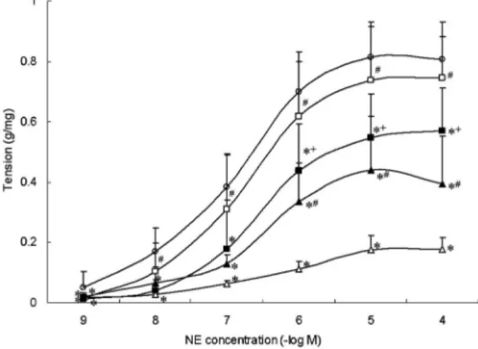
The contractile response of vascular rings to NE in the shock group was significantly decreased at all concentra-tions compared with that in the sham group (P,0.05). The vascular response to NE in the shock++drainage group was significantly higher than that of the shock group from 1610-8 to 1610-4 M NE (P,0.05). No significant difference was observed in the response of vascular rings to NE at various concentrations, except for 1610-9M in the
shock++drainage and sham groups (P.0.05, Figure 2). After the vascular rings were obtained from the shock and shock++drainage groups, they were incubated with tool agents (i.e., an angonist and an inhibitor). SP significantly enhanced the contractile response of SMAs obtained from the shock group to NE in the shock group at 1610-6, 1610-5, and 1610-4 M (P,0.05). ML-7 sig-nificantly reduced vascular reactivity of SMAs obtained from the shock group to NE in the shock++drainage group to NE at 1610-6, 1610-5, and 1610-4 M (P,0.05). However, at 1610-9, 1610-8, and 1610-7M of NE, SP,
and ML-7, no significant effect was observed on the contractile response of SMA (P.0.05; Figure 2).
In addition, Emaxand pD2of SMA to NE in the shock
group significantly decreased compared with those of the sham group, whereas Emaxin the shock++drainage group
was markedly increased when compared with that of the shock group (P,0.05). Emax of the vascular rings
response to NE of the shock group was significantly elevated by SP, but the value was still lower than that of the sham group (P,0.05). Emaxof the vascular contractile
response of the shock++drainage group was significantly
Figure 1. Effect of post-shock mesenteric lymph drainage on phospho-myosin light chain kinase (p-MLCK) level in superior mesenteric artery tissue from rats in hemorrhagic shock. Data are reported as means±SD (n=6). *P,0.05 vssham group, and #P
,0.05vsshock group (one-way ANOVA).
Figure 2.Myosin light chain kinase increases vascular reactivity on post-shock mesenteric lymph drainage in hemorrhagic-shock rats. Data are reported as means±SD (n=6). SP: substance P, an agonist of MLCK; ML-7: an inhibitor of MLCK. *P,0.05vs
sham group; #P
,0.05 vs shock group, and +P
,0.05 vs
reduced by ML-7, but the value was still higher than that of the shock group (P,0.05; Table 1).
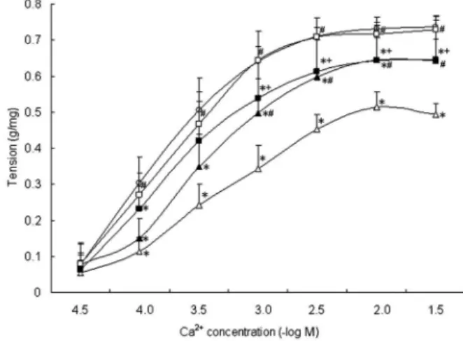
Function of MLCK on PSML drainage in increasing the vascular calcium sensitivity of hemorrhagic-shocked rats
The contractile response of SMA rings to gradient concentration of Ca2++in the shock group (from 1610-4M)
was significantly decreased compared with that in the sham group (P,0.05). The contractile responses of vascular rings to Ca2++from 1610-4M in the shock++drainage group
were significantly higher than those of the shock group (P,0.05). No significant difference was observed in vascular contractile responses to Ca2++ between the shock++drainage and sham groups (P,0.05; Figure 3).
Meanwhile, at 1610-3, 3610-3, 1610-2, and 3610-2
M Ca2++, SP significantly increased the contractile response of vascular rings compared with the shock group. ML-7 decreased the vascular response to Ca2++
compared with the shock++drainage group (P,0.05). However, at 3610-5, 1610-4, and 3610-4 M Ca2++, SP
and ML-7 had no significant effect on the contractile response of the SMA rings (P.0.05; Figure 3).
Meanwhile, Emax and pD2 of the SMA rings to the
gradient concentration of Ca2++ in the shock group significantly decreased compared to those in the sham and shock++drainage groups (P,0.05). Emax was
sig-nificantly elevated by SP, but it was still lower than that of the sham group (P,0.05). Emaxof the SMA rings to Ca2++
in the shock++drainage group was significantly decreased by ML-7, but the value was still higher than that in the shock group (P,0.05; Table 2).
Discussion
Studies have shown that the structural foundations of vascular motion are the contractile apparatus in VSMCs. The contraction of VSMC is controlled by both cytoplasmic calcium and calcium sensitivity of MLC20phosphorylation
(16). In general, agonist binding to G protein-coupled
receptors activates phospholipase Cb, which hydrolyzes
phosphatidylinositol 4,5-bisphosphate into two second messengers: inositol 1,4,5-trisphosphate (IP3) and
diacyl-glycerol. IP3binding with the receptor in the membrane of
the sarcoplasmic reticulum releases stored intracellular Ca2++ and, in turn, triggers Ca2++ influx from the extra-cellular compartment, which leads to the rapid increase of myoplasmic Ca2++. The increase in Ca2++via calmodulin
(CaM) activates MLCK, which phosphorylates MLC20.
Phosphorylated myosin cyclically binds to actin filaments producing VSMC contraction. The activation of MLCK by Ca2++/CaM is one of the key steps during VSMC
contrac-tion. This process is also referred to as the calcium-dependent mechanism of VSMC contractile regulation (22). Moreover, myosin light chain phosphatase (MLCP),
Table 1. Influence of mesenteric lymph drainage on Emax and pD2 of vascular response to norepinephrine in rats following hemorrhagic shock.
Group Emax(g/mg) pD2
Sham 0.814 ± 0.102 6.903 ± 0.355
Shock 0.179 ± 0.038* 6.198 ± 0.462*
Shock++SP 0.440 ± 0.177*# 6.528 ± 0.213 Shock++Drainage 0.744 ± 0.187# 6.801 ± 0.604 Shock++Drainage++ML-7 0.570 ± 0.143*#+ 6.587 ± 0.530
Data are reported as means±SD (n=6). SP: substance P, an agonist of MLCK; ML-7: an inhibitor of MLCK. * P,0.05vssham group;#P
,0.05vsshock group, and+P
,0.05vsshock++drainage group (one-way ANOVA).
Figure 3.Myosin light chain kinase increases vascular calcium sensitivity on post-shock mesenteric lymph drainage in hemor-rhagic-shock rats. Data are reported as means±SD (n=6). SP: substance P, an agonist of MLCK; ML-7: an inhibitor of MLCK. *P,0.05vssham group;#P
,0.05vsshock group, and+P ,0.05
vsshock++drainage group (one-way ANOVA).
Table 2. Influence of mesenteric lymph drainage on Emaxand pD2of vascular response to calcium in rats following hemorrhagic shock.
Group Emax(g/mg) pD2
Sham 0.736 ± 0.018 3.751 ± 0.109
Shock 0.515 ± 0.043* 3.228 ± 0.298*
Shock++SP 0.646 ± 0.096*# 3.446 ± 0.124* Shock++Drainage 0.729 ± 0.037# 3.626 ± 0.286# Shock++Drainage++ML-7 0.645 ± 0.056*#+ 3.607 ± 0.224#
Data are reported as means±SD (n=6). SP: substance P, an agonist of MLCK; ML-7: an inhibitor of MLCK. * P,0.05vssham group;#P
,0.05vsshock group, and+P
after its activity is inhibited by Rho kinase, protein kinase C, and so on, blunts the process of MLC20dephosphorylation.
This phenomenon maintains and strengthens the contrac-tion of VSMC, which is referred to as the calcium sensitivity mechanism of VSMC contractile regulation. The intracellular Ca2++of VSMC did not decrease with the onset of severe shock. Therefore, the mechanism of calcium sensitivity regulating VSMC contractility has been receiving more attention (7). Studies have suggested that, in a state of severe shock, the compromised activities of Rho kinase (8,9,19) and protein kinase C (18,23-26) and the elevated activity of protein kinase G (7,27) significantly increase MLCP activity, decrease p-MLCK levels, and enhance MLC20dephosphorylation, resulting in the decrease of the
vascular contractile response to NE and Ca2++. Consequently, MLCK is the key enzyme of MLC20
phos-phorylation in VSMC, and it is the critical factor responsible for vascular hyporeactivity and calcium desensitivity.
Our previous study showed that PSML is an important contributor to vascular hyporeactivity and calcium desen-sitization caused by hemorrhagic shock (15), but its mechanism is unclear. To verify the hypothesis that MLCK, a key enzyme of VSMC contraction, is related to PSML drainage improving vascular hyporeactivity induced by hemorrhagic shock, we detected p-MLCK levels in SMA tissue. We also investigated the vascular reactivity and calcium sensitivity of SMA rings incubated with tool reagents well-suited to study MLCKin vitro.
The present paper reports for the first time that the increase in p-MLCK levels may be the underlying mechanism of PSML drainage, improving vascular reactivity. Using the MLCK agonist SP and the inhibitor ML-7 as tool reagents, the contractile reactivity and calcium sensitivity of SMA rings obtained from the shock and shock++drainage groups were determined with an isometric myograph. The findings showed that SP elevated the contractile response to NE and Ca2++ of
SMA rings harvested from the shock group, and ML-7 blunted the contractile response to NE and Ca2++of SMA rings isolated from the shock++drainage group.
Notably, although SP can prompt MLCK phosphoryla-tion and improve vascular contractile activity, it is not a
specific agonist of MLCK and functions by activating the whole Ca2++-CaM-MLCK signal pathway. However, com-bined with the opposing effect of the MLCK-specific inhibitor ML-7, SP was used as an MLCK agonist to determine the role played by MLCK. SP was also selected in some related studies to activate MLCK (28). Meanwhile, some limitations exist in the present study. First, whether this model of hemorrhagic shock can completely reflect the condition in the human body and in other types of shock state is unknown. Second, the hemorrhagic shock model used in this study was controlled without fluid resuscitation to simulate the common occurrence of shock cases that do not undergo timely fluid resuscitation (29,30). Thus, further studies are needed to investigate the regulatory mechanism in a hemorrhagic shock model with fluid resuscitation. In addition, Yang et al. (31) showed that the mitogen-activated protein kinases (MAPKs) participated in the regulation of vascular reactivity during hemorrhagic shock through the MLCP pathway. However, the extracellular signal-regulated kinase and p38 MAPK were regulated mainly through an MLC20 phosphorylation-dependent
pathway. Whether MAPKs are involved in the function of PSML drainage enhancing vascular reactivity following hemorrhagic shock is unclear.
In summary, MLCK was involved in the PSML drainage effect of improving vascular reactivity and calcium sensitivity. This result provides experimental evidence on the mesenteric lymph mechanisms of vascular hyporeactivity induced by severe shock and a novel insight into the treatment of vascular hyporeactivity during the condition of severe shock. However, the behavior of other molecules related to MLCK, such as RhoA, Rho kinase, and CaM-dependent kinases, as well as MAPKs, remains to be determined.
Acknowledgments
Research supported by the National Natural Science Foundation of China (#30971203) and the National Natural Science Foundation of Hebei Province, China (#C2012405020).
References
1. Levy B, Collin S, Sennoun N, Ducrocq N, Kimmoun A, Asfar P, et al. Vascular hyporesponsiveness to vasopressors in septic shock: from bench to bedside.Intensive Care Med
2010; 36: 2019-2029, doi: 10.1007/s00134-010-2045-8. 2. Kozhevnikova LM, Avdonin PP, Sukhanova IF, Avdonin PV.
[The role of desensitization of glucocorticoid receptors in the development of vascular resistance to endogenous vaso-constrictors in traumatic shock]. Vestn Ross Akad Med Nauk2007; 6: 3-8.
3. Zhou R, Chen F, Li Q, Hu DY, Liu LM. Stimulation of the adenosine A3 receptor reverses vascular hyporeactivity
after hemorrhagic shock in rats.Acta Pharmacol Sin2010; 31: 413-420, doi: 10.1038/aps.2010.18.
4. Zhao K, Liu J, Jin C. The role of membrane potential and calcium kinetic changes in the pathogenesis of vascular hyporeactivity during severe shock.Chin Med J2000; 113: 59-64.
arteriolar smooth muscle cells is involved in the pathogen-esis of vascular hyporeactivity in severe shock.Shock2007; 28: 717-721.
7. Xu J, Liu L. The role of calcium desensitization in vascular hyporeactivity and its regulation after hemorrhagic shock in the rat.Shock2005; 23: 576-581.
8. Li T, Liu L, Xu J, Yang G, Ming J. Changes of Rho kinase activity after hemorrhagic shock and its role in shock-induced biphasic response of vascular reactivity and calcium sensitivity. Shock 2006; 26: 504-509, doi: 10.1097/01.shk.0000228796.41044.41.
9. Yang G, Liu L, Xu J, Li T. Effect of arginine vasopressin on vascular reactivity and calcium sensitivity after hemorrhagic shock in rats and its relationship to Rho-kinase.J Trauma2006; 61: 1336-1342, doi: 10.1097/01.ta.0000197928.99745.22. 10. Chen SJ, Li SY, Shih CC, Liao MH, Wu CC. NO contributes to
abnormal vascular calcium regulation and reactivity induced by peritonitis-associated septic shock in rats.Shock2010; 33: 473-478, doi: 10.1097/SHK.0b013e3181ae841b. 11. Barlos D, Deitch EA, Watkins AC, Caputo FJ, Lu Q, Abungu
B, et al. Trauma-hemorrhagic shock-induced pulmonary epithelial and endothelial cell injury utilizes different pro-grammed cell death signaling pathways.Am J Physiol Lung Cell Mol Physiol 2009; 296: L404-L417, doi: 10.1152/ ajplung.00491.2007.
12. Lu Q, Xu DZ, Davidson MT, Hasko G, Deitch EA. Hemorrhagic shock induces endothelial cell apoptosis, which is mediated by factors contained in mesenteric lymph.
Crit Care Med 2004; 32: 2464-2470, doi: 10.1097/ 01.CCM.0000147833.51214.03.
13. Dayal SD, Hauser CJ, Feketeova E, Fekete Z, Adams JM, Lu Q, et al. Shock mesenteric lymph-induced rat poly-morphonuclear neutrophil activation and endothelial cell injury is mediated by aqueous factors.J Trauma2002; 52: 1048-1055, doi: 10.1097/00005373-200206000-00005. 14. Deitch EA. Gut lymph and lymphatics: a source of factors
leading to organ injury and dysfunction.Ann N Y Acad Sci
2010; 1207 (Suppl 1): E103-E111, doi: 10.1111/j.1749-6632.2010.05713.x.
15. Zhao ZG, Niu CY, Wei YL, Zhang YP, Si YH, Zhang J. Mesenteric lymph return is an important contributor to vascular hyporeactivity and calcium desensitization after hemorrhagic shock.Shock2012; 38: 186-195, doi: 10.1097/ SHK.0b013e31825f1c9b.
16. Mizuno Y, Isotani E, Huang J, Ding H, Stull JT, Kamm KE. Myosin light chain kinase activation and calcium sensitiza-tion in smooth muscle in vivo. Am J Physiol Cell Physiol
2008; 295: C358-C364, doi: 10.1152/ajpcell.90645.2007. 17. Sandoval RJ, Injeti ER, Gerthoffer WT, Pearce WJ. Postnatal
maturation modulates relationships among cytosolic Ca2++, myosin light chain phosphorylation, and contractile tone in ovine cerebral arteries.Am J Physiol Heart Circ Physiol2007; 293: H2183-H2192, doi: 10.1152/ajpheart.00647.2007. 18. Yang G, Xu J, Li T, Ming J, Chen W, Liu L. Role of V1a
receptor in AVP-induced restoration of vascular hyporeac-tivity and its relationship to MLCP-MLC20 phosphorylation pathway. J Surg Res 2010; 161: 312-320, doi: 10.1016/ j.jss.2009.01.005.
19. Li T, Liu L, Liu J, Ming J, Xu J, Yang G, et al. Mechanisms of Rho kinase regulation of vascular reactivity following hemorrhagic shock in rats.Shock2008; 29: 65-70. 20. Injeti ER, Sandoval RJ, Williams JM, Smolensky AV, Ford
LE, Pearce WJ. Maximal stimulation-induced in situ myosin light chain kinase activity is upregulated in fetal compared with adult ovine carotid arteries.Am J Physiol Heart Circ Physiol 2008; 295: H2289-H2298, doi: 10.1152/ ajpheart.00606.2008.
21. Charles SM, Zhang L, Cipolla MJ, Buchholz JN, Pearce WJ. Roles of cytosolic Ca2++ concentration and myofilament Ca2++sensitization in age-dependent cerebrovascular myo-genic tone. Am J Physiol Heart Circ Physiol 2010; 299: H1034-H1044, doi: 10.1152/ajpheart.00214.2010. 22. Akata T. Cellular and molecular mechanisms regulating
vascular tone. Part 1: basic mechanisms controlling cytosolic Ca2++ concentration and the Ca2++-dependent regulation of vascular tone. J Anesth2007; 21: 220-231, doi: 10.1007/s00540-006-0487-5.
23. Fang Y, Li T, Fan X, Zhu Y, Liu L. Beneficial effects of activation of PKC on hemorrhagic shock in rats.J Trauma
2010; 68: 865-873.
24. Yang G, Li T, Xu J, Liu L. PKC plays an important mediated effect in arginine vasopressin induced restoration of vascular responsiveness and calcium sensitization following hemorrhagic shock in rats.Eur J Pharmacol2010; 628: 148-154, doi: 10.1016/j.ejphar.2009.11.040.
25. Xu J, Yang G, Li T, Ming J, Liu L. Involvement of Cpi-17 and zipper-interacting protein kinase in the regulation of protein kinase C-alpha, protein kinase C-epsilon on vascular calcium sensitivity after hemorrhagic shock. Shock2010; 33: 49-55, doi: 10.1097/SHK.0b013e3181a76d77. 26. Schwartz IF, Chernichovski T, Schwartz D. Aortic arginine
transport is attenuated, through post-translational modula-tion of CAT-1 by PKCalpha, in old male rats. Vasc Med
2010; 15: 55-59, doi: 10.1177/1358863X09346659. 27. Yang G, Liu L, Xu J, Li T. Effects of MCI-154 on vascular
reactivity and its mechanisms after hemorrhagic shock in rats. J Cardiovasc Pharmacol 2006; 47: 751-757, doi: 10.1097/01.fjc.0000211790.14787.e7.
28. Davis MJ, Lane MM, Davis AM, Durtschi D, Zawieja DC, Muthuchamy M, et al. Modulation of lymphatic muscle contractility by the neuropeptide substance P.Am J Physiol Heart Circ Physiol 2008; 295: H587-H597, doi: 10.1152/ ajpheart.01029.2007.
29. Radhakrishnan RS, Xue H, Weisbrodt N, Moore FA, Allen SJ, Laine GA, et al. Resuscitation-induced intestinal edema decreases the stiffness and residual stress of the intestine.
Shock2005; 24: 165-170, doi: 10.1097/01.shk.0000168873. 45283.4c.
30. Santry HP, Alam HB. Fluid resuscitation: past, present, and the future. Shock 2010; 33: 229-241, doi: 10.1097/ SHK.0b013e3181c30f0c.
31. Yang G, Li T, Xu J, Peng X, Liu L. Mitogen-activated protein kinases regulate vascular reactivity after hemorrhagic shock through myosin light chain phosphorylation pathway.