Article
Printed in Brazil - ©2016 Sociedade Brasileira de Química0103 - 5053 $6.00+0.00
*e-mail: [email protected]
Development of Surface Plasmon Resonance-Based Immunosensor for Detection of
Brucella melitensis
Fatemeh Saberi,a Mehdi Kamali,b Ramezan A. Taheri,b Mahdi F. Ramandi,a Samira Bagdelic and Reza Mirnejad*,a
aMolecular Biology Research Center and bNanobiotechnology Research Center, Baqiyatallah
University of Medical Sciences, Tehran, Iran
cDepartment of Life Science Engineering, Faculty of New Sciences & Technologies, University of
Tehran, Tehran, Iran
A label-free optical immunosensor was developed based on surface plasmon resonance (SPR) method for rapid and selective detection of Brucella melitensis. This immunosensor was constructed by immobilizing capture antibody on 11-mercaptoundecanoic acid modified gold disk. This fabricated immunosensor detected B. melitensis at a concentration range from 103 to 107 cell mL-1
(R2 = 0.998) and a detection limit of 100 cell mL-1. Additionally, the kinetic equilibrium dissociation
constant (KD) for assessment of the interaction affinity was calculated as 1.1 × 10⁻⁹ mol L-1 that
can be considered as high affinity interaction. This SPR immunosensor provided advantages in term of fast response, label free and accurate detection of B. melitensis in analytical systems.
Keywords:Brucella, surface plasmon resonance, kinetics, biosensor
Introduction
Brucellosis is one of the most common infectious diseases shared between humans and animals worldwide which is an important factor in economic losses and human troublesome damages.1,2 This disease has a worldwide
distribution due to the spread of infection in domestic and wild animals. Many countries in the Eastern Mediterranean region included among brucellosis endemic areas.3,4
According to the World Health Organization (WHO) report, about 500000 people are infected with brucellosis annually and it is estimated that even in developed countries, only 3-4% of cases with brucellosis infection are diagnosed.5,6
The causative agent is genus Brucella which belongs to the family of α-proteobacteria and includes well-known species such as B. abortus, B. melitensis, B. suis, B. ovis, B. canis,
B. neotomae, and newly discovered species in marine mammals and mice.7 Brucella caused Brucellosis or Malta
fever which is introduced with the features of abortion and fertility reduction in animals, and multisystem chronic infections with symptoms such as raging fever arthritis and osteomyelitis in human. Considering the no specificity of the clinical feature of disease, the most reliable diagnostic
way of the Brucellosis is to isolate the bacteria from blood or infected tissues. Some factors such as the type of sample, sampling time (stage of the disease) and separation method affect the success rate of cultivation method. Also, equipped laboratory and skilled personnel are required due to the low-possibility of isolating the bacteria from blood and zoonotic nature of other species of brucellosis in animals and the risk for personnel. So, researchers are highly focused on serological methods by relying on the use of lipopolysaccharide (LPS) or bacterial antigens for the enzyme-linked immunosorbent assay (ELISA) or agglutination using colorimetric, that unfortunately have low sensitivity and specificity.8,9
Molecular diagnostic methods are usually faster, with low risk and more sensitivity than serological methods.10
Polymerase chain reaction (PCR)-based techniques are growing with the target of genes, including 16SrRNA, bcsp31 and, recently, per gene.11-13
Although PCR-based methods comparing to cultivation and serological methods show many advantages, their disadvantages are not negligible. The disadvantages like sample preparation and DNA (deoxyribonucleic acid) extraction difficulties make them not possible to be used in all diagnostic laboratories.14 Hence, there is not a clearly
concentration or the existing methods are not suitable to detect bacteria and count them.
In recent years, researchers paid more attention to biosensors for clinical diagnostics. Among various biosensors, SPR-based optical biosensor is a sensitive method to follow the smallest changes in refractive index or the thickness of thin films with high speed, no labeling and real time detection. One of the advantages of this method is to detect directly the molecular interactions.15 In
fact, the method is mainly used to follow two-component interactions (e.g., interactions between ligands and receptors) that ligand binds to a stabilized component and increases the mass at the surface of chip then conversely, the ligand separation decreases the mass. The changes in mass, in turn, affect the refractive index at the surface of chip and cause a detectable signal. Non-invasive and direct detection is one of the inherent advantages in using SPR. However, in the SPR biosensor the detection is based on the optical properties. Due to the lack of light penetration into the sample, light absorption or scattering in turbid samples such as blood and milk does not interfere in detection and can be safely examined. Since this technique needs no special characteristics such as fluorescence property, spectral labels or radio signals for studied molecules or high sample volume and concentration, it can be used in advanced biological science laboratories. In total, all two-component binding reactions, which have a variety of applications in the field of drug design (protein-ligand interactions), mechanisms of membrane-associated proteins (protein-membrane binding) and DNA binding proteins, can be examined by the technique. So SPR has been used successfully in the kinetics study of antigen-antibody interaction.
This study is aimed to design and fabricate a biosensor based on SPR to identify Brucella. Brucella outer membrane proteins (OMPs) have a key role in the development of cellular immune responses. For this reason, some of the outer membrane proteins are cloned and expressed.16
Among outer membrane proteins of Brucella, molecules with molecular weights of 25, 31 and 36 kDa are the main OMPs.17 Protein Omp31 was first reported in B. melitensis,
it is a protein found in Brucella species except B. abortus
and leads to a switch in host immune response to the cellular immunity. 31 kDa cell surface protein of Brucella is highly protected in B.melitensis and it is immunogenic.16,17 In this
study, recombinant clone of Omp31 in Escherichia coli
was used, which was able to yield high level of protein. Then the antigens and the antibodies against it were used to design a diagnostic system and after examining the affinity of produced antigens and antibodies and antibody stabilization on disk, B. melitensis was detected by SPR and the detection limit was examined.
Experimental
Material
11-Mercaptoundecanoic acid (11-MUA), 1-ethyl 3-3 dimethylaminopropyl carbodiimide hydrochloride (EDC), N-hydroxysuccinimic (NHS) and HEPES (4-(2-hydroxyethyl)-1-piperazineethanesulfonic acid) buffer were purchased from Sigma-Aldrich (Steinheim, Germany). Ethanolamine was obtained from Sigma-Aldrich (Steinheim, Germany). Isopropyl β-D-1-thiogalactopyranoside (IPTG), sodium dodecyl sulfate (SDS) and HRP (horseradish peroxidase) conjugated rabbit were purchased from Sigma. Urea (CH4N2O) was purchased from Merck (New York,
USA) and anti histidine antibody was obtained from QIAGENE. The solutions were prepared in deionized double distilled water and all experiments were carried out at room temperature.
Apparatus
A double channel SPR system (Autolab ESPRIT, Ecochemie B.V., Netherlands) was applied for SPR-measurements. One channel was used to perform assay and the second one was used to act as reference measurements. The outcome of the SPR measurement was automatically monitored using data acquisition software version 4.3.1 and all kinetic data were obtained using the SPR kinetic evaluation software version 5 (Ecochemie B.V.). Also, in order to purify the proteinsMontage® antibody purification kit (Millipore, USA) and HIS-Select Nickel Affinity Gel from Sigma-Aldrich were used.
Antigen (Omp31) and antibody (Anti-Omp31) production
artery. The blood cells were isolated by centrifuging at 2500 rpm for 20 minutes, then, antibodies were purified with Montage® antibody purification kit (Millipore, USA) according to standard procedures and the concentration of purified antibody was determined. To prove the purified IgG, SDS-PAGE was performed with reducing and non-reducing sample buffer. Western blot analysis was done using lysed Brucella melitensis, which was run on SDS-PAGE, and the polyclonal antibody prepared in the previous step to prove the protein.
Antibody immobilization on gold disc
For antibody (Anti-Omp31) immobilization, firstly, the surface of gold disk was cleaned using piranha solution (3:1 v/v mixture of 98% H2SO4:30% H2O2). Gold disc
was dispersed into a 11-MUA solution (0.001 mol L-1)
overnight in order to form self-assembled monolayer on gold disk. Then, for immobilization of antibody on the MUA/gold disk, the surface was washed by HBS (hepes buffered saline). Resonance angle at this point was recorded as the baseline. In the next step, MUA/gold disk was activated by injecting 100 µL solution of freshly prepared 1:1 mixture of EDC (0.4 mol L-1) and
NHS (0.1 mol L-1) in distilled water over the disk for a
period of 300 s. To optimize Anti-Omp31 concentration for immobilization, four different concentration of anti-Omp31 were immobilized on 11-MUA modified gold disk. Therefore 50 µL of Anti-Omp31 was injected on each MUA/gold disk surface (with the concentrations of 12.5, 25, 50, and 100 µg mL-1, respectively), and after 900 s it
was washed by coupling buffer (NaAc.3H2O). Following
immobilization of Anti-Omp31, it is essential to neutralize unreacted activated ester groups on the formed 11-MUA with 1 mol L-1 ethanolamine (pH = 8.5). Immobilization
of Anti-Omp31 was done in channel 1 of the device and the channel 2 was considered as a negative control without Anti-Omp31 as an antibody. We test 3 different coupling buffer pH (8, 7 and 6) for comparing which coupling buffer pH will maximize the efficiency of amine coupling.
Interaction of antigen and bacteria on the Anti-Omp31/ MUA/gold disk
In the following, the antigen (Omp31) was passed over the Anti-Omp31/MUA/gold disk surface in the various concentrations of 5, 0.4, 0.2, 0.1, and 0.05 µg mL-1 and the
sonograms in optimized pH of coupling buffer (NaAc.3H2O)
were obtained. Also, stabilization of bacteria on the Anti-Omp31/MUA/gold disk was done in same procedure, with the serial dilutions of bacteria from 101 to 108 cell mL-1. To
evaluate the specificity of sensor, Escherichia coli O157:H7,
Vibrio cholerae, Salmonella typhi, Yersinia enterocolitica,
Klebsialla spp., shigella spp. and Staphylococcus aureus, as well as B. abortus were passed with a dilution of 104 to be
compared with the 104 dilution of B. melitensis. It should
be noted that in each SPR test, a control sample without antigen and bacteria was passed.
Results and Discussion
The preparation of Omp31 antigen and antibodies
Purified recombinant Omp31 protein with a molecular weight of 31 kDa and the concentration of 0.5 mg mL-1
and polyclonal antibody against it with a concentration of 1.5 mg mL-1 were obtained. Western blot analysis with
Omp31 antigen, lysed bacteria and the antibodies proved the 31 kDa surface protein of B. melitensis. Indirect ELISA results showed the proper interaction between the purified antigen and the antibody.
SPR characterization for immobilization of anti-Omp31
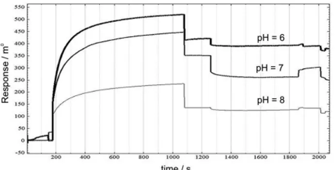
pH optimization can be a critical parameter in determining the success of immobilization of Anti-Omp31. Acetate buffer is used in the immobilization phase and the buffer pH is adjusted according to the isoelectric point of immobilized protein that should be about 0.5-0.8 less than the isoelectric point of antibody, since the carboxyl surface of sensor chip produce a negative charge at higher pH (3.5) therefore, to achieve the effective preconcentration, buffer pH should be higher than 3.5 and less than the isoelectric point of ligand. Ligand on the surface is pre-condensed through electrostatic attraction when the pH is between the isoelectric point of ligand and the surface pKa. If the pH was too lower or too high, the ligand will not be concentrated on the surface. As a result, according to the PI (isoelectric point) of antibody which is equal to 9, the pH of 8.3 for the immobilizing buffer might be appropriate, but in practice, better signals were received with immobilizing in the lower pH. Although pH 6 is more appropriate, since EDC needs uncharged amine groups and is more efficient at higher pH, pH = 7 was selected. The overlay of immobilization sensorgram at different pH is shown in Figure 1.
Affinity investigation of bacteria and antigen on Anti-Omp31/ MUA/gold disk
For evaluation of antibody affinity, initially, the various concentration of antigen (Omp31) from 9 × 10⁻⁹ to
disk surface. As shown in Figure 2, antibody-antigen interaction diagram is completely obvious, thus Anti-Omp31/MUA/gold disk has an appropriate affinity against Omp31. In the next step, Anti-Omp31-Brucella
interaction was investigated. Overlay of interaction diagram between the antibody and the bacteria with concentrations of 101 to 107 cell mL-1 were shown in
Figure 3. The SPR angle shift linearly increases with the adding of bacteria concentration.
Antibody optimization
Concentration of immobilized antibody and pH of immobilization buffer are major factors that may effect on antibody (Anti-Omp31) and antigen (Omp31) interaction.
The effect of antibody concentration on the immunosensor response was also studied in the concentration range from 25-100 µg mL-1. According to the immobilization results,
the optimum concentration of antibody for immobilization on the sensor chip was 25 µg mL-1 (Figure 4). At highly
concentrated immobilized antibody, due to spatial configuration created on the disk, Anti-Omp31/MUA/gold disk immunosensor will not have an appropriate interaction with the antigen and the resulting signals will be weak. On the other hand, at very low concentrations of immobilized antibody, sensitivity of immunosensor is low, as shown in Figure 4. Maximum response was achieved at concentration of 25 µg mL-1. Thus, it was selected as the optimal
concentration for immobilization of antibody.
Detection of B. melitensis on Ab/11-MUA/gold discs using
SPR technique
For detection of B. melitensis, designed Anti-Omp31/ MUA/gold disc sensor chip for SPR measurement was used. Figure 5 shows the relationship between the increase of the angle shift and the Brucella number.
Brucella melitensis was detected at a concentration range from 10 to 108 cell mL-1 (number of Brucellaper mL) with
a correlation coefficient of 0.998 and limit of detection (LOD) was calculated according to the equation 1. According to equation 2, LOB is equal to limit of blank.18
Actually, there is couple of equations to reach detection limit, however, LOD = 3SD/b is the most common one. Since the equations are dependent on the slope of the line, any changes in concentration unit of the sample will modify the slope and the value of the detection. But in this equation, since there is not such a dependence between the slope of the line and the detection limit, the changes of the concentration unit does not affect the detection limit value. In this regard, the detection limits of the blank samples were measured and by putting them in the respected equation, the final detection limit of the device was determined:
Figure 1. Effect of pH in antibody immobilization (25 µg mL-1) on the
immunosensor response.
Figure 2. Interacion of immobilized Anti-OMP31 (25 µg mL-1) with
various concentration of antigen (Omp31).
Figure 3. Interaction of Anti-OMP31 with various concentration of
B. melitensis.
LOD = LOB + 1.645 (SD low concentration sample) (1)
LOB = mean blank + 1.645 (SD blank) (2)
where SD is the standard deviation of the response. Based on equation 1, the detection limit was calculated to be as low as 100 cell mL-1. This value is better than those obtained
at gold nanoparticle-modified screen-printed carbon electrode,19 as well as K
D for interaction of Anti-Omp31
and Omp31was 1.1 × 10⁻⁹ mol L-1. Obtained equilibrium
dissociation constant (KD) is in the group of high affinity
interactions which is extracted by comparing the data with that of the other studies.20
For evaluation of kinetics involved in between the interaction of antigen and antibody, the affinity interactions between antigen and immobilized antibody were calculated by the equilibrium dissociation constant (KD). The data were fitted using a simple 1:1 Langmuir fit
model, A + B → AB, with molecules A and B forming the complex AB where ‘A’ is the injected analyte, ‘B’ is the immobilized ligand and ‘AB’ is the analyte-ligand complex formed during the interaction process. In the SPR system, the signal response is proportional to the amount of [AB] and the Rmax is proportional to the initial [B]. Hence, in
this study KD values were calculated for binding of Omp31
with immobilized antibody using the “kinetic evaluation software version 5.4”.21
Selectivity of the Anti-Omp31/MUA/gold immunosensor
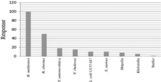
The selectivity of the clinical diagnostic methods is an important factor in analyzing biological samples that display a complex matrix. In the current study, the selectivity of the proposed immunosensor was evaluated in the presence of seven different Gram-negative and Gram-positive bacteria (such as Y. enterocolitica, S. typhi,
E. coli O157:H7, Klebsialla spp., Shigella spp., V. chlerae
and S. aureus) and B. abortus. So, these bacteria were passed with a dilution of 104 to be compared with the
104 dilution of B. melitensis. The results showed that
they have no response to Ab/11-MUA/gold disc, as means that designed immunosensor is selective enough for detection of B. melitensis and as shown in Figure 6, the signals related to the B. melitensis are higher than other bacteria.
Conclusions
The study and preparation of Brucella biosensors is very important and required for diagnostic laboratories. Because of Brucella detection is time consuming, dangerous and costly, we suggested a label-free, inexpensive, rapid and renewable B. melitensis biosensors by SPR method. Surface plasmon resonance biosensors give a unique chance to monitor B. melitensis-antibody interactions in real time without labeling requirements. The immunosensor has good linear range, low detection limit and excellent selectivity. Surface plasmon resonance instrument has been widely accepted by the research community for basic research and analytical applications. So, kinetic parameter such as KD was studied. It seems
that this system has the ability to be used as a biosensor for other pathogens, too. Given the importance of the diagnosis of B. melitensis and considering that the infectious dose of this bacteria is 102-105 CFU mL-1,22,23
and appropriate detection sensitivity obtained by the immunosensor designed in this study as well as proven specificity and the study of the absence of cross-reactivity with the other bacteria, the immunosensor based on SPR using the cell surface protein Omp31 is recommended as a rapid diagnosis option with an appropriate sensitivity and specificity.
Acknowledgments
This article has been extracted from thesis in Molecular Biology Research Center Baqiyatallah University of Medical Sciences. Authors are grateful of Nanobiotechnology Research Center, for implementation of this project.
Figure 6. Determination of SPR specificity by passing of other bacteria.
Figure 5. Calibration curve of interaction of Anti-omp31 with B. melitensis
References
1. Godfroid, J.; Cloeckaert, A.; Liautard, J. P.; Kohler, S.; Fretin, D.; Walravens, K.; Garin-Bastuji, B.; Letesson, J. J.;
Vet. Res. 2005, 36,326.
2. Pappas, G.; Papadimitriou, P.; Akritidis, N.; Christou, L.; Tsianos, E. V.; Lancet Infect. Dis. 2006, 6,91.
3. Haran, M.; Agarwal, A.; Kupfer, Y.; Seneviratne, C.; Chawla, K.; Tessler, S.; BMJ Case Rep.2011, 2011.
4. Zeinalian Dastjerdi, M.; Fadaei Nobari, R.; Ramazanpour, J.;
Public Health 2012, 126,1058.
5. Akhvlediani, T.; Clark, D. V.; Chubabria, G.; Zenaishvili, O.; Hepburn, M. J.; BMC Infect. Dis. 2010, 10, 346.
6. Dean, A. S.; Crump, L.; Greter, H.; Schelling, E.; Zinsstag, J.;
PLOS Neg. Trop. Dis. 2012, 6.
7. Foster, G.; Osterman, B. S.; Godfroid, J.; Jacques, I.; Cloeckaert, A.; Int. J. Syst. Evol. Microbiol. 2007, 57, 2688. 8. Mirnejad, R.; Mohamadi, M.; Piranfar, V.; Mortazavi, S. M.;
Kachuei, R.; Asian Pac. J. Trop. Med. 2013, 6,453.
9. Mirnejad, R.; Doust, R. H.; Kachuei, R.; Mortazavi, S. M.; Khoobdel, M.; Ahamadi, A.; Asian Pac. J. Trop. Med. 2012, 5, 24.
10. Nielsen, K.; Smith, P.; Widdison, J.; Gall, D.; Kelly, L.; Kelly, W.; Nicoletti, P.; Vet. Microbiol. 2004, 100, 25. 11. Bogdanovich, T.; Skurnik, M.; Lübeck, P. S.; Ahrens, P.;
Hoorfar, J.; J. Clin. Microbiol. 2004, 42, 2261.
12. Bounaadja, L.; Albert, D.; Chénais, B.; Hénault, S.; Zygmunt, M. S.; Poliak, S.; Garin-Bastuji, B.; Vet. Microbiol. 2009, 137, 156.
13. Baily, G.; Krahn, J.; Drasar, B.; Stoker, N.; Am. J. Trop. Med. Hyg. 1992, 95,271.
14. Piranfar, V.; Sharif, M.; Hashemi, M.; Vahdati, A. R.; Mirnejad, R.; Iran. J. Basic Med. Sci. 2015, 18, 909.
15. Homola, J.; Anal. Bioanal. Chem. 2003, 377, 528.
16. Vizcaino, N.; Cloeckaert, A.; Zygmunt, M. S.; Dubray, G.;
Infect. Immun. 1996, 64, 3744.
17. Cloeckaert, A.; Vizcaíno, N.; Paquet, J.-Y.; Bowden, R. A.; Elzer, P. H.; Vet. Microbiol. 2002, 90, 229.
18. Shrivastava, A.; Gupta, V. B.; Chron. Young Sci. 2011, 2, 21. 19. Wu, H.; Zuo, Y.; Cui, C.; Yang, W.; Ma, H.; Wang, X.; Sensors
2013, 13, 8551.
20. Sikarwar, B.; Sharma, P. K.; Srivastava, A.; Agarwal, G. S.; Boopathi, M.; Singh, B.; Jaiswal, Y. K.; Biosens. Bioelectron. 2014, 60.
21. Liu, J. T.; Chen, L. Y.; Shih, M. C.; Chang, Y.; Chen, W. Y.;
Anal. Biochem. 2008, 375, 90.
22. Wang, Z.; Wang, S. S.; Wang, G. L.; Wu, T. L.; Lv, Y. L.; Wu, Q. M.; Vet. J. 2014, 200, 116.
23. Alamian, S.; Nejad, R. B.; Jalali, H. R.; Kalantari, A.; Etemadi, A.; Vet. Sci. Dev. 2015, 5.