Influence of He-Ne laser therapy on
the dynamics of wound healing in mice
treated with anti-inflammatory drugs
1Departamento de Ciências Fisiológicas, 2Departamento de Clínica Odontológica,
Centro de Ciências da Saúde, Universidade Federal do Espírito Santo, Vitória, ES, Brasil
W.L.S. Gonçalves1,
F.M. Souza1, C.L. Conti1,
J.P. Cirqueira1, W.A. Rocha1,
J.G.P. Pires1, L.A.P. Barros2
and M.R. Moysés1
Abstract
We determined the effects of helium-neon (He-Ne) laser irradiation on wound healing dynamics in mice treated with steroidal and non-steroidal anti-inflammatory agents. Male albino mice, 28-32 g, were randomized into 6 groups of 6 animals each: control (C), He-Ne laser (L), dexamethasone (D), D + L, celecoxib (X), and X + L. D and X were injected im at doses of 5 and 22 mg/kg, respectively, 24 h before
the experiment. A 1-cm long surgical wound was made with a scalpel on the abdomens of the mice. Animals from groups L, D + L and X + L were exposed to 4 J (cm2)-1 day-1 of He-Ne laser for 12 s and were
sacrificed on days 1, 2, or 3 after the procedure, when skin samples were taken for histological examination. A significant increase of collagen synthesis was observed in group L compared with C (168 ± 20 vs 63 ± 8 mm2). The basal cellularity values on day 1 were: C = 763
± 47, L = 1116 ± 85, D = 376 ± 24, D + L = 698 ± 31, X = 453 ± 29, X + L = 639 ± 32 U/mm2. These data show that application of L
increases while D and X decrease the inflammatory cellularity com-pared with C. They also show that L restores the diminished cellularity induced by the anti-inflammatory drugs. We suggest that He-Ne laser promotes collagen formation and restores the baseline cellularity after pharmacological inhibition, indicating new perspectives for laser therapy aiming to increase the healing process when anti-inflamma-tory drugs are used.
Correspondence
M.R. Moysés
Departamento de Ciências Fisiológicas
Centro de Ciências da Saúde, UFES Av. Marechal Campos, 1468 29040-755 Vitória, ES Brasil
Fax +55-27-3335-7330 E-mail: [email protected]
Research partially supported by UFES and CAPES.
Received April 6, 2006 Accepted March 7, 2007
Key words •Photo-stimulation •Skin
•Collagen •Cyclooxygenase •Corticosteroids •Cicatrization
•Anti-inflammatory drugs
Introduction
Morphological and biochemical studies in humans have revealed a sophisticated mechanism for skin wound healing, includ-ing replacement of the affected subcutane-ous tissue with a new matrix and re-epithe-lization. Injury to the skin triggers a cascade of events including inflammation and tissue
remodeling, which eventually leads to total or partial restoration of the injured area (1,2). Various aspects of this complex process have attracted the attention of researchers over the years, particularly the factors which may hinder it.
processes intervening in the dynamics of wound repair (3,4). The repairing process begins immediately after the injury through the release of cytokines, growth factors, some hormones, and several low-molecular weight substances from plasma or activated plate-lets (1-4). The main endogenous hormone involved in the dynamics of tissue repair is cortisol, which affects the metabolism of carbohydrates and proteins and exerts im-portant anti-inflammatory effects (5,6).
Recent studies have suggested that the early events in the wound healing process are the most appropriate for useful therapeu-tic interventions, while the most important repair failures are those occurring in the initial phase (3). In this respect, we should mention studies concerning the role of stress in the wound healing process, showing that endogenous cortisol down-regulates pro-in-flammatory cytokine and chemokine expres-sion, which reduces the inflammation ede-ma, cellular recruitment and cell prolifera-tion at the injured site (7-9).
Several experimental and clinical studies have evaluated the effects of helium-neon (He-Ne) laser therapy on the process of tis-sue regeneration in areas such as skin, bone, skeletal muscle, and the nervous system (10-13), with contradictory results. However, based on clinical experience, some investi-gators have proposed that photo-stimulation with low-energy laser at certain wavelengths promotes tissue repair by releasing growth factors (2,10-15).
Taking into account these controversies, we decided to investigate the possible benefi-cial effects of He-Ne laser irradiation on the skin repair process in mice previously treated with steroidal or non-steroidal anti-inflamma-tory drugs, which possibly are able to affect the release and/or the effects of factors in-volved in the tissue repair process (9,10).
Material and Methods
Experiments were performed on adult
male albino mice, weighing 28-32 g, from our breeding stock. The animals, 6 to a group,
were housed in 0.30-m2 cages under
con-trolled conditions of 12-h light periods, tem-perature (~26ºC) and minimal noise. Ani-mals were allowed free access to filtered
water and standard rat chow. All the
experi-mental procedures adopted were in accor-dance with the International Guidelines for Animal Care.
Experimental design
The mice were randomly divided into 6 groups of 6 mice each: 1) control (C), 2) He-Ne laser (L), 3) dexamethasone (D), 4) D + L, 5) celecoxib (X), and 6) X + L. These groups were further divided into 24-, 48-and 72-h groups. D 48-and X were injected
intramuscularly (im) at doses of 5 and 22
mg/kg, respectively, 24 h before the begin-ning of the experiment.
A surgical skin wound was made with a scalpel on the abdomen of the mice who were under general anesthesia with 10 mg/
kg ketamine + 20 mg/kg xylazine, im. The
skin on the abdominal region was shaved before the procedure using an aseptic tech-nique. The surgical incision was standard-ized as follows: 1-cm length (xiphoid appen-dix as reference) and adequate depth to in-clude the epidermis, dermis and abdominal fascia. After the surgical procedure, laser irradiation was applied (see below) to the treated groups. The animals were then re-turned to their home cages.
Laser treatment
A laser device (KLD-Biosystem®, São
output were determined using the Tuner and Hode equation (16). Each animal in the laser groups received a fixed daily dose of He-Ne laser irradiation at an energy density of 4 J/
cm2 applied over a period of 12 s. The laser
probe was positioned to contact the wound. Only one session of laser was applied imme-diately after surgery.
The animals were then submitted to eu-thanasia by ethyl ether inhalation at 24, 48, and 72 h after the procedures and skin samples were removed for histological and morphometric analysis.
Histological and morphometric analysis
All skin lesion samples obtained were immediately fixed in 10% buffered forma-lin, pH 7, for at least 24 h. After fixation, the samples were gradually dehydrated in in-creasing ethanol concentrations (70 to 100%), cleared in xylene and embedded in paraffin according to routine histological methods. The paraffin-embedded fragments were cut with an “820” Spence microtome (New York, NY, USA) and 6-µm thick sections were obtained. Pairs of histological slides were kept in an incubator to dry and the sections were then stained with hematoxylin-eosin and Masson’s trichrome for histological analysis. The histology scoring was based on the degree of cellular invasion (cellularity), granulation tissue formation, vascularity, and re-epithelization. The code describing each animal’s treatment was broken after the pa-thologist completed the scoring and ranking (Table 1).
Histomorphometry was performed using images captured and evaluated by a
compu-terized Sigma-pro®image (St. Louis, MO,
USA)capture system. Images were captured
from five randomly chosen optical micro-scopic fields for each histological slide pair using the digital camera (total magnification
100 and 200X) from an Olympus®AX70
Plus microscope (Tokyo, Japan). The im-ages were stored and submitted to a count of
inflammatory cells or cellularity (i.e., cellu-lar density) and analysis of collagen forma-tion and of re-epithelizaforma-tion of the ulcerated areas of the wounds at the end of the experi-ment using digital marking (color contrast) as the discriminating parameter.
Drugs
Dexamethasone phosphate salt (Dex-azona™, Bunker, São Paulo, SP, Brazil) and celecoxib (COX-2 inhibitor; a kind donation from Pfizer, Guarulhos, SP, Brazil) were used. Doses were chosen according to those usually reported in the literature. Ketamine hydrochloride (Ketamin™, 5% solution) was purchased from Cristalia (São Paulo, SP, Brazil). A commercial veterinary prepara-tion of xylazine hydrochloride (Rompun™, Bayer, Rio de Janeiro, RJ, Brazil) was used. The two drugs were placed in the same syringe for anesthesia.
Statistical analysis
The nature of the variables studied or the variability of the means was considered
us-ing the biostatistics software GraphPad Prism
4. Unless otherwise stated, data were ana-lyzed statistically by ANOVA followed by the Tukey test for multiple comparisons or the Mann-Whitney U-test for independent samples, as appropriate. The level of signif-icance was set at P < 0.05.
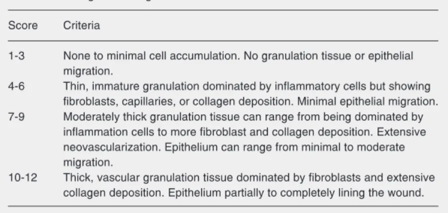
Table 1. Histological scoring.
Score Criteria
1-3 None to minimal cell accumulation. No granulation tissue or epithelial migration.
4-6 Thin, immature granulation dominated by inflammatory cells but showing fibroblasts, capillaries, or collagen deposition. Minimal epithelial migration. 7-9 Moderately thick granulation tissue can range from being dominated by
inflammation cells to more fibroblast and collagen deposition. Extensive neovascularization. Epithelium can range from minimal to moderate migration.
Results
Clinical inspection of skin lesion samples showed a regular amount of humid clot on the surface and the presence of many blood vessels in the wound area in the C and D groups, but not in the other four groups (L, D + L, X, X + L; data not shown).
The morphological criteria used for scor-ing the material are presented in Table 1. Histological analysis showed that specimens from the laser groups (L, D + L, X + L) had fewer blood clots and displayed more accel-erated re-epithelization, granulation tissue formation and capillary proliferation than
the other groups, such as C (Figure 1). Based on these morphological criteria (Table 1), it was observed that the L group displayed important healing dynamics after 24 h of treatment compared to the C group. Laser-treated animals showed significantly higher histological scores than the respective
con-trols, in the first 24 h (7.16 vs 1.38; P <
0.0001, Mann-Whitney U-test).
As shown in Figure 2A, the epithelium
samples from the D (7.3 ± 0.7 mm2) and X
(17.1 ± 0.8 mm2) groups showed expected
significant differences during the first 24 h when compared to the C group (12.6 ± 0.8
mm2). D significantly reduced
re-epitheliza-tion, while, surprisingly, X increased it. In the D + L group, laser irradiation restored the re-epithelization phenomenon (15.0 ±
0.4 mm2), whereas in the X + L group, laser
application further decreased the
phenome-non (11.0 ± 0.4 mm2). In addition, the L
group showed that laser irradiation produced
a significant increase (17.3 ± 0.8 mm2) in the
wound re-epithelization (Figure 2A). Collagen synthesis was significantly
higher in the L (168 ± 20 mm2), D + L (189
± 11 mm2), and X + L (185 ± 9 mm2) groups
(Figure 2B) and slightly but significantly attenuated in the D and X groups compared with C animals (56 ± 6, 55 ± 5, and 62 ± 8
mm2, respectively).
As expected, D or X caused a significant reduction in cellularity compared with C during the first 24 h (375 ± 24, 452 ± 29, and
763 ± 48 U/mm2 for D, X, and C,
respective-ly). Laser therapy restored cellularity to the baseline level, as seen in the D + L (688 ± 31
U/mm2) and X + L (639 ± 32 U/mm2) groups
(Figure 2C). In these two groups, laser appli-cation also increased the migration and mo-bility of leukocytes and fibroblasts/myoblasts (Figure 3).
Discussion
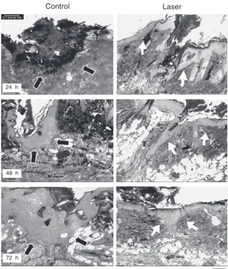
Low-intensity laser therapy is recognized as an effective therapeutic method by the Figure 1. Photomicrograph of the skin wound of control (left, black arrows) and laser
therapy (right, white arrows) groups at 24, 48, and 72 h. The white arrows show an accelerated healing process induced by He-Ne laser in the experimental groups (Masson’s trichrome; scale bar: 0.1 mm at 10X).
Control Laser
24 h
48 h
FDA, particularly to improve tissue healing
(3-6). A large body of evidence from in vitro
and in vivo studies has suggested that LILT enhances collagen synthesis (4-6,10), in-creases the motility of keratinocytes (17), releases growth factors (15,17), and pro-motes the transformation of fibroblasts into myofibroblasts (4-6,15,17-19). On the other hand, the idea of investigating the early phase of the skin repair process under the influence of anti-inflammatory agents (a corticoster-oid or a cyclooxygenase (COX-2) inhibitor) was based on the known pharmacological properties of such drugs and on their broad clinical use and side-effects (20-24).
Studies have shown that during normal wound healing the inflammatory phase lasts up to two days (3,24-26). Cell proliferation, epithelization, granulation tissue formation, and wound contraction occur during the pro-liferative phase. Growth of new epithelial cells across the surface of the wound and collagen remodeling occur during the matu-ration phase of wound healing, which lasts for months or even longer. The present re-sults showed that He-Ne laser irradiation of the skin of mice produced beneficial effects on the macroscopic aspects of the surgical wound, such as reduction of humidity, ap-parent vascular modulation and effective control of the amount of clot in the injured area, during the first 24 h. These data there-fore, suggest that laser therapy increased the healing dynamics compared to control.
Werner and Grose (26) described three
different stages of wound repair: i)12-24 h
after injury the wounded area is filled with a blood clot which is invaded by neutrophils; ii) at days 3-7 after injury, most neutrophils have undergone apoptosis whereas macro-phages are abundant in the wound tissue at this stage of repair. Endothelial cells migrate into the clot, proliferate and form new blood vessels. Fibroblasts migrate into the wound tissue, where they proliferate and deposit extracellular matrix. The new tissue is called granulation tissue. Keratinocytes proliferate
Figure 2. Determinations of the volumetric fractions of the epi-thelium (A), collagen (B), and cellularity (C; neutrophils and fibroblasts/myofibroblasts) of mouse skin 24 h after treat-ments. C = control; L = He-Ne laser; D = steroidal anti-inflam-matory drug dexamethasone; D+L = dexamethasone plus la-ser; X = non-steroidal anti-in-flammatory drug celecoxib; X+L = celecoxib plus laser. Data are reported as mean ± SEM. *P < 0.01 vs C; **P < 0.01 vs D+L; +P
< 0.05 vs X+L (Tukey test).
Figure 3. Photomicrography of the skin wound in the dexamethasone (A) and dexametha-sone + laser groups (B) at 24 h. The white arrows point to increased cellularity with the intense presence of mononuclear cells in the infiltrate, predominantly fibroblasts and neutrophils in the skin wound margins (H&E; scale bar: 0.1 mm at 20X).
at the wound edge and migrate down the injured dermis and above the provisional matrix; iii) 1-2 weeks after injury the wound is completely filled with granulation tissue. Fibroblasts have transformed into myofi-broblasts, leading to wound contraction and collagen deposition. The wound is then com-pletely covered with a neoepidermis.
The time course of the present results obtained from histological analysis of the C group fit with the picture described by Werner and Grose (26). However, morphological analysis of the laser therapy groups indicates that significant differences do exist in these groups, suggesting an acceleration of the healing dynamics during the first days caused by laser application.
Similar results were obtained by Maiya et al. (15) in diabetic rats, showing that laser-treated animals healed faster and better than controls. Other studies (11,12,14,18,22) us-ing He-Ne low-energy laser have indicated that it is mainly the laser energy at 633 nm wavelength that affects the healing dynam-ics, producing changes in the early phase of the repair process, i.e., the inflammatory phase.
In the first hours of healing, the repair events are directed towards preventing sub-sequent blood loss (hemostasis) and towards the formation of a fibrin plug that supplies the preliminary matrix for the subsequent processes in which platelets adhere to the collagen in the perivascular space. Such con-tact activates platelets, which releases bioactive factors that accelerate the migra-tion and proliferamigra-tion of fibroblasts, a key cell in this process (11,12).
The suggestion that laser affects the early events in the dynamics of wound healing was partially based on the observed low-intensity laser therapy-induced attenuation of reactive oxygen species production by neutrophils in inflammatory models. This was initially suggested by the study of Fujimaki et al. (20), who described a dimin-ished oxidative stress-induced apoptosis of
neutrophils in acute inflammation (20,27-28). Nevertheless, the basic aspects of oxi-dative stress and the mechanisms by which reactive oxygen species modulate physiologi-cal and pathologiphysiologi-cal processes, with empha-sis on wound healing, are still motive of debate.
As stated before, the early events in wound healing, with special reference to the inflammatory phase, are crucial for the suc-cess of the prosuc-cess, as suggested by studies on the effects of anti-inflammatory drugs (3). It is well known that corticosteroids down-regulate pro-inflammatory proteins and affect gene expression, interfering with almost all phases of the inflammatory pro-cess (7-9). On the other hand, controlled laboratory trials have reported that laser photostimulation can reduce inflammation through inhibition of inducible COX-2, lead-ing to a reduction in prostanoid levels (28,29). Additionally, experiments involving vari-ous cell culture stages have shown that laser irradiation at early stages significantly stimu-lates cell proliferation, alkaline phosphatase activity and osteocalcin gene expression, indicating that laser photostimulation
en-hances bone formation in vitro (3,11,19).
produced by He-Ne irradiation in the pres-ence of D or X towards the maintenance of inflammatory cells at the “baseline” level (Figure 2C). It should be mentioned that recent studies (30,31) have shown that He-Ne laser irradiation can effectively acceler-ate the expression of tumor growth factor ß1 and facilitate changes in leukocyte activity
and accumulation of lipids byoxidation
prod-ucts. Furthermore, tumor growth factor ß1 is a chemoattractant for neutrophils, macro-phages and fibroblasts (18) and is also asso-ciated with the wound healing defect seen in glucocorticoid-treated animals (26).
Thus, the present findings suggest that the stimulatory effects of laser photostimu-lation are related to specific events during the first two phases of wound healing, i.e., the inflammatory phase and the proliferative phase, indicating that the time of interven-tion may be critical and also suggesting that satellite cells are major irradiation-respon-sive candidates. The present results show that He-Ne laser therapy can influence the
behavior of many inflammatory cell types, and that multiple effects can occur simulta-neously and accelerate healing dynamics in the presence of corticosteroids or non-ste-roidal anti-inflammatory drugs. Addition-ally, our results suggest that He-Ne laser irradiation modulates the early phases of the
repair process in vivo. We speculate that this
could be due to biochemical events in the mitochondrial oxidation process and/or to accumulation of lipid by oxidation products. However, much more research using selec-tive inhibitors and markers will be need to elucidate the exact mechanisms of action of laser photostimulation at the cellular and molecular levels.
Acknowledgments
The authors thank Dr. C.A. Redins, Dr. M.A.S. Novaes and Mrs. L. Bressoni (UFES, Vitória) for help with the techniques and morphological analysis.
References
1. Ishida Y, Watanabe H. Structural study of wound healing in mouse skin with special reference to quantitative changes of cellular con-stituents. Yamagata Med J 1999; 17: 332-342.
2. Werner S, Grose R. Regulation of wound healing by growth factors and cytokines. Physiol Rev 2003; 83: 835-870.
3. Reddy GK. Photobiological basis and clinical role of low-intensity lasers in biology and medicine. J Clin Laser Med Surg 2004; 22: 141-150.
4. Medrado AR, Pugliese LS, Reis SR, Andrade ZA. Influence of low level laser therapy on wound healing and its biological action upon myofibroblasts. Lasers Surg Med 2003; 32: 239-244.
5. Campana V, Moya M, Gavotto A, Juri H, Palma JA. Effects of diclofenac sodium and He:Ne laser irradiation on plasmatic fibrino-gen levels in inflammatory processes. J Clin Laser Med Surg 1998; 16: 317-320.
6. Pessoa ES, Melhado RM, Theodoro LH, Garcia VG. A histologic assessment of the influence of low-intensity laser therapy on wound healing in steroid-treated animals. Photomed Laser Surg 2004; 22: 199-204.
7. Mercado AM, Padgett DA, Sheridan JF, Marucha PT. Altered kinet-ics of IL-1 alpha, IL-1 beta, and KGF-1 gene expression in early wounds of restrained mice. Brain Behav Immun 2002; 16: 150-162. 8. Sheridan JF, Padgett DA, Avitsur R, Marucha PT. Experimental
models of stress and wound healing. World J Surg 2004; 28: 327-330.
9. Pruzanski W, Vadas P. Phospholipase A2 - a mediator between proximal and distal effectors of inflammation. Immunol Today 1991; 12: 143-146.
10. Posten W, Wrone DA, Dover JS, Arndt KA, Silapunt S, Alam M. Low-level laser therapy for wound healing: mechanism and efficacy.
Dermatol Surg 2005; 31: 334-340.
11. de Carvalho PT, Mazzer N, Barbieri CH, Siqueira JFR. Morphomet-ric analysis of the percentage of collagen and number of macro-phages highlighted by immunohistochemistry, in cutaneous wound in diabetic and non-diabetics rats treated through He-Ne laser.
Lasers Med Sci 2003; 18 (Suppl 1): S0167 (Abstract).
12. de Carvalho PT, Mazzer N, dos Reis FA, Belchior AC, Silva IS. Analysis of the influence of low-power He-Ne laser on the healing of skin wounds in diabetic and non-diabetic rats. Acta Cir Bras 2006; 21: 177-183.
13. Amaral AC, Parizotto NA, Salvini TF. Dose-dependency of low-energy HeNe laser effect in regeneration of skeletal muscle in mice.
Lasers Med Sci 2001; 16: 44-51.
15. Maiya GA, Kumar P, Rao L. Effect of low intensity helium-neon (He-Ne) laser irradiation on diabetic wound healing dynamics. Photomed Laser Surg 2005; 23: 187-190.
16. Tuner J, Hode L. It’s all in the parameters: a critical analysis of some well-known negative studies on low-level laser therapy. J Clin Laser Med Surg 1998; 16: 245-248.
17. Rood PA, Haas AF, Graves PJ, Wheeland RG, Isseroff RR. Low-energy helium neon laser irradiation does not alter human keratino-cyte differentiation. J Invest Dermatol 1992; 99: 445-448.
18. Rocha AM Jr, Oliveira RG, Farias RE, Andrade LCR, Aarestrup FM. Modulation of fibroblast proliferation and inflammatory response by low-intensity laser in tissue repair process. An Bras Dermatol 2006; 81: 150-156.
19. Hawkins DH, Abrahamse H. The role of laser fluence in cell viability, proliferation, and membrane integrity of wounded human skin fibro-blasts following helium-neon laser irradiation. Lasers Surg Med
2006; 38: 74-83.
20. Fujimaki Y, Shimoyama T, Liu Q, Umeda T, Nakaji S, Sugawara K. Low-level laser irradiation attenuates production of reactive oxygen species by human neutrophils. J Clin Laser Med Surg 2003; 21: 165-170.
21. Jiang Y. Thirty cases of sub-healthy state regulated by acupuncture and He-Ne laser vascular irradiation. J Tradit Chin Med 2006; 26: 102-103.
22. Lan CC, Wu CS, Chiou MH, Hsieh PC, Yu HS. Low-energy helium-neon laser induces locomotion of the immature melanoblasts and promotes melanogenesis of the more differentiated melanoblasts: recapitulation of vitiligo repigmentation in vitro. J Invest Dermatol
2006; 126: 2119-2126.
23. Simunovic Z, Ivankovich AD, Depolo A. Wound healing of animal and human body sport and traffic accident injuries using low-level laser therapy treatment: a randomized clinical study of seventy-four patients with control group. J Clin Laser Med Surg 2000; 18: 67-73. 24. Limpanichkul W, Godfrey K, Srisuk N, Rattanayatikul C. Effects of low-level laser therapy on the rate of orthodontic tooth movement.
Orthod Craniofac Res 2006; 9: 38-43.
25. Singer AJ, Clark RA. Cutaneous wound healing. N Engl J Med 1999; 341: 738-746.
26. Werner S, Grose R. Regulation of wound healing by growth factors and cytokines. Physiol Rev 2003; 83: 835-870.
27. Rojkind M, Dominguez-Rosales JA, Nieto N, Greenwel P. Role of hydrogen peroxide and oxidative stress in healing responses. Cell Mol Life Sci 2002; 59: 1872-1891.
28. Muller-Decker K. Cyclooxygenases in the skin. J Dtsch Dermatol Ges 2004; 2: 668-675.
29. Sakurai Y, Yamaguchi M, Abiko Y. Inhibitory effect of low-level laser irradiation on LPS-stimulated prostaglandin E2 production and cy-clooxygenase-2 in human gingival fibroblasts. Eur J Oral Sci 2000; 108: 29-34.
30. Sun XH, Wang R, Zhang XY. Effects of He-Ne laser irradiation on the expression of transforming growth factor beta1 during experi-mental tooth movement in rabbits. Shanghai Kou Qiang Yi Xue
2006; 15: 52-57.