A
RTIGOC
IENTÍFICO©Revista Brasileira de Fisioterapia
Analysis of low-level laser therapy doses
in Brazilian equipment
Análise da dose do laser de baixa potência em equipamentos nacionais
Fukuda TY1,3, Malfatti CA2
Abstract
Introduction: Low-level laser therapy is becoming more popular and there is a growing interest in its effects, as reflected in the increased number of articles published about the subject. Many therapists and researchers have used a laser dose definition based on energy density (∆E). However, the variety of laser equipments may lead to differences in the therapeutic results found, since the parameters supplied by these equipments vary according to the manufacturer. Objective: To analyze the final energy transmitted to the tissue when applying the same ∆E using equipment of different Brazilian brands. Material and methods: Seven brands of Brazilian equipment with different mean power (Pm) were evaluated by means of simulations. ∆E of 1J/cm2 was applied using each brand of equipment, in
order to evaluate possible differences in the final energy. Results: The same ∆E applied using different brands of Brazilian equipment supplied final energy that ranged from 10 to 90mJ. This variation in the energy was mainly due to differences in Pm. These values ranged between 5.4 and 75mW. Conclusions: This variability in the final energy that is transmitted to the tissue indicates that ∆E may not be the best parameter for describing the dose to be used. In addition to ∆E, the final energy needs also to be stated, in order to establish the dose for obtaining the best therapeutic results.
Key words: low-level laser therapy; dose; parameters.
Resumo
Contextualização: A laserterapia de baixa potência vem sendo cada vez mais utilizada, e o crescente interesse por seus efeitos relaciona-se com a grande quantidade de publicações científicas. Muitos terapeutas e pesquisadores têm-se baseado na definição da dose do laser pela densidade energética (∆E); porém, a grande variedade de equipamentos de laser pode levar a diferença nos resultados terapêuticos encontrados, por fornecerem parâmetros que variam de acordo com o fabricante. Objetivo: Analisar a energia final transmitida ao tecido ao aplicar-se a mesma ∆Eem equipamentos de diferentes marcas nacionais. Materiais e métodos: Foram avaliados sete equipamentos nacionais, com potência média(Pm)diferentes, e foram realizadas simulações aplicando ∆E de 1J/cm2 em
cada aparelho, para avaliar possíveis diferenças na energia final. Resultados: A mesma ∆E aplicada em diferentes aparelhos nacionais forneceu energia final que variou entre 10 e 90mJ. Esta variação na energia deveu-se principalmente a diferenças na Pm, sendo encontrados valores entre 5,4 e 75mW. Conclusão: Esta variabilidade na energia final, que é transmitida ao tecido, indica que a ∆E parece não ser o parâmetro que melhor descreve a dose a ser utilizada. É preciso mencionar não só a ∆E, mas também a energia final, para que se possa estabelecer a dose para obtenção do melhor resultado terapêutico.
Palavras-chave: laser de baixa potência; dose; parâmetros.
Recebido: 27/11/2006 – Revisado: 23/07/2007 – Aceito: 8/11/2007
1 Department of Physical Therapy, Centro Universitário São Camilo, São Paulo (SP), Brazil 2 Research Institute, Hospital Israelita Albert Einstein, São Paulo (SP), Brazil
3 Physical Therapy Sector, Irmandade Santa Casa de Misericórdia de São Paulo, São Paulo (SP), Brazil
Correspondece to: Thiago Yukio Fukuda, Rua Flavio de Melo, 156, apto. 32, Vila Mariana, CEP 04117-130, São Paulo (SP), Brazil, e-mail: [email protected]
71
Introduction
Low-level laser therapy has been investigated and used in clinical practice for approximately 20 years. The initial studies were done in Europe by Mester1,2 at the beginning of
the 1970s. There has been growing interest in the effects of laser energy, as shown by the significant quantity of scien-tific publications, with controlled experiments on both ani-mals and humans3,4.
However, researchers and therapists have questioned the clinical benefits of laser energy because of divergences in the encountered results, due to the lack of methodologi-cal standardization of the studies1,2. While some researchers
have defended the idea that laser energy has therapeutic effects, others have contested such properties, thus highli-ghting the need for cautious interpretation of results when clinically reproduced.
In this sense, when characterizing a laser application, all its parameters need to be described in detail, such as wavelength, energy emitted to the tissue, energy density, beam area, duration of application, peak power, mean power (in the case of pulsed applications) and power density1,5,6. This complete description of the parameters
has the function of helping the professional who is applying the laser therapy to clinically reproduce the findings from experimental trials.
One of the most important aspects of laser applications, and where the greatest divergences are found, is the dose, which is deined as the quantity of radiation emitted to the tissue. he ideal dose to be used is based on research in the literature describing successful laboratory practices, and is es-timated according to the tissue to be irradiated, and adjusted according to the energy absorbed by each tissue, the duration of irradiation and the size of the afected area7.
Contributing towards this diiculty in dose standardiza-tion, is that the various models of laser equipment provide parameters that may vary according to the manufacturer. Considering that the parameter most described in the
literature is the energy density (∆E), the same value used in diferent models of equipment may lead to variations in total energy absorbed by the tissue, which may give rise to a variety of efects and may even be harmful to the tissue to which it was applied7-12.
In view of this, the present study had the objective of analyzing whether there are any diferences in inal energy transmitted to the tissue when applying the same ∆E using diferent equipment options available in Brazil.
Materials and methods
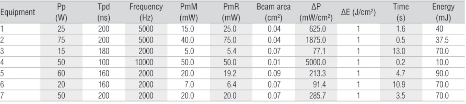
First, data relating to the infrared laser apparatus, with wa-velengths of 904 or 905 nm, were gathered from the instruction manuals provided by the companies. Since some of the infor-mation was not found in the manuals, contacts were also made by telephone and/or the internet. he models and brands of the equipment were numbered as follows (Table 1): 1. Laser Plus Microcontrolled Communicator 904-25W (KW Equip. Eletr.); 2. Laser Plus Microcontrolled Communicator 904-75W (KW Equip. Eletr.); 3. Laserpulse (Ibramed); 4. Laser Endophoton LLT-IR (KLD Biosistemas); 5. Lasermed 4090 – 60W (Carci); 6. Lasermed 4090 – 20W (Carci); 7. Laser 904 (HTM Eletronica).
Seven companies with nine models of laser therapy equipment were originally selected. However, the equi-pment of “Kroman” and “Bioset” brands were excluded because of difficulties in obtaining complete information regarding their parameters.
he parameters investigated were: Peak power (Pp), pulse duration (Tpd), frequency (f), mean power provided by the manufacturer (PmM) and beam area. Using these data, the real mean power (PmR) and the irradiancy or power density (∆P) were obtained. PmR was compared with PmM. For these calculations, the following equations were used1,13:
PmR (W) = Pp (W) x Tpd (s) x f (Hz) ΔP (W/cm2) = Pm
R (W) / beam area (cm 2)
Equipment Pp
(W)
Tpd (ns)
Frequency (Hz)
PmM (mW)
PmR (mW)
Beam area (cm2)
∆P
(mW/cm2) ∆E (J/cm
2) Time
(s)
Energy (mJ)
1 25 200 5000 15.0 25.0 0.04 625.0 1 1.6 40
2 75 200 5000 40.0 75.0 0.04 1875.0 1 0.5 37.5
3 15 180 2000 5.0 5.4 0.07 77.1 1 13.0 70.0
4 50 100 10000 50.0 50.0 0.01 5000.0 1 0.2 10.0
5 60 160 2000 20.0 19.2 0.09 213.3 1 4.7 90.0
6 20 160 2000 7.0 6.4 0.07 91.4 1 10.9 70.0
7 50 200 2000 20.0 20.0 0.07 285.7 1 3.5 70.0
Table 1. Technical characteristics of the equipment studied and their respective parameters: peak power (Pp), pulse duration (Tpd); frequency;
72
After calculating the above values, a simulation was done using an energy density (∆E) of 1 J/cm2 in all the equipment models. With
the data obtained, the following equations7 were used to show
whether the energy emitted to the tissue (E) through the point of application would be equal in equipment with diferent Pm:
ΔE (J/cm2) = ΔP (W/cm2) x t (s)
E (J) = PmR (W) x t (s)
Results and discussion
In addition to the data obtained from the manufacturers, the calculations for each model of equipment are presented in Table 1. It was seen that the parameters obtained and calcula-ted varied greatly according to the diferent types of equipment. Diferences were found in the following parameters: peak power (15-60 W), pulse duration (100-200 ns) and frequency (2,000 to 10,000 Hz), which caused variations in the PmR calculations for each model of equipment (5.4 – 75 mW).
Comparing PmR with PmM, it was observed that in two of the seven models of equipment there was disagreement in the obtained values (equipment “1” and “2”), which could indicate a deiciency in the information generated by the manufacturers of the respective models. It is emphasized that, in this study,
PmR was considered to represent the most reliable aspect of the data for each model of equipment.
he beam area obtained was slightly diferent between the investigated equipment models. When added to the great va-riations found in the calculated PmR, this gave rise to a large diference in the ∆P calculation (77.1 - 5000 mW/cm2).hus, the
duration of the application needed to reach the selected energy density (1 J/cm2) was directly inluenced, since the equipment
with lower ∆P needed longer application times per point. When applying the same ∆E to the analyzed equipment, the variations in the duration of application and PmRled us to ob-tain diferent energy quantities for each type of equipment, with a range from 10 to 90 mJ for each application point.
Although the Pm of equipment “4” was relatively high (50 mW), the short duration of application needed to reach 1 J/cm2 (0.2 s) contributed towards the low inal energy that
was obtained (10 mJ). Likewise, equipment “3”, with a low
Pm (5.4 mW) and a long application time (13 s), provided a relatively high inal energy level (70 mJ). his would lead us to conclude that the energy is inluenced mainly by the duration of the application. However, equipment “5” demonstrated the highest energy level (90 mJ), although it did not provide a prolonged application time (4.7 s), thus showing the need to specify all of the parameters used, and not only one parameter to characterize the selected dose.
Due to the great variety of tissue types exposed to laser treatments that have been described in the literature, some experimental indings have been correlated with the results of this present study. Most studies on laser application for cicatrizing skin wounds show positive efects, as observed through the proliferation of ibroblasts and endothelial cells, and increased deposition of collagen and keratin4,12-15.
Howe-ver, there is great variation in relation to the ∆E used, and the values found ranged from 1 to 21.4 J/cm2 (12,15). It is worth
remembering that the calculated inal energy levels in these studies were 1 and 1.5 J, respectively. hus, diferent ∆Es were observed to produce similar inal energy levels and physiolo-gical results. Correlating the previous data with that from the present study, it was observed that applying an ∆E of 21.4 J/cm2 with equipment “4” and “5” would produce inal
energy levels of between 0.2 J and 1.9 J, respectively. his di-ference in the inal dose may not be numerically signiicant, but it may have a therapeutic inluence, if it is considered that there is a therapeutic window for anti-inlammatory, analge-sic and cicatrizing efects for each tissue.
In evaluating cell growth and collagen synthesis in ibro-blast cultures, Pereira et al.16 concluded that an ∆E of 3 or 4 J/cm2
produced better results than did 5 J/cm2. In analyzing these
data, it can be seen that the inal energy levels obtained in their study were 2.9 J, 3.9 J and 4.8 J, respectively. Bjordal et al.7 stated
that doses over 4 J for each point might inhibit ibroblast acti-vity. hese studies show that high energy doses do not seem to provide the best efects for tissue repair.
Also with regard to the efects of doses with speciic the-rapeutic aims, Matera et al.4 stated that the ∆E recommended
in laser therapy to promote increased numbers of ibroblasts and collagen ibers, and increased vascularization and reepi-thelialization, should be between 1 and 5 J/cm2. In their study,
they concluded that 2 J/cm2 demonstrated better results than
4 J/cm2.
In the same way, Pugliese et al.17 observed the inluences
of the GaAlAs laser on the biomodulation of elastic ibers and collagen in skin wounds in rats, concluding that 4 J/cm2 was
superior to 8 J/cm2. However, neither their study nor that of
Matera et al.4 stated the parameters needed to arrive at the
inal energy, although Matera pointed out the importance of giving details about the dose.
Contradicting the indings that pointed towards a probable therapeutic window for lasers with ∆E below 5 J/cm2, Hopkins
et al.13 evaluated changes in experimental human wounds using
an 820-nm laser at 8 J/cm2. From two skin abrasions produced
73 a systemic efect. It is worth remembering that the inal energy
used in their study was 1.8 J for each point of application. To reach this inal energy of 1.8 J in the laser equipment that we analyzed, ive minutes and thirty seconds would be needed for each application point using equipment “3”, with a Pm of 5.4 mW, and four minutes and 40 seconds would be needed using equip-ment “6”, which demonstrated a Pm of 6.4 mW. We put forward the idea that it may be important to use the Brazilian equipment with greater Pm, in order to decrease the duration of each laser therapy application, thus facilitating the clinical applicability.
It could be seen that ∆E alone did not seem to be the ideal pa-rameter to be followed for studies to be reproduced clinically. he scientiic evidence is contradictory, principally because of the lack of details on the dose used, thus making it diicult to identify the inal energy transmitted to the tissue. he diversity of the subjects exposed to irradiation, i.e., humans or experimentation animals, also contributes towards obtaining diferent results.
In analyzing some studies on the efects of laser energy for cicatrizing tendons, these also were found to show great variety in their choice of parameters. In a study on the use of the GaAs laser for tendon cicatrization in rats, Tavares et al.18 stated that
the ∆E responsible for the cicatrizing efect must be around 3 to 6 J/cm2. For this reason, they used 4 J/cm2, and this generated
satisfactory results. It is worth emphasizing that their study does not mention other parameters, such as the duration of applica-tion and the beam area, and that their Pm does not correspond to the calculated Pm based on the parameters used.
Other studies19,20 also used ∆E within this range, i.e., 3.6 and
5 J/cm2,obtained positive results from cicatrization with diferent
energy levels: 5.4 J and 1.5 J, respectively. Demir et al.21 chose an ∆E
of 1 J/cm2 for tendon repair in rats, which was outside of the range
proposed by Tavares et al.18, thereby obtained success with a inal
energy level of 0.36 J. By correlating this result with our study, and by using equipment “4” with the same ∆E (1 J/cm2) for the same
period of time (60s) that was applied by Demir et al.21, we would
obtain a inal energy level of 3 J. Likewise, if we wanted to reach a inal energy of 0.36 J using 1 J/cm2 with the same equipment, only
0.3s of application would be required. herefore, it is presumed that the use of the same ∆E levelin equipment with diferent Pm ofers divergent physiological results, which could be explained by the large diferences in the inal energy levels transmitted to the tissue.
he applicability of laser energy to nerve tissues seems to be one of the most controversial areas of phototherapy22. In the
study by Chen et al.23, inhibition of nerve regeneration in rats
occurred with energy densities between 2 and 15 J/cm2 and
energy levels, approximately, between 1.6 and 6.5 J. In disagree-ment with these indings, Miloro et al. 24 showed positive results
with 6 J/cm2 and 6.3 J of emitted energy in a synthetic tube. It
may be presumed that there were diferences in the obtained results because the samples were not identical. However, Bagis
et al.25 also studied the efect of laser energy on the nerve tissue
of rats and did not obtain signiicant results, using ∆Es between 0.31 and 19 J/cm2 and energy levels between 0.09 and 5.3 J. To
reach these inal energy values, the authors used a prolonged application time (900s) for their equipment with a low Pm (0.02-0.08 mW). As can be seen, there is a need for more clinical trials with better descriptions of the characteristics of the laser and the biological efects of phototherapy on nerve regeneration22.
hese and other studies reinforce the doubts that exist when establishing the laser dose through the total energy: do the best therapeutic efects obtained through laser irradiation on the tissue occur when a high power is emitted for a short time or a lower power is emitted for a more prolonged time?
In addition to the parameters needed for establishing an ideal dose for low-level laser applications, some other ques-tions still need to be answered. One of these is in relation to the use of laser energy on infected tissue. Infection has always been considered to be an absolute contraindication for the applica-tion of phototherapy because the efects of laser energy on the growth of bacteria still remain obscure.
In an in vitro study26, application of red laser induced the
de-ath of the photosensitive organisms Staphylococcus aureus and
Pseudomonas aeruginosa using doses of 0.1, 0.2 and 0.4 J/cm2 with
an HeNe laser and 2.5, 5 and 10 J/cm2 with an InGaAl laser. Several
parameters that were used are mentioned in the study, making it possible to calculate the inal energy levels: 0.028, 0.057 and 0.114 J with the HeNe laser and 0.15, 0.3 and 0.6 J with the InGaAl laser.
However, Navratil et al.27 reported that GaAs laser
applica-tions might stimulate bacterial growth with ∆E of 0.33 J/cm2,
si-milar to what was used in the preceding study. Nonetheless, they did not report the inal energy transmitted. For laser energy to have a bactericidal efect, their data concur with our hypothesis that ∆E cannot be the only way to establish the dose.
he analysis of these various studies leads us to highlight the need for equipment in Brazil that not only provides the calculation of the ∆E dose, but also informs the inal energy levels emitted to the tissue. In this way, the parameters used could be better described, which would facilitate the clinical reproduction of successful trials.
Conclusion
74
References
1. Kitchen SS, Partridge CJ. A review of low level laser therapy. Physioter. 1991;77(3):161-8.
2. Gam AN, Thorsen H, Lonnberg F. The effect of low level laser therapy on musculoskeletal pain: a meta-analysis. Pain. 1993;52:63-6.
3. Karu T.I. Molecular mechanism of low-power lasertherapy. Lasers Life Sci. 1998;2:53-74.
4. Matera JM, Dagli MLZ, Pereira DB. Efeitos da radiação soft-laser (diodo) sobre o processo de cicatrização cutânea em felinos. Braz J Vet Res Anim Sci. 1994;31(1):43-8.
5. Baker KG, Robertson VJ, Duck FA. A review of therapeutic ultrasound: biophysical effects [review]. Phys Ther. 2001;81:1351-8.
6. Enwemeka CS, Parker JC, Dowdy DS, Harkness EE, Sanford LE, Woodruff LD. The effect of laser phototherapy on tissue repair and pain control: a meta-analysis of the literature. Photomed Lasers Surg. 2004;22:323-9.
7. Bjordal JM, Couppé C, Chow RT, Tunér J, Ljunggren EA. A systematic review of low level laser therapy with location-specific doses for pain from chronic disorders. Austr J Physioter. 2003;49:107-16.
8. Oliveira NM, Parizotto NA, Salvini TF. GaAs (904nm) laser irradiation does not affect muscle regeneration in mouse skeletal muscle. Lasers Surg Med. 1999;25:13-21.
9. Amaral AC, Parizotto NA, Salvini TF. Dose-dependency of low-energy HeNe laser effect in regeneration of skeletal muscle mice. Lasers Med Sci. 2001;16:44-51.
10. Rochkind S, Rousso M, Nissan M, Villarreal M, Barr-Nea L, Rees DG. Systemic effects of low-power laser irradiation on the peripheral and central nervous system, cutaneous wound, and burns. Lasers Surg Med. 1989;9:174-82.
11. Okuno E, Caldas IL, Chow C. Física para ciências biológicas e biomédicas. 1° edição.São Paulo: Harbra; 1982.
12. Lucas C, Gemert MJC, Haan RJ. Efficacy of low-level laser therapy in the management of stage III decubitus ulcers: a prospective, observer-blinded multicentre randomized clinical trial. Lasers Med Sci. 2003;18:72-7.
13. Hopkins JT, McLoda TA, Seegmiller JG, Baxter D. Low-level laser therapy facilitates superficial wound healing in humans: a triple-blind, sham-controlled study. J Athl Train. 2004;39(3):223-9.
14. Tatarunas AC, Matera JM, Dagli MLZ. Estudo clínico e anatomopatológico da cicatrização cutânea no gato doméstico. Utilização do laser de baixa potência GaAs (904nm). Acta Cir Bras. 1998;13(2):86-93.
15. Noronha L, Chin EWK, Kimura LY, Graf R. Estudo morfométrico e morfológico da cicatrização após uso do laser erbium: YAG em tecidos cutâneos de ratos. J Bras Patol Med. 2004;40(1):41-8.
16. Pereira AN, Eduardo CP, Matson E, Marques MM. Effect of low power laser irradiation on cell growth and procollagen synthesis of cultured fibroblasts. Lasers Surg Med. 2002;31:263-67.
17. Pugliese LS, Medrado AP, Reis SRA, Andrade ZA. The influence of low-level laser therapy on biomodulation of collagen and elastic fibers. Pesqui Odontol Bras. 2003; 17(4):307-13.
18. Tavares MR, Mazzer N, Pastorello M. Efeito do laser terapêutico na cicatrização tendinosa: estudo experimental em ratos. Fisioter Bras. 2005;6(2):96-100.
19. Bjordal JM, Lopes-Martins RAB, Iversen VV. A randomized, placebo controlled trial of low level laser therapy for activated Achilles tendonitis with microdialysis measurement of peritendinous prostaglandin E concentrations. Br J Sports Med. 2006;40:76-80.
20. Fillipin LI, Mauriz JL, Vedovelli K, Moreira AJ, Zettler CG, Lech O, et al. Low-level laser therapy (LLLT) prevents oxidative stress and reduces fibrosis in rat traumatized Achilles tendon. Lasers Surg Med. 2005;37:293-300.
21. Demir H, Mentu P, Kirnap M, Calir M, Ikizali I. Comparison of the effects of laser, US and combinated laser + US treatments in experimental tendon healing. Lasers Surg Med. 2004;35:84-9.
22. Gigo-Benato D, Geuna S, Rochkind S. Phototherapy for enhancing peripheral nerve repair: a review of the literature. Muscle Nerve. 2005;31(6):694-701.
23. Chen YS, Hsu SF, Chiu CW, Lin JG, Chen CTC, Yao CH. Effect of low power pulsed laser on peripheral nerve regeneration in rats. Microsurg. 2005;25:83-9.
24. Miloro M, Halkias LE, Mallery S, Travers S, Rashid RG. Low-level laser effect on neural regeneration in Gore-Tex tubes. Oral Surg Oral Med Oral Pathol Oral Radiol Endod. 2002;93:27-34.
25. Bagis S, Comelekoglu U, Coskun B, Milean A, Buyukakilli B, Sahin G, et al. No effect of GaAs (904nm) laser irradiation on the intact skin of the injured rat sciatic nerve. Lasers Med Sci. 2003;18:83-8.
26. DeSimone N, Christiansen C, Dore D. Bactericidal effect of 0,95mW helium-neon and 5mW indium-gallium-aluminum-phosphate laser irradiation at exposure times of 30, 60 and 120 seconds on photosensitized
Staphylococcus aureus and Pseudomonas aeruginosa in vitro. Phys Ther.
1999;79(9):839-46.