www.bjournal.com.br
www.bjournal.com.br
Institutional Sponsors
The Brazilian Journal of Medical and Biological Research is partially financed by
Volume 43 (5) 409-521 May 2010
Braz J Med Biol Res, May 2010, Volume 43(5) 460-466
Expression and purification of the immunogenically active
fragment B of the Park Williams 8 Corynebacterium diphtheriae
strain toxin
D.V. Nascimento, E.M.B. Lemes, J.L.S. Queiroz, J.G. Silva Jr., H.J. Nascimento, E.D. Silva,
R. Hirata Jr., A.A.S.O. Dias, C.S. Santos, G.M.B. Pereira, A.L. Mattos-Guaraldi and
Expression and purification of the
immunogenically active fragment B of
the Park Williams 8
Corynebacterium
diphtheriae
strain toxin
D.V. Nascimento
1,4,
E.M.B. Lemes
1, J.L.S. Queiroz
1, J.G. Silva Jr.
1,
H.J. Nascimento
1, E.D. Silva
1, R. Hirata Jr.
4, A.A.S.O. Dias
3, C.S. Santos
4,
G.M.B. Pereira
2,4, A.L. Mattos-Guaraldi
4and G.R.G. Armoa
2 1Instituto de Tecnologia em Imunobiológicos, 2Instituto Oswaldo Cruz, 3Instituto Nacional de Controle de Qualidade em Saúde, Fundação Oswaldo Cruz, Rio de Janeiro, RJ, Brasil4Faculdade de Ciências Médicas, Universidade do Estado do Rio de Janeiro, Rio de Janeiro, RJ, Brasil
Abstract
The construction of a hexahistidine-tagged version of the B fragment of diphtheria toxin (DTB) represents an important step in the study of the biological properties of DTB because it will permit the production of pure recombinant DTB (rDTB) in less time and with higher yields than currently available. In the present study, the genomic DNA of the Corynebacterium diphtheriae Park
Williams 8 (PW8) vaccine strain was used as a template for PCR amplification of the dtb gene. After amplification, the dtb gene
was cloned and expressed in competent Escherichia coli M15™ cells using the expression vector pQE-30™. The lysate obtained
from transformed E. coli cells containing the rDTBPW8 was clarified by centrifugation and purified by affinity chromatography.
The homogeneity of the purified rDTBPW8 was confirmed by immunoblotting using mouse polyclonal anti-diphtheria toxoid anti
-bodies and the immune response induced in animals with rDTBPW8 was evaluated by ELISAand dermonecrotic neutralization
assays. The main result of the present study was an alternative and accessible method for the expression and purification of immunogenically reactive rDTBPW8 using commercially available systems. Data also provided preliminary evidence that rabbits
immunized with rDTBPW8 are able to mount a neutralizing response against the challenge with toxigenic C. diphtheriae.
Key words: Fragment B; Diphtheria toxin; Diphtheria; dtb gene; E. coli gene expression; Immobilized metal affinity
Introduction
Correspondence: A.L. Mattos-Guaraldi, Faculdade de Ciências Médicas, UERJ, Av. 28 de Setembro, 87 - Fundos, 3º andar,
20551-030 Rio de Janeiro, RJ, Brasil. Fax: +55-21-2587-6476. E-mail: guaraldi@pq.cnpq.br; guaraldi@uerj.br
Received September 29, 2009. Accepted April 6, 2010. Available online April 22, 2010. Published May 14, 2010.
Diphtheria toxin (DT) is an A-B type protein toxin pro
-duced by Corynebacterium diphtheriae (1-4). The B fragment
(DTB) binds to the receptor on the host cell surface and mediates the translocation of the A fragment (DTA) through the cell membrane, which inactivates the protein synthesis elongation factor 2 in some mammalian cells (5-9). The expression of recombinant DTB (rDTB) in other prokaryotic organisms is necessary to understand the role of DT in the development and severity of toxemic infectious processes (10-12). The expression of rDTB in bacteria was initially con
-sidered difficult, due in part to the fact that DTB without DTA was found to be rapidly degraded during the process (13,14). In the early experiments that succeeded in producing rDTB
in Escherichia coli,only low yields were achieved (15). Later,
Spilsberg et al. (16) constructed a hexahistidine-tagged version of a modified rDTB that was expressed in higher levels by E. coli BL21. Attempts to produce immunogenically
reactive rDTB in bacteria in a more accessible form using newer expression systems are of interest. The objective of the present study was to express the dtb gene of the Park
Williams 8 (PW8) C. diphtheriae vaccinestrain to produce
the immunogenically reactive rDTBPW8 using commercially
available expression and purification systems.
Material and Methods
Amplification and cloning of the dtb gene
E. coli vector-mediated DTB cloning process 461
genomic DNA extracted from the C. diphtheriae PW8 ATCC
13812 strain was used as a template for PCR amplification of the dtb gene using 5’- GGG ATC CTA GAA GGT AGC
TCA TTG -3’ as the forward primer and 5’- CCC GGG TGA CCC CAC TAC CTT TCA G -3’ as the reverse primer. After purification with the Gene-Clean® gel extraction kit (BIO 101,
USA), the dtb gene was cloned into the expression vector
pQE-30™ of the QIAexpress System based on standard methods described by the manufacturer (Qiagen, USA).
Transformation of E. coli M15™ cells and expression
of the dtb gene
The hexahistidine-tagged-fused DTBPW8 protein was
successfully expressed in competent E. coli M15™ cells.
During this procedure,E. coli M15™cells were transformed
with the pQE-30/dtb construct and a selected transformant
was grown for 12 h at 37°C in 300 mL Luria-Bertani medium containing 25 µg/mL ampicillin to 0.6 absorbance at 600 nm. Subsequently, transformants were induced with 0.2 mM isopropyl-β, D-thiogalactoside (Promega, USA) for 4 h, collected by centrifugation (10,000 g, 10 min, 4°C),
resuspended in 2.0 mL lysis buffer [20 mM Tris-HCl, 0.1% Triton X-100, 0.5 mM phenylmethylsulfonyl fluoride (Sigma, USA), 1 µg/mL lysozyme], and incubated for 1 h at 4°C. After 10X 10-s ultrasonic pulses, the suspension was centrifuged
(10,000 g, 20 min, 4°C) and the clarified lysateadded to
a 2-mL suspension of a 50% Superflow Ni-NTA slurry and rotated overnight at 22°C. The mixture was transferred to a 5-mL gravity column and beads were washed twice with 4 mL washing buffer [20 mM Tris-HCl, 0.5 M NaCl, 5 mM imidazole in phosphate-buffered saline (PBS; Sigma)]. The protein was finally eluted with 4X 0.5 mL elution buffer [20 mM Tris-HCl, 0.5 M NaCl, 0.5 M imidazole, 8 M urea and 5 mM dithiothreitol (DTT; Sigma)] (16).
SDS-PAGE and densitometric analysis
The material eluted from the Ni-NTA column was ana
-lyzed by 12% SDS-PAGE under denaturing conditions (17). Fractions containing the highest concentration of rDTBPW8
were dialyzed overnight against 5X PBS containing 0.3 M urea at 4°C. Protein concentration was determined using a BioRad Protein Assay™ Kit (USA), based on the method of Bradford, and densitometric analysis was performed with an Image Master 1-D densitometer (GE Healthcare, USA) as follows. The crude rDTBPW8 preparation was chromato
-graphed through a previously equilibrated affinity column containing a nickel streamline chelating matrix. Elution was performed using 20 mM Tris-HCl buffer containing 8 M urea, 0.5 M NaCl, 5 mM DTT and 0.5 M imidazole. The affinity chromatography fractions were analyzed by 12% SDS-PAGE. The crude rDTBPW8 preparation was also submitted
to sieving exclusion chromatography on a Superdex 200 column (Pharmacia, USA) previously equilibrated with 50 mM Tris-HCl. Effluent absorbing at 220 nm was combined and analyzed by SDS-PAGE as described (18,19).
Western blotting and localization of the heterologous
protein in E. coli M15™ cells
Western blotting analysis of rDTBPW8 fractions was
performed using standard procedures (20). Proteins were blotted onto a 0.45-µm nitrocellulose membrane and blocked overnight with 5% skim milk/PBS/0.1% Tween 20 (PBS-T). On the next day, the blocked membrane was incubated with 1:1000 mouse polyclonal antibody against the antidiphtheria toxoid produced in house and then with alkaline phosphatase-conjugated anti-mouse IgG (Sigma) diluted in PBS-T. Blots were further developed with nitroblue tetrazolium chloride and 5-bromo-4-chloro-3-indolylphosphate p-toluidine salt
(Sigma).
Immunization of mice and rabbits
Immunization experiments with rDTBPW8 were con
-ducted on 4- to 6-week-old male BALB/c mice and New Zealand rabbits in compliance with the Ethical Principles in Animal Experimentation established by the Brazilian College of Animal Experimentation and approved by the Fundação Instituto Oswaldo Cruz - Animal Use Ethics Committee - CEUA (P0163-03) under protocol #CEUA L00034-07.
Mice (N = 5) were immunized intraperitoneally with 20 µg of the recombinant protein rDTBPW8 in a 100-µL PBS
(20%) emulsified in complete Freund’s adjuvant. A booster was given 15 days after immunization. Blood samples were collected from the retro-orbital plexus before immunization and 15 and 60 days thereafter.
A New Zealand rabbit was immunized intradermally in the thigh with 2 µg rDTBPW8 in 0.1-mL PBS supplemented
with complete Freund’s adjuvant and boosted with the same formulation 21 days later. Blood samples were collected from saphena or ear veins before challenge at 21 and 28 days thereafter.
Detection of rDTBPW8-specific antibodies by ELISA
ELISA was performed for the detection and quantifica
-tion of both anti-rDTBPW8 mouse and rabbit antibodies (21).
Wells of Maxisorp plates were coated with 0.1 µg DT (Sigma) in 100 µL PBS. After overnight incubation at 4°C, microplates were washed with PBS-T, 100 µL goat anti-rabbit IgG or goat anti-mouse IgG (1:4000) conjugated with horseradish peroxidase (both from Sigma) was added to each well and the plates were incubated for 1 h at 37°C. After washing, reactions were observed 10 min after incubation at room temperature and in the absence of light with 10 mg/mL 3, 3’,5,5’ tetramethyl benzidine in 100 µL citrate phosphate buffer and 0.01% hydrogen peroxide as substrate. Finally, the reaction was stopped by the addition of 50 µL 2 N sulfuric acid, and theabsorbance at 450 nmof the yellow-orange color was measured with a spectrophotometer.
DT dermonecrotic neutralization test in rabbits
In order to evaluate the in vivo dermonecrotic neu
we used an adaptation of Fraser’s protocol (22). Seven days after the booster dose with DTBPW8, an animal was
challenged on the shaved back with injections of 0.1 mL bacterial supernatants adjusted to a concentration of ap
-proximately 3 x 10⁸ CFU toxigenic PW8 vaccine strain
and nontoxigenic ATCC 27010 type strain. The efficacy of DT neutralization was monitored visuallyand sized when possible at the site of injection after 24, 48, and 120 h, for the absence or presence of dermoreactions to the bacte
-rial challenges. Similarly, a non-immunized rabbit was also challenged on the shaved back with the supernatants of both bacterial strains.
Results
Amplification of the dtb gene of the C. diphtheriae PW8 vaccine strain by PCR
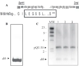
Following the purification step with the Gene-Clean Extraction kit the 1051-bp amplicon was cloned in frame within the BamHI and SmaI restriction sites of the pQE-30™
expression vector to create a fusion product with the hexa
-histidine tag-coding sequence as shown in Figure 1A. Com
-petent E. coli M15™ cells were successfully transformed
with the ligation product and the details of the plasmid and the inserted DTB are presented in Figure 1B and C.
Expression of the rDTBPW8 recombinant protein
Expression of rDTBPW8 recombinant protein in E. coli
M15™cells was demonstrated by SDS-PAGE and immu
-noblot analysis (Figure 2). The clarified rDTBPW8 lysate was
purified in one step using nickel-coated agarose beads and the eluted material was submitted to analysis by SDS-PAGE. A final protein concentration of 0.38 mg/mL was observed in the pool of rDTBPW8-containing fractions. rDTBPW8
(lane 7) was recognized by anti-DT antibodies in Western blotting assays, confirming that the purified product was a fragment of DT protein.
Densitometric analysis
SDS-PAGE of the crude rDTBPW8 preparation revealed
various polypeptide bands suggestive of minor polypep
-tide contaminants. Densitometric analysis of these bands showed the relative abundance of each polypeptide as well as their estimated molecular weight (Figure 3A and B). As expected, the highest peak (84.8%) corresponded to the fraction containing the target protein rDTBPW8. On the other
hand, weshowedthat Superdex-200 chromatography of the same preparation can be used prior to affinity chromatog
-raphy to improve the fractionation in immobilized metal ion affinity chromatography columns and to improve the moni
-toring of the progress of the protein in the chromatograms. Such progress was indicated by the increase in intensity of the peptide peaks. The highest peak corresponded to the fraction containing the target protein rDTBPW8 according
to SDS-PAGE analysis (Figure 3C).
Humoral immune response of mice and rabbits immunized with rDTBPW8
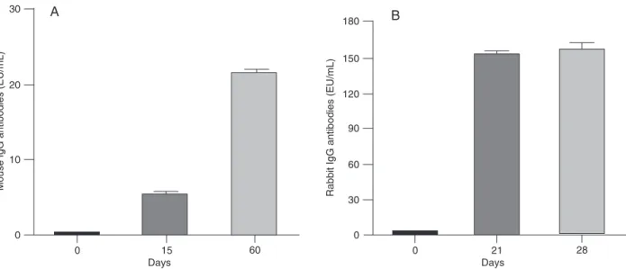
The results presented in Figure 4 indicate significant differences between the antigenic and immunogenic prop
-Figure 1. Amplification and cloning of the Corynebacterium diph-theriae Park Williams 8 (PW8) 1051-bp dtb gene. A, Restriction
sites are indicated in bold and the translated amino acid se-quences from the N-terminal to the C-terminal end are shown in upper case in a one-letter code; the DTBPW8 amino acid se-quence is boxed and the hexahistidine tag (HisTag) is shown as 6X HisTag. B, dtb gene from C. diphtheriae PW8 vaccine strain
amplified by PCR. C, Agarose gel electrophoresis (0.8%) of three
pQE-30 DTBPW8 clones after digestion with BamHI and SmaI.
MW = molecular weight markers.
Figure 2. Expression of the B fragment of recombinant diphthe
-ria toxin Park Williams 8 (rDTBPW8) in Escherichia coli M15™ cells. A, SDS-PAGE (12%) of fractions obtained during purifi
-cation with Ni-NTA Superflow. Polypeptide bands were stained with the Coomassie brilliant blue R-350 reagent: Lane 1, MW =
molecular weight markers; lane 2, flow-through fraction; lanes 3
and 4, wash fractions. B, Lane 5, eluted and dialyzed fraction; lane 6, diphtheria toxin (positive-control). C, Immunoblotting us
-ing mouse polyclonal antibody against the antidiphtheria toxoid
E. coli vector-mediated DTB cloning process 463
erties of rDTBPW8 in mice (Figure 4A) and rabbits (Figure
4B). Both types of animal responded to the purified protein. However, while an antibody response of approximately 20
ELISA units/mL was observed in mice 60 days after im
-munization, a higher response (150 ELISA units/mL) was observed in rabbits starting at day 21 post-immunization.
Figure 3. Densitometric analysis of recombinant diphtheria toxin Park Williams 8 (rDTBPW8) protein bands. A, SDS-PAGE gel stained
with Coomassie brilliant blue R-350. MW = molecular weight markers. B, Table displaying the relative abundance of polypeptide-stained
bands and their estimated molecular mass. C, Crude preparation of diphtheric protein subunit B chromatographed onto Superdex-200.
The highest peak with a molecular mass near 40 kDa corresponds to the fraction containing the target protein (rDTBPW8).
The IgG response to the rDTBPW8 protein was much stron
-ger in the rabbitthan in mice.
DT dermonecrotic neutralization test in rabbits immunized with rDTBPW8
The reading for the non-vaccinated animal at the site challenged with the toxigenic PW8 strain revealed a local of necrosis at 24 h, which increased up to 1 cm in diameter at 120 h, as illustrated in Figure 5A. On the other hand, the rabbit vaccinated with rDTBPW8 (Figure 5B) remained
unharmed after 120 h and presented no more than a slight induration at the site of the challenge with the virulent strain. As expected, no reactions were detected at the sites of injection with the non-virulent strain in either animal spe
-cies. Animals were sacrificed 1 week after the challenge and no evidence of bacterial dissemination in their organs was detected.
Discussion
In the present study, genomic DNA from the C. diph-theriae PW8 vaccine strain was used as a template for
PCR amplification of the dtb gene. The amplicon obtained
was sequenced and found to contain the entire B fragment DNA sequence from nucleotide 888 to 1939, as reported previously (23).
rDTB has been produced by different laboratories in the last few years (16,24). While Johnson et al. (24) used the pGEMEX expression plasmid from Promega™, Spilsberg et al. (16) used the pET21d+ vector from Novagen™. However, neither group studied the neutralization potency of the immune response induced by DTB. In our study, we produced the DTB fragment using the pQE-30 expression vector from Quiagen™. The major feature of this system is the expression of the recombinant protein fused to a
hexahistidine tag, which is important for purification. In the present study, despite the high level of expression in E. coli
M15™ the yield of the purified rDTBPW8 was low (0.38 mg/
mL) because most of the protein was insoluble (>70%). However, this yield can be improved by optimization of the production and purification protocols.
The densitometric analysis of the bands separated by electrophoresis of the crude rDTBPW8 preparation showed,
as expected, that the highest peak (84.8%) contained the target protein rDTBPW8. We also demonstrated for the same
preparation that Superdex-200 chromatography can be used prior to affinity chromatography to improve the reso
-lution of the target-protein in immobilized metal ion affinity chromatograph columns. Such progress was indicated by the increase in intensity of the peptide peaks.
The humoral immune response against rDTBPW8 was
evaluated in mice and rabbits by ELISA. Both species of animals responded to immunization with the DTB fragment; however, rabbits mounted a much stronger IgG antibody response than mice. In fact, it should be pointed out that in our study rabbits were immunized with ten times less antigen than mice. These results suggest that mice are less sensitive to the DT than rabbits. For this reason, we decided to evaluate the neutralization capacity of rDTBPW8
in the rabbit model.
The first in vivo assay for the determination of the
virulence of diphtheria bacilli was developed by Fraser (22) in 1931. The test is very sensitive and is based on the estimation of antitoxin levels on the skin of rabbits im
-munized or not with the antidiphtheritic vaccine and on the demonstration of the presence/absence of typical reactions at the site of injection of the bacterial challenge. Fraser’s original assay was performed in rabbits and was recom
-mended whenever results from the guinea pigs and Elek tests were negative (23). We also used rabbits to assay the
Figure 5. DT dermonecrotic neutralization test in rabbits immunized with rDTBPW8. A, At 120 h post-challenge with the supernatant of
the toxigenic PW8 strain (approximately 3 x 10⁸ CFU), the readings over the shaved back of the non-vaccinated animal revealed an
area of necrosis (up to 1 cm in diameter). B, The vaccinated animal remained unharmed and presented no more than a slight indura
-tion at the site of challenge.
E. coli vector-mediated DTB cloning process 465
1. Pappenheimer AM Jr. Diphtheria toxin. Annu Rev Biochem
1977; 46: 69-94.
2. Mattos-Guaraldi AL, Moreira LO, Damasco PV, Hirata Junior R. Diphtheria remains a threat to health in the developing world - an overview. Mem Inst Oswaldo Cruz 2003; 98:
987-993.
3. Hansmeier N, Chao TC, Kalinowski J, Puhler A, Tauch A. Mapping and comprehensive analysis of the extracellular and cell surface proteome of the human pathogen Coryne-bacterium diphtheriae. Proteomics 2006; 6: 2465-2476.
4. Collier RJ. Understanding the mode of action of diphtheria toxin: a perspective on progress during the 20th century.
Toxicon 2001; 39: 1793-1803.
5. Collier RJ. Effect of diphtheria toxin on protein synthesis: inactivation of one of the transfer factors. J Mol Biol 1967;
25: 83-98.
6. Sandvig K, Olsnes S. Rapid entry of nicked diphtheria toxin into cells at low pH. Characterization of the entry process and effects of low pH on the toxin molecule. J Biol Chem
1981; 256: 9068-9076.
7. Sandvig K, Olsnes S. Diphtheria toxin-induced channels in Vero cells selective for monovalent cations. J Biol Chem
1988; 263: 12352-12359.
8. Stenmark H, Olsnes S, Sandvig K. Requirement of specific receptors for efficient translocation of diphtheria toxin A frag
-ment across the plasma membrane. J Biol Chem 1988; 263:
13449-13455.
9. Stenmark H, Afanasiev BN, Ariansen S, Olsnes S. Associa
-tion between diphtheria toxin A- and B-fragment and their fusion proteins. Biochem J 1992; 281 (Part 3): 619-625.
10. Zanen J, Muyldermans G, Beugnier N. Competitive antago
-nists of the action of diphtheria toxin in HeLa cells. FEBS Lett 1976; 66: 261-263.
11. Cabiaux V, Brasseur R, Wattiez R, Falmagne P, Ruysschaert JM, Goormaghtigh E. Secondary structure of diphtheria toxin and its fragments interacting with acidic liposomes studied by polarized infrared spectroscopy. J Biol Chem 1989; 264:
4928-4938.
12. Spilsberg B, Hanada K, Sandvig K. Diphtheria toxin translo
-cation across cellular membranes is regulated by sphingolip -ids. Biochem Biophys Res Commun 2005; 329: 465-473.
13. Yamaizumi M, Uchida T, Takamatsu K, Okada Y. Intracellular stability of diphtheria toxin fragment A in the presence and absence of anti-fragment A antibody. Proc Natl Acad Sci U S A 1982; 79: 461-465.
14. Papini E, Rappuoli R, Murgia M, Montecucco C. Cell pen
-etration of diphtheria toxin. Reduction of the interchain disulfide bridge is the rate-limiting step of translocation in the cytosol. J Biol Chem 1993; 268: 1567-1574.
15. Cabiaux V, Phalipon A, Wattiez R, Falmagne P, Ruysschaert JM, Kaczorek M. Expression of a biologically active diphthe
-ria toxin fragment B in Escherichia coli. Mol Microbiol 1988;
2: 339-346.
16. Spilsberg B, Sandvig K, Walchli S. Reconstitution of active diphtheria toxin based on a hexahistidine tagged version of the B-fragment produced to high yields in bacteria. Toxicon
2005; 46: 900-906.
17. Laemmli UK. Cleavage of structural proteins during the as
-sembly of the head of bacteriophage T4. Nature 1970; 227:
680-685.
18. Wang H, Del Grosso AV, May JC. Determination of benze
-thonium chloride in anthrax vaccine adsorbed by HPLC.
Biologicals 2006; 34: 257-263.
19. Azarkan M, Huet J, Baeyens-Volant D, Looze Y, Vanden
-bussche G. Affinity chromatography: a useful tool in pro
-teomics studies. J Chromatogr B Analyt Technol Biomed Life Sci 2007; 849: 81-90.
20. Towbin H, Staehelin T, Gordon J. Electrophoretic transfer of proteins from polyacrylamide gels to nitrocellulose sheets: procedure and some applications. Proc Natl Acad Sci U S A
1979; 76: 4350-4354.
21. Gor DO, Ding X, Li Q, Greenspan NS. Genetic fusion of three tandem copies of murine C3d sequences to diphtheria toxin fragment B elicits a decreased fragment B-specific antibody
response. Immunol Lett 2006; 102: 38-49.
22. Fraser DT. The technique of a method for the quantitative determination of diphtheria antitoxin by a skin test in rabbits.
Transact Royal Soc Canada 1931; 5: 175-181.
References
protective effect (neutralization) of rDTBPW8 because, in
addition to their ability to mount a superior IgG response after immunization with the purified B fragment, rabbits are much larger animals and therefore are more appropriate for the skin dermonecrotic neutralization test designed by Fraser. Our results demonstrated that the rabbit immu
-nized with rDTBPW8 was capable of mounting an effective
neutralization response against the virulent challenge of the PW8 strain of C. diphtheriae, as opposed to the
non-immunized animal and, consequently, that rDTBPW8 is
able to induce a potent neutralizing response against DT in immunized rabbits.
The present study has provided additional evidence about an alternative and accessible method for the ex
-pression and purification of the immunogenically reac
-tive fragment B (rDTBPW8) of diphtheria toxin from the
C. diphtheriae PW8 vaccine strain using a commercially
available expression system. More importantly, we also provide preliminary data about the protective potential of the DTB fragment against the challenge with toxigenic corynebacteria in rabbits.
Acknowledgments
This study was carried out in partial fulfillment of the re
-quirements of a PhD thesis for D.V. Nascimento, Faculdade de Ciências Médicas (PGCM), Universidade do Estado
do Rio de Janeiro, Rio de Janeiro, RJ, Brazil. Research
supported by Bio-Manguinhos/FIOCRUZ, PAPES II/FI
23. Ratti G, Rappuoli R, Giannini G. The complete nucleotide sequence of the gene coding for diphtheria toxin in the Cory-nephage omega (tox+) genome. Nucleic Acids Res 1983; 11:
6589-6595.

