Vojnosanit Pregl 2013; 70(7): 697–699. VOJNOSANITETSKI PREGLED Strana 697
Correspondence to: Radmila Spariý, Clinic of Gynecology and Obsterics, Clinical Center of Serbia, Višegradska 26, 11 000 Belgrade, Serbia. Phone.: +381 11 366 3583, Fax.: +381 11 361 5603. E-mail: radmila@rcub. bg.ac.rs
C A S E R E P O R T UDC: 618.1-089-06::>616.62-007.4-089:618.15-007.43-089 DOI: 10.2298/VSP1307699S
Paravesical haematoma following placement of an isolated anterior
mesh for cystocele repair
Paravezikalni hematom posle umetanja izolovane prednje mrežice radi
korekcije cistokele
Radmila Spariü*, Rajka Argiroviü*†, Snežana Buzadžiü*, Milica Berisavac*†
*Clinic of Gynecology and Obstetrics, Clinical Center of Serbia, Belgrade, Serbia; †
Faculty of Medicine, University of Belgrade, Belgrade, Serbia
Abstract
Introduction. Pelvic organ prolapse is a substantial health problem for women around the world. Given the limita-tions of traditional surgery in the reconstruction of normal vaginal anatomy and function in genitourinary prolapse, various synthetic implants have been developed for surgi-cal repair. Mesh procedures are gaining in popularity, en-couraged by preliminary data. Although minimally invasive and relatively safe, serious complications following these procedures have been described. Case report. We pre-sented a patient who had underwent an isolated anterior mesh procedure and developed postoperative haematoma which required surgical intervention. Conclusion. This report suggests that minimally invasive urogynecological procedures could result in significant complications. Thus, surgeons should be familiar with effective interventions in order to manage them.
Key words:
cystocele; surgical mesh; surgical procedures minimally invasive; postoperative complications; hematoma; treatment outcome.
Apstrakt
Uvod. Prolaps pelviÿnih organa predstavlja znaÿajan zdrav-stveni problem žena širom sveta. Imajuýi u vidu ograniÿenja tradicionalne hirurgije u rekonstrukciji normalnog anatom-skog izgleda vagine i njene funkcije kod prolapsa genitalnih i urinarnih organa, razvijeni su razliÿiti sintetski implantati koji se koriste pri hirurškom leÿenju. Postupci s mrežicom se sve ÿešýe primenjuju, a preliminarni podaci su ohrabruju-ýi. Iako su ovi postupci minimalno invazivni i relativno bez-bedni, opisane su i teške komplikacije nakon njih. Prikaz bolesnika. Prikazana je 71-godišnja žena kojoj je ugraĀena izolovana prednja mrežica, posle ÿega je došlo do nastanka postoperativnog hematoma koji je bilo potrebno hirurški zbrinuti. Zakljuÿak. Ovaj sluÿaj ukazuje na ÿinjenicu da minimalno invazivne uroginekološke procedure mogu biti praýene znaÿajnim komplikacijama. Važno je da hirurzi, koji vrše ove operacije, budu obuÿeni za intervencije kojima se uspešno zbrinjavaju nastale komplikacije.
Kljuÿne reÿi:
cistokela; hirurška mreža; hirurgija, minimalno-invazivne procedure; postoperativne komplikacije; hematom; leÿenje, ishod.
Introduction
Pelvic organ prolapse is a substantial health problem for women around the world. Studies show that 50% of parous women lose pelvic support that results in prolapse 1. The pathogenesis of genital prolapse is the result of the weakness of any or all of the pelvic support structures, which include levator ani muscle, connective tissue, uterosacral and cardi-nal ligaments, and rectovagicardi-nal fascia. It has a negative im-pact on these women’s quality of life due to the associated urinary, anorectal, as well as coital dysfunction.
The choice of treatment depends on the patient’s gen-eral health status, symptoms, quality of life impairment and prolapse type and grade. Surgical treatments aim at restoring the physiological anatomy of the vagina, alleviating symp-toms and preserving lower urinary tract, bowel and sexual functions.
Strana 698 VOJNOSANITETSKI PREGLED Volumen 70, Broj 7
Spariý R, et al. Vojnosanit Pregl 2013; 70(7): 697–699. genitourinary prolapse, various synthetic implants have been
developed for surgical repair 1. They are used to substitute or augment supportive tissue, thus improving surgical success and increasing the longevity of repairs 2, 3. In the era of pro-moting minimally invasive surgery with the aim to decrease morbidity and hospitalization costs, the vaginal approach using synthetic mesh appears to be more attractive than con-ventional procedures 1.
Prolapse recurrence is most common in the anterior compartment, and traditional repair by anterior colporrhaphy has been associated with up to 32% failure rate 4. Thus, mesh procedures, such as isolated anterior mesh, are gaining in popularity and preliminary data are encouraging. This proce-dure is a unique way of placing a prolene mesh between the vaginal mucosa and the prolapsed organ, thus recreating support for weakened pelvic structures. The surgeon ap-proaches the repair vaginally, passing a specially designed trocars through pelvic landmarks. Trocar placement for ante-rior vaginal wall repair involves traversing the obturator membrane and the arcus tendineus fascia pelvis near the is-chial spine.
Although minimally invasive and relatively safe, seri-ous complications following an isolated anterior mesh pro-cedure have been described, such as haemorrhage and the need for blood concentrate, bladder injuries, urinary reten-tion, urinary tract infecreten-tion, de novo urinary incontinence, in-fection, fever, buttock and groin pain, fistula formation, mesh shrinkage and erosion, and dyspareunia 1, 3, 5–7. Hae-matoma formation has been cited as a possible complication of this procedure 3, 5, 6, 8.
Case report
A 71-year-old woman (G7, P2) presented with bearing-down sensation and incomplete urinary bladder emptying. Her history revealed no complaints of urinary incontinence. Vaginal examination in the dorsal lithotomy position re-vealed descent of the anterior compartment, while medial and posterior compartments were well-supported. The pro-lapse was classified according to Pelvic Organ Propro-lapse Quantification (POP-Q) classification. The patient had an isolated cystocele (Ba = +3), and during Valsava maneuver there was no descensus of the uterine cervix in relation to the hymen level (C = -7, D = -8, Bp = -2). Stress test in the su-pine and standing positions was negative.
The patient underwent an isolated anterior mesh proce-dure (Prolift® system, Ethicon, Somerville, NJ, USA) under general anaesthesia. It was performed according to the French TVM group technique, without difficulty, by the sur-geon experienced in urogynaecological surgery. No cysto-copy was performed, and the total operative time was 40 min. Intraoperative blood loss was average for our experi-ence. A Foley catheter and vaginal packing were introduced for 24 h. The patient voided spontaneously and completely after cathether removal. The patient received intravenous an-tibiotic (ceftriaxon 2 g per day) and low-molecular-weight heparin prophylaxis for 3 days postoperatively. Her postop-erative hemoglobin level was 118 g/L, and coagulation
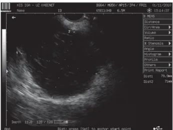
pro-file was normal. The postoperative course was uneventful, allowing the patient to void normally. The patient was stable at discharge and went home on the postoperative day 4. On the day 42 after the surgery the patient felt pain in the right hypogastric region of the abdomen. She was circulatory sta-ble, but an 8-cm mass was palpated to the right of the uterus, indicating paravesical haematoma. The patient did not report any use of aspirin, anticoagulants or risk medications. Ultra-sound scan revealed a hypoechogenic mass to the right of the urinary bladder of 80 u 71 mm (Figure 1). Surgical revision
was initiated, clotted blood was evacuated from the paravesi-cal space, just above the superior ramus of the right pubic bone, and a drain was inserted. No active bleeding site was found. The mesh could not be seen in the operative field, but was palpable in place. A total hysterectomy and a bilateral salpingoophorectomy were performed. Intravenous antibiot-ics (ceftriaxon 2 g per day and metronidazole 500 mg/8h) were administered for prophylactic reasons. Preoperative hemoglobin was 126 g/L, and 122 g/L after the procedure. A coagulation profile was repeatedly normal. The drain was removed on the second postoperative day. A repeat ultra-sound scan showed no fluid collection in the pelvis, and the clinical examination revealed appropriate position of the mesh without displacement. Further postoperative course was without complications, and the patient was discharged on the postoperative day 7. One year after the surgery the patient was continent, and without complaints. On pelvic exam, the anterior vaginal wall remained well supported with no recurrence of her symptoms.
Discussion
New approaches to pelvic organ prolapse have been evolving rapidly with little data reported on safety issues. As more novel approaches to pelvic organ prolapse are intro-duced, a new set of complications may evolve. Any new sur-gical procedure also raises the question of the associated anatomical risks, especially when a part of the procedure is performed blindly.
Volumen 70, Broj 7 VOJNOSANITETSKI PREGLED Strana 699
Spariý R, et al. Vojnosanit Pregl 2013; 70(7): 697–699.
The Prolift® procedure is a technique that incorporates mesh to compensate for areas of pelvic weakness. Operating in a highly vascularized, confined space, the surgeon may encounter complications that later may be challenging to manage. The placement of troacars near highly vascularized areas creates the possibility of haematoma formation as an operative complication. It is a well-known possibility in pel-vic organ prolapse surgery, and therefore, this complication is not unique to the isolated anterior mesh application. Re-ports from the manufacturer indicate a risk of 1.75% surgery-derived hematomas 6.
If abnormal abdominal pain appears after those proce-dures, it is necessary to perform both vaginal and ultrasound examination. Patients should also be carefully examined if other complications occur, like buttock or groin pain, signs of shock, brisk vaginal bleeding and urinary retention. Most of the haematomas are asymptomatic or produce only minor symptoms. These are haematomas with small volume and usually no intervention is necessary. In contrast to that, hae-matomas with a greater volume provoke moderate to severe problems, like abdominal pain, urge symptoms, dysuria or circulatory disturbances. In such cases operative manage-ment of the haematoma is indicated 5. Therefore, the decision if a patient should be treated conservatively or surgically must be made for each patient individually and with their consent.
A possible cause of haematoma formation following the insertion of an isolated anterior mesh is the injury of corona mortis, which refers to vascular connections between the external iliac and obturator systems in the obturator canal. These connections may be arterial, venous or both. It is known to hernia and orthopedic surgeons, but probably less well known to gynaecological surgeons 3. With the increase in surgery of the anterior pelvic ring, many investigators have started to study the detailed anatomy of the retropubic vascular system. The incidence of communicating vascular channels has been reported to be 83% 3. The name `crown of death` testifies to the importance of this feature, as signifi-cant haemorrhage may occur from its accidental lesion. This
bleeding could be either arterial or venous in nature. The slow onset and late presentation of the haematoma in the pre-sented case are not consistent with a corona mortis lesion. The presented patient had no recognized risk factors for postoperative bleeding, except the postoperative use of low-molecular-weight heparin, which might be a factor increas-ing the likelihood of postoperative haemorrhage. We assume venous source of haematoma. The haematoma was self-tamponaded and resolved after the surgical intervention.
This report illustrates that minimally invasive urogyne-cological procedures are not without significant complica-tions. Various mesh kits are being heavily marketed, but there is a concern regarding a lack of information on their safety and efficacy 6. With the number of mesh implants growing, there is always a concern for new complications that may arise whether from the kit itself or the use of the kit by those less experienced than the investigators who publish their data. It is equally important for the patient and the sur-geon to be aware of different complications that may occur with these new procedures. Surgeons should counsel women about the complications that may occur when using these procedures, particularly those related to the use of mesh and the possibility that their management might necessitate sur-gical intervention under general anaesthesia. It is also im-portant for surgeons to be familiar with effective interven-tions to manage them. Future research should be directed to-wards well-conducted and adequately powered randomized control trials, comparing vaginal mesh procedures with tra-ditional surgeries with respect to surgical complications rate and how surgeons should manage device-related complica-tions.
Conclusion
The prevalence of corona mortis and its anatomical re-lation to the pubic bone is important and should be consid-ered when introducing new surgical approaches in pelvic surgery, thus decreasing the incidence of surgical complica-tions and improving the results of operacomplica-tions.
R E F E R E N C E S
1. Argiroviý R, Gudoviý A, Baboviý I, Berisavac M. Transvaginal repair of genital prolapse with polypropylene mesh using a tension-free technique. Eur J Obstet Gynecol Reprod Biol 2010; 153(1): 104î7.
2. Fatton B, Ambalard J, Debodinance P, Cosson M, Jacquetin B. Transvaginal repair of genital prolapse: preliminary results of a new tension-free vaginal mesh (Prolift technique)-a case series multicentric study. Int Urogynecol J Pelvic Floor Dysfunct J 2007; 18(7): 743î52.
3. Ignjatovic I, Stosic D. Retrovesical haematoma after anterior Pro-lift procedure for cystocele correction. Int Urogynecol J Pelvic Floor Dysfunct 2007; 18(12): 1495î7.
4. LaSala CA, Schimpf MO. Occurrence of postoperative hema-tomas after prolapse repair using a mesh augmentation system. Obstet Gynecol 2007; 109(2 Pt2): 569î72.
5. Darmanis S, Lewis A, Mansoor A, Bircher M. Corona mortis: an anatomical study with clinical implications in approaches to the pelvis and acetabulum. Clin Anat 2007; 20(4): 433î9. 6. Hiltunen R, Nieminen K, Takala T, Heiskanen E, Merikari M,
Niemi K, et al. Low-weight polypropylene mesh for anterior vaginal wall prolapse: a randomized controlled trial. Obstet Gynecol 2007; 110(2 Pt2): 455î62.
7. von Theobald P. Place of mesh in vaginal surgery, including its removal and revision. Best Pract Res Clin Obstet Gynaecol 2011; 25(2): 197î203.
8. Simon M, Debodinance P. Vaginal prolapse using the Prolift™ kit: a registry of 100 successive cases. Eur J Obstet Gynecol Reprod Biol 2011; 158(1): 104î9.